
Anti-PDI, Mouse-Mono(RL90) | GeneTex International Corporation
掲載日情報:2026/06/03 現在Webページ番号:125355

GeneTex International CorporationのAnti-PDI, Mouse-Mono(RL90)商品情報ページです。
※本製品は研究用です。研究用以外には使用できません。
カートに商品を
追加しました。
追加しました。
価格
[在庫・価格 :2026年07月16日 18時55分現在]
※ 表示されている納期は弊社に在庫が無く、取り寄せた場合の納期目安となります。
| 詳細 | 商品名 |
|
文献数 | ||||||||||||||||||||||||||||||||||||||||||||||||||||||||||||||||||||||||||
|---|---|---|---|---|---|---|---|---|---|---|---|---|---|---|---|---|---|---|---|---|---|---|---|---|---|---|---|---|---|---|---|---|---|---|---|---|---|---|---|---|---|---|---|---|---|---|---|---|---|---|---|---|---|---|---|---|---|---|---|---|---|---|---|---|---|---|---|---|---|---|---|---|---|---|---|---|---|
|
Anti-PDI, Mouse-Mono(RL90) |
|
1 | |||||||||||||||||||||||||||||||||||||||||||||||||||||||||||||||||||||||||||
|
|||||||||||||||||||||||||||||||||||||||||||||||||||||||||||||||||||||||||||||


[在庫・価格 :2026年07月16日 18時55分現在]
※ 表示されている納期は弊社に在庫が無く、取り寄せた場合の納期目安となります。
Anti-PDI, Mouse-Mono(RL90)
文献数: 1
- 商品コード:GTX22792
- メーカー:GNT
- 包装:100μl
- 価格:¥103,000
- 在庫:1個
- 納期:2~3週間 ※※ 表示されている納期は弊社に在庫がなく、取り寄せた場合の目安納期となります。
- 法規制等:
| 説明文 | 別名:prolyl 4-hydroxylase subunit beta,PDI,PDIR クローン:RL90 Genbank No: 25506 |
||
|---|---|---|---|
| 法規制等 | |||
| 保存条件 | -20℃ | 法規備考 | |
| 抗原種 | Rat | 免疫動物 | Mouse |
| 交差性 | Dog/Hamster/Human/Mouse/Pig/Primate/Rat/Rhesus Monkey | 適用 | FCM,IC,IF,IHC,IP,Immunomicroscopy,Western Blot |
| 標識 | Unlabeled | 性状 | Protein A/G Affinity Purified |
| 吸収処理 | クラス | IgG | |
| クロナリティ | Monoclonal | フォーマット | |
| 掲載カタログ |
|
||
| 製品記事 | 小胞体ストレス関連抗体 小胞体(Endoplasmic Reticulum:ER)ストレス研究用製品特集 小胞体、ゴルジ体、リポソーム、ドラッグデリバリーシステムの研究に!細胞内輸送特集 脂質過酸化研究用製品特集 |
||
| 関連記事 | GeneTex社における抗体の品質管理 |
||
カートに商品を
追加しました。
追加しました。
画像
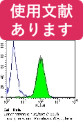
![GTX22792 FACS Image<br>Flow cytometry analysis of PDI in HeLa cells compared to an isotype control (blue). Cells were harvested, adjusted to a concentration of 1-5x10^6 cells/ml, fixed with 2% paraformaldehyde and washed with PBS. Cells were blocked with a 2% solution of BSA-PBS for 30 min at room temperature and incubated with PDI antibody [RL90] at a dilution of 1 ug/test for 60 min at room temperature. Cells were then incubated for 40 min at room temperature in the dark using a proper secondary antibody and re-suspended in PBS for FACS analysis.](/domestic/data/graphics/GNT/graphics/GTX22792_FACS.jpg)
GTX22792 FACS Image
Flow cytometry analysis of PDI in HeLa cells compared to an isotype control (blue). Cells were harvested, adjusted to a concentration of 1-5x10^6 cells/ml, fixed with 2% paraformaldehyde and washed with PBS. Cells were blocked with a 2% solution of BSA-PBS for 30 min at room temperature and incubated with PDI antibody [RL90] at a dilution of 1 ug/test for 60 min at room temperature. Cells were then incubated for 40 min at room temperature in the dark using a proper secondary antibody and re-suspended in PBS for FACS analysis.
Flow cytometry analysis of PDI in HeLa cells compared to an isotype control (blue). Cells were harvested, adjusted to a concentration of 1-5x10^6 cells/ml, fixed with 2% paraformaldehyde and washed with PBS. Cells were blocked with a 2% solution of BSA-PBS for 30 min at room temperature and incubated with PDI antibody [RL90] at a dilution of 1 ug/test for 60 min at room temperature. Cells were then incubated for 40 min at room temperature in the dark using a proper secondary antibody and re-suspended in PBS for FACS analysis.
![GTX22792 WB Image<br>Western blot analysis of PDI in 25 ug of HepG2, HeLa and NIH-3T3 cell lysates. Proteins were transferred to a PVDF membrane and blocked at 4C overnight. The membrane was probed with PDI antibody [RL90] at a dilution of 1:1000 overnight at 4C, washed in TBST, and probed with an HRP-conjugated secondary antibody. Chemiluminescent detection was performed.](/domestic/data/graphics/GNT/graphics/GTX22792_WB.jpg)
GTX22792 WB Image
Western blot analysis of PDI in 25 ug of HepG2, HeLa and NIH-3T3 cell lysates. Proteins were transferred to a PVDF membrane and blocked at 4C overnight. The membrane was probed with PDI antibody [RL90] at a dilution of 1:1000 overnight at 4C, washed in TBST, and probed with an HRP-conjugated secondary antibody. Chemiluminescent detection was performed.
Western blot analysis of PDI in 25 ug of HepG2, HeLa and NIH-3T3 cell lysates. Proteins were transferred to a PVDF membrane and blocked at 4C overnight. The membrane was probed with PDI antibody [RL90] at a dilution of 1:1000 overnight at 4C, washed in TBST, and probed with an HRP-conjugated secondary antibody. Chemiluminescent detection was performed.
![GTX22792 ICC/IF Image<br>Immunofluorescent analysis of Phalloidin (blue) and PDI (green) in NIH 3T3 cells. Formalin fixed cells were permeabilized with 0.1% Triton X-100 in PBS for 10 minutes at room temperature and blocked with 2% BSA in PBS + 0.1% Triton X-100 for 30 minutes at room temperature. Cells were probed with PDI antibody [RL90] at a dilution of 1:75 for at least 1 hour at room temperature, washed with PBS, and incubated with a proper secondary antibody. Actin was stained with Dylight 350 Phalloidin and nuclei (red) were stained with DRAQ5. Images were taken at 20X magnification.](/domestic/data/graphics/GNT/graphics/GTX22792_ICC%20IF.jpg)
GTX22792 ICC/IF Image
Immunofluorescent analysis of Phalloidin (blue) and PDI (green) in NIH 3T3 cells. Formalin fixed cells were permeabilized with 0.1% Triton X-100 in PBS for 10 minutes at room temperature and blocked with 2% BSA in PBS + 0.1% Triton X-100 for 30 minutes at room temperature. Cells were probed with PDI antibody [RL90] at a dilution of 1:75 for at least 1 hour at room temperature, washed with PBS, and incubated with a proper secondary antibody. Actin was stained with Dylight 350 Phalloidin and nuclei (red) were stained with DRAQ5. Images were taken at 20X magnification.
Immunofluorescent analysis of Phalloidin (blue) and PDI (green) in NIH 3T3 cells. Formalin fixed cells were permeabilized with 0.1% Triton X-100 in PBS for 10 minutes at room temperature and blocked with 2% BSA in PBS + 0.1% Triton X-100 for 30 minutes at room temperature. Cells were probed with PDI antibody [RL90] at a dilution of 1:75 for at least 1 hour at room temperature, washed with PBS, and incubated with a proper secondary antibody. Actin was stained with Dylight 350 Phalloidin and nuclei (red) were stained with DRAQ5. Images were taken at 20X magnification.
カートに商品を
追加しました。
追加しました。
商品情報
| 抗原動物 | Rat |
|---|---|
| 交差性 | Dog/Hamster/Human/Mouse/Pig/Primate/Rat/Rhesus Monkey |
| 免疫動物 | Mouse |
| 性状 | Protein A/G Affinity Purified |
| 適用 | FCM, IC, IF, IHC, IP, Immunomicroscopy, Western Blot |
| クラス | IgG |
| クローン名 | RL90 |
| 標識 | Unlabeled |
| クロナリティ | Monoclonal |
| 別名 | prolyl 4-hydroxylase subunit beta, PDI, PDIR |
| Genbank No | 25506 |
| データシート | データシート |
| メーカーサイト | メーカーサイト |
| 使用文献 | 使用文献 |
| 保存条件 | -20℃ |
カートに商品を
追加しました。
追加しました。
製品情報は掲載時点のものですが、価格表内の価格については随時最新のものに更新されます。お問い合わせいただくタイミングにより製品情報・価格などは変更されている場合があります。
表示価格に、消費税等は含まれていません。一部価格が予告なく変更される場合がありますので、あらかじめご了承下さい。