
Anti-GPC1, Rabbit-Poly | GeneTex International Corporation
掲載日情報:2026/06/03 現在Webページ番号:547430

GeneTex International CorporationのAnti-GPC1, Rabbit-Poly商品情報ページです。
※本製品は研究用です。研究用以外には使用できません。
カートに商品を
追加しました。
追加しました。
価格
[在庫・価格 :2026年07月17日 00時01分現在]
※ 表示されている納期は弊社に在庫が無く、取り寄せた場合の納期目安となります。
| 詳細 | 商品名 |
|
文献数 | ||||||||||||||||||||||||||||||||||||||||||||||||||||||||||||||||||||||||||||||||||
|---|---|---|---|---|---|---|---|---|---|---|---|---|---|---|---|---|---|---|---|---|---|---|---|---|---|---|---|---|---|---|---|---|---|---|---|---|---|---|---|---|---|---|---|---|---|---|---|---|---|---|---|---|---|---|---|---|---|---|---|---|---|---|---|---|---|---|---|---|---|---|---|---|---|---|---|---|---|---|---|---|---|---|---|---|---|
|
Anti-GPC1, Rabbit-Poly |
|
11 | |||||||||||||||||||||||||||||||||||||||||||||||||||||||||||||||||||||||||||||||||||
|
|||||||||||||||||||||||||||||||||||||||||||||||||||||||||||||||||||||||||||||||||||||


[在庫・価格 :2026年07月17日 00時01分現在]
※ 表示されている納期は弊社に在庫が無く、取り寄せた場合の納期目安となります。
Anti-GPC1, Rabbit-Poly
文献数: 11
- 商品コード:GTX104557
- メーカー:GNT
- 包装:25μl
- 価格:¥30,000
- 在庫:1個
- 納期:10日程度 ※※ 表示されている納期は弊社に在庫がなく、取り寄せた場合の目安納期となります。
- 法規制等:
| 説明文 | レビューあり。KO/KDバリデーション済み抗体。 別名:glypican 1,glypican Genbank No: 2817 |
||||||
|---|---|---|---|---|---|---|---|
| 別包装品 | 別包装品あり | ||||||
| 法規制等 | |||||||
| 保存条件 | -20℃ | 法規備考 | |||||
| 抗原種 | Human | 免疫動物 | Rabbit | ||||
| 交差性 | Human/Mouse/Rat | 適用 | ELISA,FCM,IC,IF,IHC,IP,Western Blot | ||||
| 標識 | Unlabeled | 性状 | Purified | ||||
| 吸収処理 | クラス | IgG | |||||
| クロナリティ | Polyclonal | フォーマット | |||||
| 掲載カタログ |
|
||||||
| 製品記事 | Glypican-1 Antibody GeneTex社 抗腫瘍マーカー抗体 -膵臓がん- GeneTex社 がん研究用抗体特集 使いっきり抗体 |
||||||
| 関連記事 | GeneTex社における抗体の品質管理 |
||||||
カートに商品を
追加しました。
追加しました。
ラインナップ
カートに商品を
追加しました。
追加しました。
画像
![GTX104557 ICC/IF Image<br>Glypican 1 antibody [N3C3] detects Glypican 1 protein at cytoplasm by immunofluorescent analysis.<br>Sample: MCF7 cells were fixed in ice-cold MeOH for 5 min.<br>Green: Glypican 1 protein stained by Glypican 1 antibody [N3C3] (GTX104557) diluted at 1:500.<br>Blue: Hoechst 33342 staining.<br>](/domestic/data/graphics/GNT/graphics/GTX104557_42697_20170524_IFA.jpg)
GTX104557 ICC/IF Image
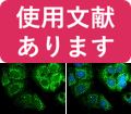
Glypican 1 antibody [N3C3] detects Glypican 1 protein at cytoplasm by immunofluorescent analysis.
Sample: MCF7 cells were fixed in ice-cold MeOH for 5 min.
Green: Glypican 1 protein stained by Glypican 1 antibody [N3C3] (GTX104557) diluted at 1:500.
Blue: Hoechst 33342 staining.
Glypican 1 antibody [N3C3] detects Glypican 1 protein at cytoplasm by immunofluorescent analysis.
Sample: MCF7 cells were fixed in ice-cold MeOH for 5 min.
Green: Glypican 1 protein stained by Glypican 1 antibody [N3C3] (GTX104557) diluted at 1:500.
Blue: Hoechst 33342 staining.
![GTX104557 WB Image<br>Various whole cell extracts (30 ug) were separated by 7.5% SDS-PAGE, and the membranes were blotted with Glypican 1 antibody [N3C3] (GTX104557) diluted at 1:500 and competitor's antibody (sc-365000) diluted at 1:500. The HRP-conjugated anti-rabbit IgG antibody (GTX213110-01) was used to detect the primary antibody.</br>](/domestic/data/graphics/GNT/graphics/GTX104557_42326_20171110_WB_competitor_watermark.jpg)
GTX104557 WB Image
Various whole cell extracts (30 ug) were separated by 7.5% SDS-PAGE, and the membranes were blotted with Glypican 1 antibody [N3C3] (GTX104557) diluted at 1:500 and competitor's antibody (sc-365000) diluted at 1:500. The HRP-conjugated anti-rabbit IgG antibody (GTX213110-01) was used to detect the primary antibody.
Various whole cell extracts (30 ug) were separated by 7.5% SDS-PAGE, and the membranes were blotted with Glypican 1 antibody [N3C3] (GTX104557) diluted at 1:500 and competitor's antibody (sc-365000) diluted at 1:500. The HRP-conjugated anti-rabbit IgG antibody (GTX213110-01) was used to detect the primary antibody.
![GTX104557 IHC-P Image<br>Immunohistochemical microscopy analysis of paraffin-embedded human normal pancreas (left) or pancreatic adenocarcinoma (grade II) (right) tissues using Glypican-1 antibody [N3C3] (GTX104557) at a 1:250 dilution.](/domestic/data/graphics/GNT/graphics/GTX104557_42326_20150505_IHC-P.jpg)
GTX104557 IHC-P Image
Immunohistochemical microscopy analysis of paraffin-embedded human normal pancreas (left) or pancreatic adenocarcinoma (grade II) (right) tissues using Glypican-1 antibody [N3C3] (GTX104557) at a 1:250 dilution.
Immunohistochemical microscopy analysis of paraffin-embedded human normal pancreas (left) or pancreatic adenocarcinoma (grade II) (right) tissues using Glypican-1 antibody [N3C3] (GTX104557) at a 1:250 dilution.
![GTX104557 WB Image<br>Non-transfected (?) and transfected (+) MCF-7 whole cell extracts (50 ug) were separated by 7.5% SDS-PAGE, and the membrane was blotted with Glypican 1 antibody [N3C3] (GTX104557) diluted at 1:1500. The HRP-conjugated anti-rabbit IgG antibody (GTX213110-01) was used to detect the primary antibody.](/domestic/data/graphics/GNT/graphics/GTX104557_42326_20160509_WB_shRNA_watermark.jpg)
GTX104557 WB Image
Non-transfected (?) and transfected (+) MCF-7 whole cell extracts (50 ug) were separated by 7.5% SDS-PAGE, and the membrane was blotted with Glypican 1 antibody [N3C3] (GTX104557) diluted at 1:1500. The HRP-conjugated anti-rabbit IgG antibody (GTX213110-01) was used to detect the primary antibody.
Non-transfected (?) and transfected (+) MCF-7 whole cell extracts (50 ug) were separated by 7.5% SDS-PAGE, and the membrane was blotted with Glypican 1 antibody [N3C3] (GTX104557) diluted at 1:1500. The HRP-conjugated anti-rabbit IgG antibody (GTX213110-01) was used to detect the primary antibody.
![GTX104557 WB Image<br>Untreated (?) and treated (+) MDA-MB-231 whole cell extracts (30 ug) were separated by 7.5% SDS-PAGE, and the membrane was blotted with Glypican 1 antibody [N3C3] (GTX104557) diluted at 1:1500. The HRP-conjugated anti-rabbit IgG antibody (GTX213110-01) was used to detect the primary antibody.](/domestic/data/graphics/GNT/graphics/GTX104557_42326_20160509_WB_Tunicamycin.jpg)
GTX104557 WB Image
Untreated (?) and treated (+) MDA-MB-231 whole cell extracts (30 ug) were separated by 7.5% SDS-PAGE, and the membrane was blotted with Glypican 1 antibody [N3C3] (GTX104557) diluted at 1:1500. The HRP-conjugated anti-rabbit IgG antibody (GTX213110-01) was used to detect the primary antibody.
Untreated (?) and treated (+) MDA-MB-231 whole cell extracts (30 ug) were separated by 7.5% SDS-PAGE, and the membrane was blotted with Glypican 1 antibody [N3C3] (GTX104557) diluted at 1:1500. The HRP-conjugated anti-rabbit IgG antibody (GTX213110-01) was used to detect the primary antibody.
![GTX104557 WB Image<br>U87-MG whole cell and membrane extracts (30 ug) were separated by 7.5% SDS-PAGE, and the membrane was blotted with Glypican 1 antibody [N3C3] (GTX104557) diluted at 1:1500. The HRP-conjugated anti-rabbit IgG antibody (GTX213110-01) was used to detect the primary antibody.](/domestic/data/graphics/GNT/graphics/GTX104557_42326_20160509_WB_Fraction.jpg)
GTX104557 WB Image
U87-MG whole cell and membrane extracts (30 ug) were separated by 7.5% SDS-PAGE, and the membrane was blotted with Glypican 1 antibody [N3C3] (GTX104557) diluted at 1:1500. The HRP-conjugated anti-rabbit IgG antibody (GTX213110-01) was used to detect the primary antibody.
U87-MG whole cell and membrane extracts (30 ug) were separated by 7.5% SDS-PAGE, and the membrane was blotted with Glypican 1 antibody [N3C3] (GTX104557) diluted at 1:1500. The HRP-conjugated anti-rabbit IgG antibody (GTX213110-01) was used to detect the primary antibody.
![GTX104557 WB Image<br>U87-MG whole cell extract and conditioned medium (30 ug) were separated by 7.5% SDS-PAGE, and the membrane was blotted with Glypican 1 antibody [N3C3] (GTX104557) diluted at 1:1500. The HRP-conjugated anti-rabbit IgG antibody (GTX213110-01) was used to detect the primary antibody.](/domestic/data/graphics/GNT/graphics/GTX104557_42326_20160509_WB_Fraction_2.jpg)
GTX104557 WB Image
U87-MG whole cell extract and conditioned medium (30 ug) were separated by 7.5% SDS-PAGE, and the membrane was blotted with Glypican 1 antibody [N3C3] (GTX104557) diluted at 1:1500. The HRP-conjugated anti-rabbit IgG antibody (GTX213110-01) was used to detect the primary antibody.
U87-MG whole cell extract and conditioned medium (30 ug) were separated by 7.5% SDS-PAGE, and the membrane was blotted with Glypican 1 antibody [N3C3] (GTX104557) diluted at 1:1500. The HRP-conjugated anti-rabbit IgG antibody (GTX213110-01) was used to detect the primary antibody.
![GTX104557 WB Image<br>Mouse tissue extracts (50 ug) were separated by 7.5% SDS-PAGE, and the membrane was blotted with Glypican 1 antibody [N3C3] (GTX104557) diluted at 1:1500. The HRP-conjugated anti-rabbit IgG antibody (GTX213110-01) was used to detect the primary antibody.](/domestic/data/graphics/GNT/graphics/GTX104557_42326_20160509_WB_M_brain.jpg)
GTX104557 WB Image
Mouse tissue extracts (50 ug) were separated by 7.5% SDS-PAGE, and the membrane was blotted with Glypican 1 antibody [N3C3] (GTX104557) diluted at 1:1500. The HRP-conjugated anti-rabbit IgG antibody (GTX213110-01) was used to detect the primary antibody.
Mouse tissue extracts (50 ug) were separated by 7.5% SDS-PAGE, and the membrane was blotted with Glypican 1 antibody [N3C3] (GTX104557) diluted at 1:1500. The HRP-conjugated anti-rabbit IgG antibody (GTX213110-01) was used to detect the primary antibody.
GTX104557 ELISA Image
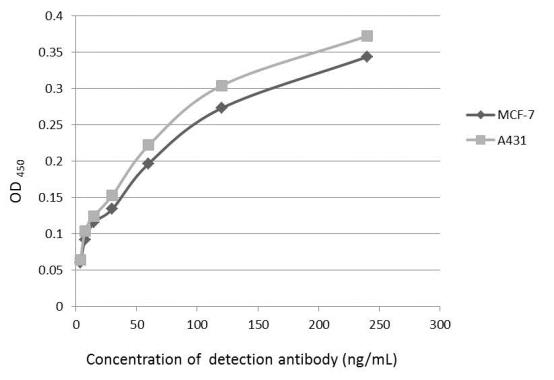
An ELISA plate is coated with MCF-7 and A431 cells. The coated cells are detected with Glypican 1 antibody (GTX104557) at concentration ranged from 7.5 to 240 ng/mL.
An ELISA plate is coated with MCF-7 and A431 cells. The coated cells are detected with Glypican 1 antibody (GTX104557) at concentration ranged from 7.5 to 240 ng/mL.
![GTX104557 WB Image<br>Various whole cell extracts (30 ug) were separated by 7.5% SDS-PAGE, and the membrane was blotted with Glypican 1 antibody [N3C3] (GTX104557) diluted at 1:1500.](/domestic/data/graphics/GNT/graphics/GTX104557_42697_20161215_WB.jpg)
GTX104557 WB Image
Various whole cell extracts (30 ug) were separated by 7.5% SDS-PAGE, and the membrane was blotted with Glypican 1 antibody [N3C3] (GTX104557) diluted at 1:1500.
Various whole cell extracts (30 ug) were separated by 7.5% SDS-PAGE, and the membrane was blotted with Glypican 1 antibody [N3C3] (GTX104557) diluted at 1:1500.
![GTX104557 WB Image<br>Untreated (?) and treated (+) A431 whole cell extracts (30 ug) were separated by 7.5% SDS-PAGE, and the membrane was blotted with Glypican 1 antibody [N3C3] (GTX104557) diluted at 1:3000. The HRP-conjugated anti-rabbit IgG antibody (GTX213110-01) was used to detect the primary antibody.](/domestic/data/graphics/GNT/graphics/GTX104557_42326_20160728_WB_Benzyl%20glucosinolate_Tunicamycin.jpg)
GTX104557 WB Image
Untreated (?) and treated (+) A431 whole cell extracts (30 ug) were separated by 7.5% SDS-PAGE, and the membrane was blotted with Glypican 1 antibody [N3C3] (GTX104557) diluted at 1:3000. The HRP-conjugated anti-rabbit IgG antibody (GTX213110-01) was used to detect the primary antibody.
Untreated (?) and treated (+) A431 whole cell extracts (30 ug) were separated by 7.5% SDS-PAGE, and the membrane was blotted with Glypican 1 antibody [N3C3] (GTX104557) diluted at 1:3000. The HRP-conjugated anti-rabbit IgG antibody (GTX213110-01) was used to detect the primary antibody.
![GTX104557 FACS Image<br>Glypican 1 antibody [N3C3] (GTX104557) detects Glypican 1 protein by flow cytometry analysis.<br>Sample: A431 cell.<br>Black: Unlabelled sample was used as a control.<br>Red: Glypican 1 antibody [N3C3] (GTX104557) dilution: 1:100.<br>Acquisition of 20,000 events were collected using a Dylight 488-conjugated secondary antibody for FACS analysis.](/domestic/data/graphics/GNT/graphics/GTX104557_42326_20160407_FACS.jpg)
GTX104557 FACS Image
Glypican 1 antibody [N3C3] (GTX104557) detects Glypican 1 protein by flow cytometry analysis.
Sample: A431 cell.
Black: Unlabelled sample was used as a control.
Red: Glypican 1 antibody [N3C3] (GTX104557) dilution: 1:100.
Acquisition of 20,000 events were collected using a Dylight 488-conjugated secondary antibody for FACS analysis.
![GTX104557 IP Image<br>Immunoprecipitation of Glypican 1 protein from MCF-7 whole cell extracts using 5 ug of Glypican 1 antibody [N3C3] (GTX104557).<br>Western blot analysis was performed using Glypican 1 antibody [N3C3] (GTX104557).<br>EasyBlot anti-Rabbit IgG (GTX221666-01) was used as a secondary reagent.</br></br></br>](/domestic/data/graphics/GNT/graphics/GTX104557_42326_20160429_IP.jpg)
GTX104557 IP Image
Immunoprecipitation of Glypican 1 protein from MCF-7 whole cell extracts using 5 ug of Glypican 1 antibody [N3C3] (GTX104557).
Western blot analysis was performed using Glypican 1 antibody [N3C3] (GTX104557).
EasyBlot anti-Rabbit IgG (GTX221666-01) was used as a secondary reagent.
Immunoprecipitation of Glypican 1 protein from MCF-7 whole cell extracts using 5 ug of Glypican 1 antibody [N3C3] (GTX104557).
Western blot analysis was performed using Glypican 1 antibody [N3C3] (GTX104557).
EasyBlot anti-Rabbit IgG (GTX221666-01) was used as a secondary reagent.
![GTX104557 WB Image<br>Untreated (?) and treated (+) A431 whole cell extracts (30 ug) were separated by 7.5% SDS-PAGE, and the membrane was blotted with Glypican 1 antibody [N3C3] (GTX104557) diluted at 1:500. The HRP-conjugated anti-rabbit IgG antibody (GTX213110-01) was used to detect the primary antibody.](/domestic/data/graphics/GNT/graphics/GTX104557_42326_20160509_WB_PNGase%20F.jpg)
GTX104557 WB Image
Untreated (?) and treated (+) A431 whole cell extracts (30 ug) were separated by 7.5% SDS-PAGE, and the membrane was blotted with Glypican 1 antibody [N3C3] (GTX104557) diluted at 1:500. The HRP-conjugated anti-rabbit IgG antibody (GTX213110-01) was used to detect the primary antibody.
Untreated (?) and treated (+) A431 whole cell extracts (30 ug) were separated by 7.5% SDS-PAGE, and the membrane was blotted with Glypican 1 antibody [N3C3] (GTX104557) diluted at 1:500. The HRP-conjugated anti-rabbit IgG antibody (GTX213110-01) was used to detect the primary antibody.
カートに商品を
追加しました。
追加しました。
商品情報
| 商品説明 | レビューあり。KO/KDバリデーション済み抗体 |
|---|---|
| 抗原動物 | Human |
| 交差性 | Human/Mouse/Rat |
| 免疫動物 | Rabbit |
| 性状 | Purified |
| 適用 | ELISA, FCM, IC, IF, IHC, IP, Western Blot |
| クラス | IgG |
| 標識 | Unlabeled |
| クロナリティ | Polyclonal |
| 別名 | glypican 1, glypican |
| Genbank No | 2817 |
| データシート | データシート |
| メーカーサイト | メーカーサイト |
| 使用文献 | 使用文献 |
| 保存条件 | -20℃ |
カートに商品を
追加しました。
追加しました。
製品情報は掲載時点のものですが、価格表内の価格については随時最新のものに更新されます。お問い合わせいただくタイミングにより製品情報・価格などは変更されている場合があります。
表示価格に、消費税等は含まれていません。一部価格が予告なく変更される場合がありますので、あらかじめご了承下さい。