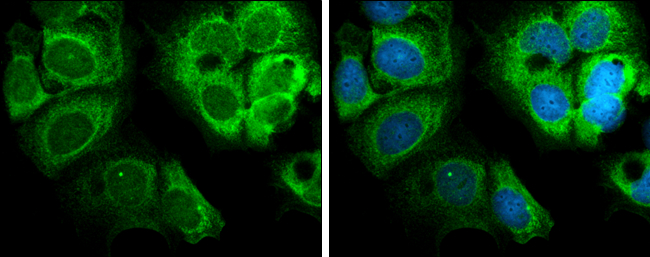
GTX104557 ICC/IF Image
Glypican 1 antibody [N3C3] detects Glypican 1 protein at cytoplasm by immunofluorescent analysis.
Sample: MCF7 cells were fixed in ice-cold MeOH for 5 min.
Green: Glypican 1 protein stained by Glypican 1 antibody [N3C3] (GTX104557) diluted at 1:500.
Blue: Hoechst 33342 staining.
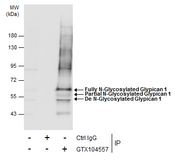
GTX104557 IP Image
Immunoprecipitation of Glypican 1 protein from MCF-7 whole cell extracts using 5 ug of Glypican 1 antibody [N3C3] (GTX104557).
Western blot analysis was performed using Glypican 1 antibody [N3C3] (GTX104557).
EasyBlot anti-Rabbit IgG (GTX221666-01) was used as a secondary reagent.
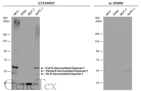
GTX104557 WB Image
Various whole cell extracts (30 ug) were separated by 7.5% SDS-PAGE, and the membranes were blotted with Glypican 1 antibody [N3C3] (GTX104557) diluted at 1:500 and competitor's antibody (sc-365000) diluted at 1:500. The HRP-conjugated anti-rabbit IgG antibody (GTX213110-01) was used to detect the primary antibody.
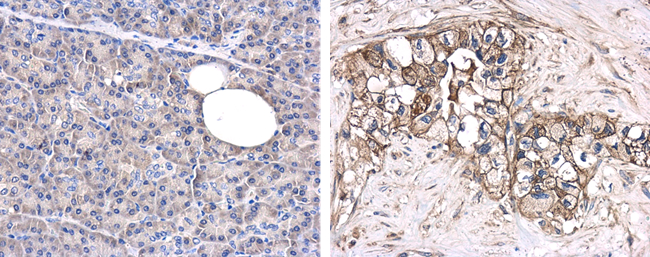
GTX104557 IHC-P Image
Immunohistochemical microscopy analysis of paraffin-embedded human normal pancreas (left) or pancreatic adenocarcinoma (grade II) (right) tissues using Glypican-1 antibody [N3C3] (GTX104557) at a 1:250 dilution.
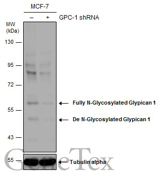
GTX104557 WB Image
Non-transfected (?) and transfected (+) MCF-7 whole cell extracts (50 ug) were separated by 7.5% SDS-PAGE, and the membrane was blotted with Glypican 1 antibody [N3C3] (GTX104557) diluted at 1:1500. The HRP-conjugated anti-rabbit IgG antibody (GTX213110-01) was used to detect the primary antibody.

GTX104557 WB Image
Untreated (?) and treated (+) A431 whole cell extracts (30 ug) were separated by 7.5% SDS-PAGE, and the membrane was blotted with Glypican 1 antibody [N3C3] (GTX104557) diluted at 1:500. The HRP-conjugated anti-rabbit IgG antibody (GTX213110-01) was used to detect the primary antibody.
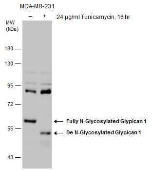
GTX104557 WB Image
Untreated (?) and treated (+) MDA-MB-231 whole cell extracts (30 ug) were separated by 7.5% SDS-PAGE, and the membrane was blotted with Glypican 1 antibody [N3C3] (GTX104557) diluted at 1:1500. The HRP-conjugated anti-rabbit IgG antibody (GTX213110-01) was used to detect the primary antibody.
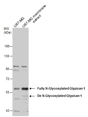
GTX104557 WB Image
U87-MG whole cell and membrane extracts (30 ug) were separated by 7.5% SDS-PAGE, and the membrane was blotted with Glypican 1 antibody [N3C3] (GTX104557) diluted at 1:1500. The HRP-conjugated anti-rabbit IgG antibody (GTX213110-01) was used to detect the primary antibody.
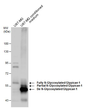
GTX104557 WB Image
U87-MG whole cell extract and conditioned medium (30 ug) were separated by 7.5% SDS-PAGE, and the membrane was blotted with Glypican 1 antibody [N3C3] (GTX104557) diluted at 1:1500. The HRP-conjugated anti-rabbit IgG antibody (GTX213110-01) was used to detect the primary antibody.
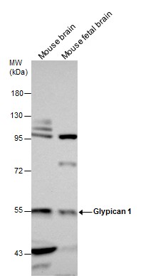
GTX104557 WB Image
Mouse tissue extracts (50 ug) were separated by 7.5% SDS-PAGE, and the membrane was blotted with Glypican 1 antibody [N3C3] (GTX104557) diluted at 1:1500. The HRP-conjugated anti-rabbit IgG antibody (GTX213110-01) was used to detect the primary antibody.
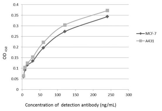
GTX104557 ELISA Image
An ELISA plate is coated with MCF-7 and A431 cells. The coated cells are detected with Glypican 1 antibody (GTX104557) at concentration ranged from 7.5 to 240 ng/mL.
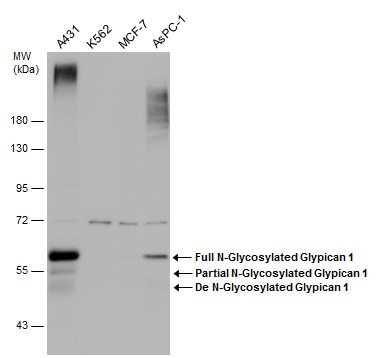
GTX104557 WB Image
Various whole cell extracts (30 ug) were separated by 7.5% SDS-PAGE, and the membrane was blotted with Glypican 1 antibody [N3C3] (GTX104557) diluted at 1:1500.

GTX104557 WB Image
Untreated (?) and treated (+) A431 whole cell extracts (30 ug) were separated by 7.5% SDS-PAGE, and the membrane was blotted with Glypican 1 antibody [N3C3] (GTX104557) diluted at 1:3000. The HRP-conjugated anti-rabbit IgG antibody (GTX213110-01) was used to detect the primary antibody.
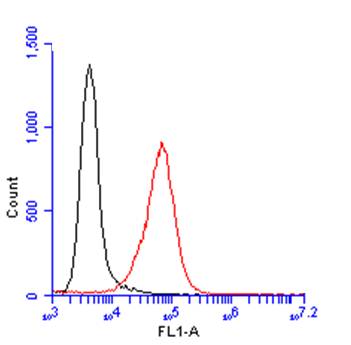
GTX104557 FACS Image
Glypican 1 antibody [N3C3] (GTX104557) detects Glypican 1 protein by flow cytometry analysis.
Sample: A431 cell.
Black: Unlabelled sample was used as a control.
Red: Glypican 1 antibody [N3C3] (GTX104557) dilution: 1:100.
Acquisition of 20,000 events were collected using a Dylight 488-conjugated secondary antibody for FACS analysis.