
Anti-PARP1, Rabbit-Poly | GeneTex International Corporation
掲載日情報:2026/06/03 現在Webページ番号:35176

GeneTex International CorporationのAnti-PARP1, Rabbit-Poly商品情報ページです。
※本製品は研究用です。研究用以外には使用できません。
カートに商品を
追加しました。
追加しました。
価格
[在庫・価格 :2026年06月09日 09時55分現在]
※ 表示されている納期は弊社に在庫が無く、取り寄せた場合の納期目安となります。
| 詳細 | 商品名 |
|
文献数 | ||||||||||||||||||||||||||||||||||||||||||||||||||||||||||||||||||||||||||||||||||
|---|---|---|---|---|---|---|---|---|---|---|---|---|---|---|---|---|---|---|---|---|---|---|---|---|---|---|---|---|---|---|---|---|---|---|---|---|---|---|---|---|---|---|---|---|---|---|---|---|---|---|---|---|---|---|---|---|---|---|---|---|---|---|---|---|---|---|---|---|---|---|---|---|---|---|---|---|---|---|---|---|---|---|---|---|---|
|
Anti-PARP1, Rabbit-Poly |
|
80 | |||||||||||||||||||||||||||||||||||||||||||||||||||||||||||||||||||||||||||||||||||
|
|||||||||||||||||||||||||||||||||||||||||||||||||||||||||||||||||||||||||||||||||||||


[在庫・価格 :2026年06月09日 09時55分現在]
※ 表示されている納期は弊社に在庫が無く、取り寄せた場合の納期目安となります。
Anti-PARP1, Rabbit-Poly
文献数: 80
- 商品コード:GTX100573
- メーカー:GNT
- 包装:100μl
- 価格:¥85,000
- 在庫:1個
- 納期:10日程度 ※※ 表示されている納期は弊社に在庫がなく、取り寄せた場合の目安納期となります。
- 法規制等:
| 説明文 | レビューあり。KO/KDバリデーション済み抗体。抗原:aa 151~385 別名:poly(ADP-ribose) polymerase 1,ADPRT,ADPRT 1,ADPRT1,ARTD1,PARP,PARP-1,PPOL,pADPRT-1 Genbank No: 142 |
||||||
|---|---|---|---|---|---|---|---|
| 別包装品 | 別包装品あり | ||||||
| 法規制等 | |||||||
| 保存条件 | -20℃ | 法規備考 | |||||
| 抗原種 | Human | 免疫動物 | Rabbit | ||||
| 交差性 | Human/Mouse/Rat | 適用 | ChIP,IC,IF,IHC,IP,Western Blot | ||||
| 標識 | Unlabeled | 性状 | Purified | ||||
| 吸収処理 | クラス | IgG | |||||
| クロナリティ | Polyclonal | フォーマット | |||||
| 掲載カタログ |
|
||||||
| 製品記事 | GeneTex社 DNA損傷/修復関連抗体 細胞周期・微小管研究用製品特集 |
||||||
| 関連記事 | GeneTex社における抗体の品質管理 |
||||||
カートに商品を
追加しました。
追加しました。
ラインナップ
カートに商品を
追加しました。
追加しました。
画像
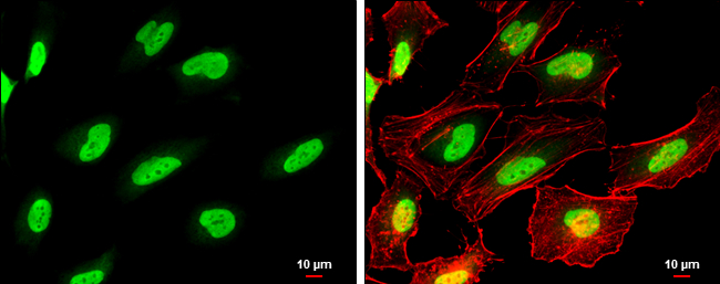
GTX100573 ICC/IF Image
PARP antibody detects PARP protein at nucleus by immunofluorescent analysis.
Sample: HeLa cells were fixed in 4% paraformaldehyde at RT for 15 min.
Green: PARP protein stained by PARP antibody (GTX100573) diluted at 1:500.
Red: Phalloidin, a cytoskeleton marker, diluted at 1:100.
Scale bar = 10 um.
PARP antibody detects PARP protein at nucleus by immunofluorescent analysis.
Sample: HeLa cells were fixed in 4% paraformaldehyde at RT for 15 min.
Green: PARP protein stained by PARP antibody (GTX100573) diluted at 1:500.
Red: Phalloidin, a cytoskeleton marker, diluted at 1:100.
Scale bar = 10 um.
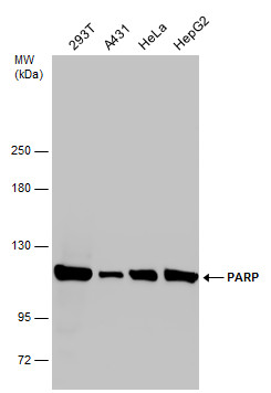
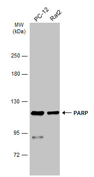
GTX100573 WB Image
Various whole cell extracts (30 ug) were separated by 5% SDS-PAGE, and the membrane was blotted with PARP antibody (GTX100573) diluted at 1:2000.
Various whole cell extracts (30 ug) were separated by 5% SDS-PAGE, and the membrane was blotted with PARP antibody (GTX100573) diluted at 1:2000.
GTX100573 WB Image
Untreated (?) and treated (+) HeLa whole cell extracts (30 ug) were separated by 7.5% SDS-PAGE, and the membrane was blotted with PARP antibody (GTX100573) diluted at 1:2000. The HRP-conjugated anti-rabbit IgG antibody (GTX213110-01) was used to detect the primary antibody.
Untreated (?) and treated (+) HeLa whole cell extracts (30 ug) were separated by 7.5% SDS-PAGE, and the membrane was blotted with PARP antibody (GTX100573) diluted at 1:2000. The HRP-conjugated anti-rabbit IgG antibody (GTX213110-01) was used to detect the primary antibody.
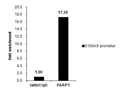
GTX100573 ChIP assay Image
Cross-linked ChIP was performed with Raji chromatin extract and 5 ug of either control rabbit IgG or anti-PARP1 antibody. The precipitated DNA was detected by PCR with primer set targeting to S100A9 promoter.
Cross-linked ChIP was performed with Raji chromatin extract and 5 ug of either control rabbit IgG or anti-PARP1 antibody. The precipitated DNA was detected by PCR with primer set targeting to S100A9 promoter.
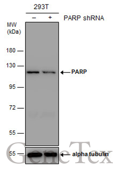
GTX100573 WB Image
Non-transfected (?) and transfected (+) 293T whole cell extracts (30 ug) were separated by 7.5% SDS-PAGE, and the membrane was blotted with PARP antibody (GTX100573) diluted at 1:2000. The HRP-conjugated anti-rabbit IgG antibody (GTX213110-01) was used to detect the primary antibody.
Non-transfected (?) and transfected (+) 293T whole cell extracts (30 ug) were separated by 7.5% SDS-PAGE, and the membrane was blotted with PARP antibody (GTX100573) diluted at 1:2000. The HRP-conjugated anti-rabbit IgG antibody (GTX213110-01) was used to detect the primary antibody.
GTX100573 WB Image
Various whole cell extracts (30 ug) were separated by 5% SDS-PAGE, and the membrane was blotted with PARP antibody (GTX100573) diluted at 1:1000. The HRP-conjugated anti-rabbit IgG antibody (GTX213110-01) was used to detect the primary antibody.
Various whole cell extracts (30 ug) were separated by 5% SDS-PAGE, and the membrane was blotted with PARP antibody (GTX100573) diluted at 1:1000. The HRP-conjugated anti-rabbit IgG antibody (GTX213110-01) was used to detect the primary antibody.
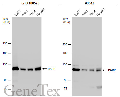
GTX100573 WB Image
Various whole cell extracts (30 ug) were separated by 5% SDS-PAGE, and the membranes were blotted with PARP antibody (GTX100573) diluted at 1:2000 and competitor's antibody (#9542) diluted at 1:500. The HRP-conjugated anti-rabbit IgG antibody (GTX213110-01) was used to detect the primary antibody.
*The competitor is not affiliated with GeneTex and does not endorse this product.
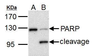
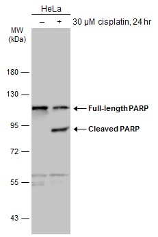
GTX100573 WB Image
PARP1 antibody detects PARP1 protein by western blot analysis.
A. 30 ug HCT116 whole cell lysate/extract (untreated)
B. 30 ug HCT116 whole cell lysate/extract (30 uM cisplatin treatment for 24hr)
7.5% SDS-PAGE
PARP1 antibody (GTX100573) dilution: 1:1000
The HRP-conjugated anti-rabbit IgG antibody (GTX213110-01) was used to detect the primary antibody.
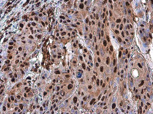
GTX100573 IHC-P Image
PARP antibody detects PARP protein at nucleus in human oral carcinoma by immunohistochemical analysis.
Sample: Paraffin-embedded human oral carcinoma.
PARP antibody (GTX100573) diluted at 1:500.
PARP antibody detects PARP protein at nucleus in human oral carcinoma by immunohistochemical analysis.
Sample: Paraffin-embedded human oral carcinoma.
PARP antibody (GTX100573) diluted at 1:500.
カートに商品を
追加しました。
追加しました。
商品情報
| 商品説明 | レビューあり。KO/KDバリデーション済み抗体 |
|---|---|
| 抗原 | aa 151~385 |
| 抗原動物 | Human |
| 交差性 | Human/Mouse/Rat |
| 免疫動物 | Rabbit |
| 性状 | Purified |
| 適用 | ChIP, IC, IF, IHC, IP, Western Blot |
| クラス | IgG |
| 標識 | Unlabeled |
| クロナリティ | Polyclonal |
| 別名 | poly(ADP-ribose) polymerase 1, ADPRT, ADPRT 1, ADPRT1, ARTD1, PARP, PARP-1, PPOL, pADPRT-1 |
| Genbank No | 142 |
| データシート | データシート |
| メーカーサイト | メーカーサイト |
| 使用文献 | 使用文献 |
| 保存条件 | -20℃ |
カートに商品を
追加しました。
追加しました。
製品情報は掲載時点のものですが、価格表内の価格については随時最新のものに更新されます。お問い合わせいただくタイミングにより製品情報・価格などは変更されている場合があります。
表示価格に、消費税等は含まれていません。一部価格が予告なく変更される場合がありますので、あらかじめご了承下さい。