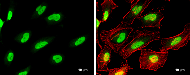
GTX100573 ICC/IF Image
PARP antibody detects PARP protein at nucleus by immunofluorescent analysis.
Sample: HeLa cells were fixed in 4% paraformaldehyde at RT for 15 min.
Green: PARP protein stained by PARP antibody (GTX100573) diluted at 1:500.
Red: Phalloidin, a cytoskeleton marker, diluted at 1:100.
Scale bar = 10 um.
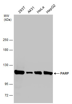
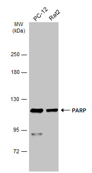
GTX100573 WB Image
Various whole cell extracts (30 ug) were separated by 5% SDS-PAGE, and the membrane was blotted with PARP antibody (GTX100573) diluted at 1:2000.
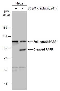
GTX100573 WB Image
Untreated (?) and treated (+) HeLa whole cell extracts (30 ug) were separated by 7.5% SDS-PAGE, and the membrane was blotted with PARP antibody (GTX100573) diluted at 1:2000. The HRP-conjugated anti-rabbit IgG antibody (GTX213110-01) was used to detect the primary antibody.
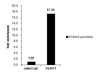
GTX100573 ChIP assay Image
Cross-linked ChIP was performed with Raji chromatin extract and 5 ug of either control rabbit IgG or anti-PARP1 antibody. The precipitated DNA was detected by PCR with primer set targeting to S100A9 promoter.
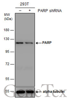
GTX100573 WB Image
Non-transfected (?) and transfected (+) 293T whole cell extracts (30 ug) were separated by 7.5% SDS-PAGE, and the membrane was blotted with PARP antibody (GTX100573) diluted at 1:2000. The HRP-conjugated anti-rabbit IgG antibody (GTX213110-01) was used to detect the primary antibody.
GTX100573 WB Image
Various whole cell extracts (30 ug) were separated by 5% SDS-PAGE, and the membrane was blotted with PARP antibody (GTX100573) diluted at 1:1000. The HRP-conjugated anti-rabbit IgG antibody (GTX213110-01) was used to detect the primary antibody.
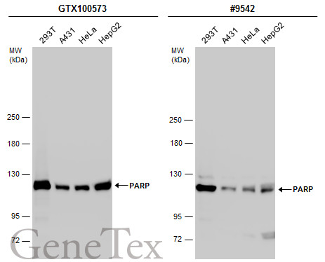
GTX100573 WB Image
Various whole cell extracts (30 ug) were separated by 5% SDS-PAGE, and the membranes were blotted with PARP antibody (GTX100573) diluted at 1:2000 and competitor's antibody (#9542) diluted at 1:500. The HRP-conjugated anti-rabbit IgG antibody (GTX213110-01) was used to detect the primary antibody.
*The competitor is not affiliated with GeneTex and does not endorse this product.
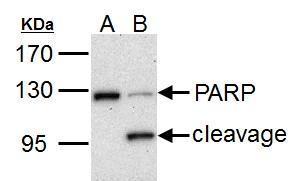
GTX100573 WB Image
PARP1 antibody detects PARP1 protein by western blot analysis.
A. 30 ug HCT116 whole cell lysate/extract (untreated)
B. 30 ug HCT116 whole cell lysate/extract (30 uM cisplatin treatment for 24hr)
7.5% SDS-PAGE
PARP1 antibody (GTX100573) dilution: 1:1000
The HRP-conjugated anti-rabbit IgG antibody (GTX213110-01) was used to detect the primary antibody.
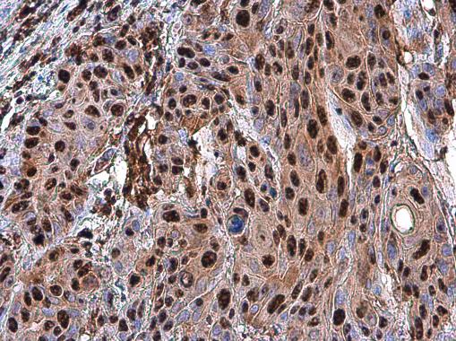
GTX100573 IHC-P Image
PARP antibody detects PARP protein at nucleus in human oral carcinoma by immunohistochemical analysis.
Sample: Paraffin-embedded human oral carcinoma.
PARP antibody (GTX100573) diluted at 1:500.