
抗NDP52 [GT422]抗体 | Anti-NDP52 [GT422] antibody
掲載日情報:2020/04/02 現在Webページ番号:141808

GeneTex社の抗NDP52 [GT422]抗体 | Anti-NDP52 [GT422] antibodyです。
※ 本製品は研究用です。研究用以外には使用できません。
追加しました。
特長
- 高品質の抗体です。
- 幅広い研究分野に関連する抗体を取り揃えています。
- 使用されたアプリケーションや動物種などの情報が充実しています。
- 多数の論文で使用実績がある信頼性の高い抗体です。
追加しました。
価格
[在庫・価格 :2026年04月01日 00時00分現在]
| 詳細 | 商品名 |
|
文献数 | ||||||||||||||||||||||||||||||||||||||||||||||||||||||||||||||||||||||||||||||||||
|---|---|---|---|---|---|---|---|---|---|---|---|---|---|---|---|---|---|---|---|---|---|---|---|---|---|---|---|---|---|---|---|---|---|---|---|---|---|---|---|---|---|---|---|---|---|---|---|---|---|---|---|---|---|---|---|---|---|---|---|---|---|---|---|---|---|---|---|---|---|---|---|---|---|---|---|---|---|---|---|---|---|---|---|---|---|
|
Anti-NDP52, Mouse-Mono(GT422) |
|
2 | |||||||||||||||||||||||||||||||||||||||||||||||||||||||||||||||||||||||||||||||||||
|
|||||||||||||||||||||||||||||||||||||||||||||||||||||||||||||||||||||||||||||||||||||
|
Anti-NDP52, Mouse(GT422) |
|
2 | |||||||||||||||||||||||||||||||||||||||||||||||||||||||||||||||||||||||||||||||||||
|
|||||||||||||||||||||||||||||||||||||||||||||||||||||||||||||||||||||||||||||||||||||


[在庫・価格 :2026年04月01日 00時00分現在]
Anti-NDP52, Mouse-Mono(GT422)
文献数: 2
- 商品コード:GTX630396
- メーカー:GNT
- 包装:25μl
- 価格:¥30,000
- 在庫:無(未発注)
- 納期:10日程度 ※※ 表示されている納期は弊社に在庫がなく、取り寄せた場合の目安納期となります。
- 法規制等:
| 説明文 | KO/KDバリデーション済み抗体。 別名:calcium binding and coiled-coil domain 2,NDP52 クローン:GT422 Genbank No: 10241 |
||||||
|---|---|---|---|---|---|---|---|
| 別包装品 | 別包装品あり | ||||||
| 法規制等 | |||||||
| 保存条件 | -20℃ | 法規備考 | |||||
| 抗原種 | Human | 免疫動物 | Mouse | ||||
| 交差性 | Human | 適用 | IC,IF,IP,Western Blot | ||||
| 標識 | Unlabeled | 性状 | Protein A/G Affinity Purified | ||||
| 吸収処理 | クラス | IgG | |||||
| クロナリティ | Monoclonal | フォーマット | |||||
| 掲載カタログ |
|
||||||
| 製品記事 | 使いっきり抗体 マイトファジー関連抗体(GeneTex社) ミトコンドリア(Mitochondria)研究用製品特集 |
||||||
| 関連記事 | GeneTex社における抗体の品質管理 |
||||||
Anti-NDP52, Mouse(GT422)
文献数: 2
- 商品コード:GTX630396
- メーカー:GNT
- 包装:100μl
- 価格:¥85,000
- 在庫:無(未発注)
- 納期:10日程度 ※※ 表示されている納期は弊社に在庫がなく、取り寄せた場合の目安納期となります。
- 法規制等:
| 説明文 | KO/KDバリデーション済み抗体。 別名:calcium binding and coiled-coil domain 2,NDP52 クローン:GT422 Genbank No: 10241 |
||||||
|---|---|---|---|---|---|---|---|
| 別包装品 | 別包装品あり | ||||||
| 法規制等 | |||||||
| 保存条件 | -20℃ | 法規備考 | |||||
| 抗原種 | Human | 免疫動物 | Mouse | ||||
| 交差性 | Human | 適用 | IC,IF,IP,Western Blot | ||||
| 標識 | Unlabeled | 性状 | Protein A/G Affinity Purified | ||||
| 吸収処理 | クラス | IgG | |||||
| クロナリティ | Monoclonal | フォーマット | |||||
| 掲載カタログ |
|
||||||
| 製品記事 | マイトファジー関連抗体(GeneTex社) ミトコンドリア(Mitochondria)研究用製品特集 |
||||||
| 関連記事 | GeneTex社における抗体の品質管理 |
||||||
追加しました。
DATA IMAGES
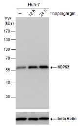 | NDP52 antibody detects NDP52 protein by western blot analysis. Un-treated (-) and treated (+, Thapsigargin treatment for 12hrs and 24hrs) Huh-7 whole cell extracts (30 μg) were separated by 10% SDS-PAGE, and the membrane was blotted with NDP52 antibody (GTX630396) diluted by 1:500.The ACTB was used as internal control (GTX110564, 1:50000) shown at the bottom panel. |
 | NDP52 antibody [GT422] detects NDP52 protein at autophagosome by immunofluorescent analysis.Sample: Mock and treated HeLa cells were fixed in 4% paraformaldehyde at RT for 15 min.Green: NDP52 stained by NDP52 antibody [GT422] (GTX630396) diluted at 1:1000.Red: phalloidin, a cytoskeleton marker, diluted at 1:200.Blue: Fluoroshield with DAPI (GTX30920).Scale bar= 10 μm. |
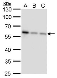 | NDP52 antibody [GT422] detects NDP52 protein by western blot analysis.A. 30 μg Jurkat whole cell lysate/extractB. 30 μg Raji whole cell lysate/extractC. 30 μg NCI-H929 whole cell lysate/extract 10 % SDS-PAGENDP52 antibody [GT422] (GTX630396) dilution: 1:1000 |
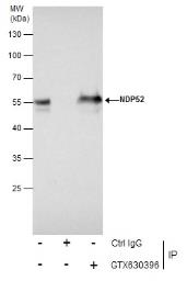 | Immunoprecipitation of NDP52 protein from Jurkat whole cell extracts using 5 μg of NDP52 antibody [GT422] (GTX630396).Western blot analysis was performed using NDP52 antibody [GT422] (GTX630396).EasyBlot anti-Mouse IgG (GTX221667-01) was used as a secondary reagent. |
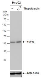 | NDP52 antibody detects NDP52 protein by western blot analysis. Un-treated (-) and treated (+, Thapsigargin treatment for 12hrs) HepG2 whole cell extracts (30 μg) were separated by 10% SDS-PAGE, and the membrane was blotted with NDP52 antibody (GTX630396) diluted by 1:500.The ACTB was used as internal control (GTX110564, 1:50000) shown at the bottom panel. |
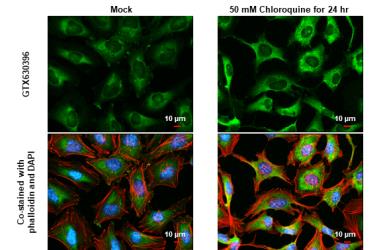
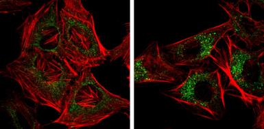 | NDP52 antibody [GT422] detects NDP52 protein at autophagosome by immunofluorescent analysis. Samples: HeLa cells mock (left) and treated with 50μM Chloroquine for 24 hr (right) were fixed in 4% paraformaldehyde at RT for 15 min.Green: NDP52 protein stained by NDP52 antibody [GT422] (GTX630396) diluted at 1:1000.Red: Phalloidin, a F-actin marker. |
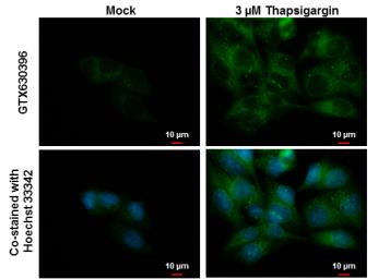 | NDP52 antibody [GT422] detects NDP52 protein at autophagosome by immunofluorescent analysis. Samples: Hep G2 cells mock (left) and treated with 3 μM Thapsigargin for 16 hrs (right) were fixed in ice-cold MeOH for 10 min and permeabilized with 100% MeOH for 30 sec.Green: NDP52 protein stained by NDP52 antibody [GT422] (GTX630396) diluted at 1:1000.Blue: Hoechst 33342 staining.Scale bar = 10 μm. |
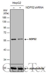 | Non-transfected (–) and transfected (+) HepG2 whole cell extracts (30 μg) were separated by 10% SDS-PAGE, and the membrane was blotted with NDP52 antibody [GT422] (GTX630396) diluted at 1:1000. |
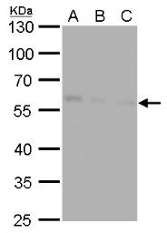 | NDP52 antibody [GT422] detects NDP52 protein by western blot analysis.A. 30 μg Huh7 whole cell lysate/extractB. 30 μg Hep3B whole cell lysate/extractC. 30 μg HepG2 whole cell lysate/extract10 % SDS-PAGENDP52 antibody [GT422] (GTX630396) dilution: 1:1000 |
追加しました。
製品情報
| Host | Mouse |
|---|---|
| Clonality | Monoclonal |
| Clone Name | GT422 |
| Isotype | IgG1 |
| Application | WB, ICC/IF, IP |
| Reactivity | Human |
追加しました。
APPLICATION
Application Note
*Optimal dilutions/concentrations should be determined by the researcher.| Application | Dilution |
|---|---|
| WB | 1:500-1:3000 |
| ICC/IF | 1:100-1:1000 |
| IP | 1:100-1:500 |
| Calculated MW | 52 kDa. ( Note ) |
|---|---|
| Positive Control | Jurkat , Raji , NCI-H929 , Huh-7 , Hep3B , HepG2 , Huh-7 (untreated) , Huh-7 (3 μM Thapsigargin treatment for 12 hr) , Huh-7 (3 μM Thapsigargin treatment for 24 hr) , HepG2 (untreated) , HepG2 (3 μM Thapsigargin treatment for 12 hr) |
| Predict Reactivity | Chimpanzee(>80% identity) |
追加しました。
PROPERTIES
| Form | Liquid |
|---|---|
| Buffer | PBS |
| Storage | Store as concentrated solution. Centrifuge briefly prior to opening vial. For short-term storage (1-2 weeks), store at 4ºC. For long-term storage, aliquot and store at -20ºC or below. Avoid multiple freeze-thaw cycles. |
| Concentration | 1 mg/ml (Please refer to the vial label for the specific concentration.) |
| Antigen Species | Human |
| Immunogen | Recombinant protein encompassing a sequence within the center region of human NDP52. The exact sequence is proprietary. |
| Purification | Affinity purified by Protein G. |
| Conjugation | Unconjugated |
| Note | For laboratory use only. Not for any clinical, therapeutic, or diagnostic use in humans or animals. Not for animal or human consumption. |
追加しました。
TARGET
| Synonyms | calcium binding and coiled-coil domain 2 , NDP52 |
|---|---|
| Cellular Localization | Cytoplasm , perinuclear region , Golgi apparatus , cytoskeleton |
| Background | The protein encoded by this gene is a subunit of nuclear domain 10 (ND10) bodies. ND10 bodies are nuclear domains appearing immunohistochemically as ten dots per nucleus. They are believed to be associated with the nuclear matrix on the basis of their resistance to nuclease digestion and salt extraction. ND10 proteins are removed from the nucleus by herpes simplex virus-1 infection and may have a role in viral life cycles. [provided by RefSeq] |
| Database | ・ Gene ID: 10241 CALCOCO2 ・ UniProt: Q13137 CALCOCO2 |
追加しました。
REFERENCEE(抜粋)
| Application Reference | Application/Reactivity |
|---|---|
| Prabakaran T et al. EMBO J 2018; (Epub) Attenuation of cGAS-STING signaling is mediated by a p62/SQSTM1-dependent autophagy pathway activated by TBK1. | Application : WB Reactivity : Human |
追加しました。
製品情報は掲載時点のものですが、価格表内の価格については随時最新のものに更新されます。お問い合わせいただくタイミングにより製品情報・価格などは変更されている場合があります。
表示価格に、消費税等は含まれていません。一部価格が予告なく変更される場合がありますので、あらかじめご了承下さい。