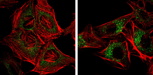
GTX630396 ICC/IF Image
NDP52 antibody [GT422] detects NDP52 protein at autophagosome by immunofluorescent analysis.
Samples: HeLa cells mock (left) and treated with 50uM Chloroquine for 24 hr (right) were fixed in 4% paraformaldehyde at RT for 15 min.
Green: NDP52 protein stained by NDP52 antibody [GT422] (GTX630396) diluted at 1:1000.
Red: Phalloidin, a F-actin marker.
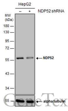
GTX630396 WB Image
Non-transfected (?) and transfected (+) HepG2 whole cell extracts (30 ug) were separated by 10% SDS-PAGE, and the membrane was blotted with NDP52 antibody [GT422] (GTX630396) diluted at 1:1000.
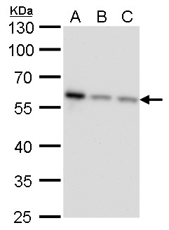
GTX630396 WB Image
NDP52 antibody [GT422] detects NDP52 protein by western blot analysis.
A. 30 ug Jurkat whole cell lysate/extract
B. 30 ug Raji whole cell lysate/extract
C. 30 ug NCI-H929 whole cell lysate/extract
10 % SDS-PAGE
NDP52 antibody [GT422] (GTX630396) dilution: 1:1000
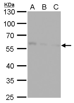
GTX630396 WB Image
NDP52 antibody [GT422] detects NDP52 protein by western blot analysis.
A. 30 ug Huh7 whole cell lysate/extract
B. 30 ug Hep3B whole cell lysate/extract
C. 30 ug HepG2 whole cell lysate/extract
10 % SDS-PAGE
NDP52 antibody [GT422] (GTX630396) dilution: 1:1000
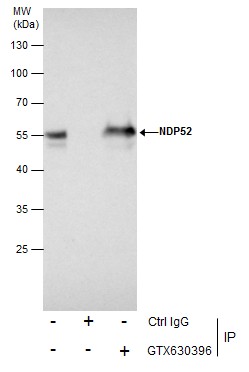
GTX630396 IP Image
Immunoprecipitation of NDP52 protein from Jurkat whole cell extracts using 5 ug of NDP52 antibody [GT422] (GTX630396).
Western blot analysis was performed using NDP52 antibody [GT422] (GTX630396).
EasyBlot anti-Mouse IgG (GTX221667-01) was used as a secondary reagent.
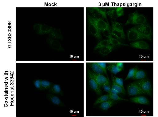
GTX630396 ICC/IF Image
NDP52 antibody [GT422] detects NDP52 protein at autophagosome by immunofluorescent analysis.
Samples: Hep G2 cells mock (left) and treated with 3 uM Thapsigargin for 16 hrs (right) were fixed in ice-cold MeOH for 10 min and permeabilized with 100% MeOH for 30 sec.
Green: NDP52 protein stained by NDP52 antibody [GT422] (GTX630396) diluted at 1:1000.
Blue: Hoechst 33342 staining.
Scale bar = 10 um.
GTX630396 WB Image
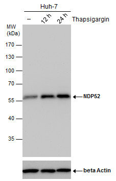
NDP52 antibody detects NDP52 protein by western blot analysis. Un-treated (-) and treated (+, Thapsigargin treatment for 12hrs and 24hrs) Huh-7 whole cell extracts (30 ug) were separated by 10% SDS-PAGE, and the membrane was blotted with NDP52 antibody (GTX630396) diluted by 1:500.
The ACTB was used as internal control (GTX110564, 1:50000) shown at the bottom panel.
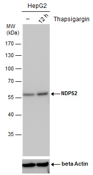
GTX630396 WB Image
NDP52 antibody detects NDP52 protein by western blot analysis. Un-treated (-) and treated (+, Thapsigargin treatment for 12hrs) HepG2 whole cell extracts (30 ug) were separated by 10% SDS-PAGE, and the membrane was blotted with NDP52 antibody (GTX630396) diluted by 1:500.
The ACTB was used as internal control (GTX110564, 1:50000) shown at the bottom panel.