HOME>
Products>
Protein / Enzyme>
Protein Labeling Reagents>
ER-Protein Capture Kit | Funakoshi
HOME>
Products>
Protein / Enzyme>
Other reagent>
ER-Protein Capture Kit | Funakoshi
Kit for Specially Labeling, Extracting, and Purifying Endoplasmic Reticulum (ER)-Localized Proteins ER-Protein Capture Kit | Funakoshi
Date:August 07 2020Web Page No:81520

Funakoshi Co.,Ltd.
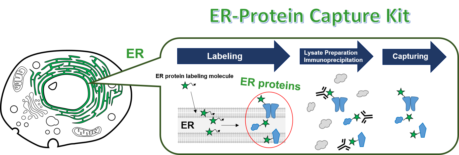
ER-Protein Capture Kit labels ER-associated proteins specifically and enables to isolate them by immunoprecipitation. This kit is a powerful tool for global analysis of various ER proteins.
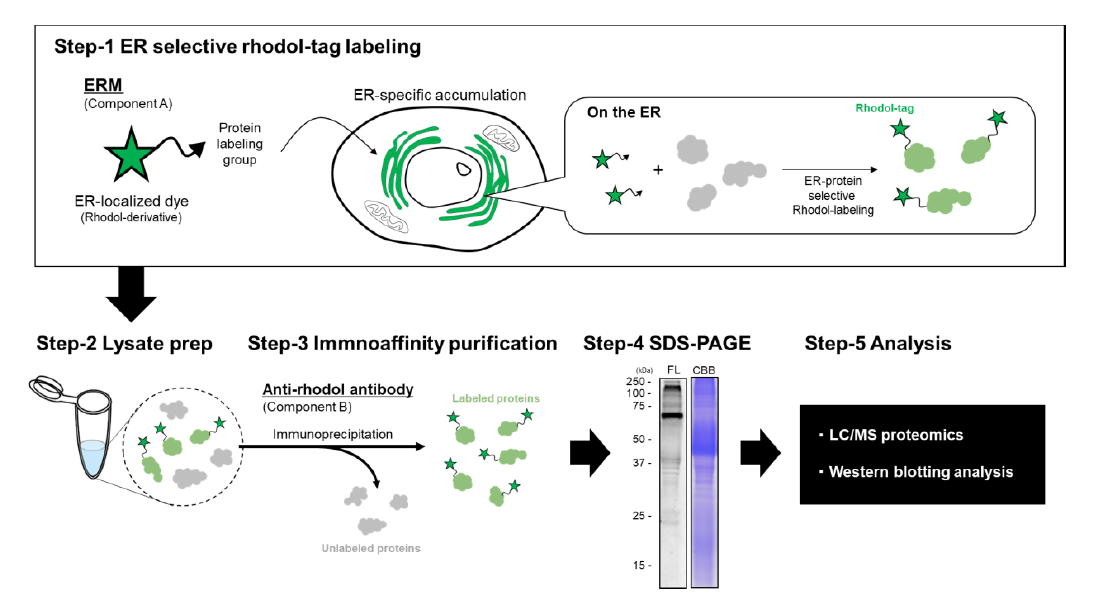
Principle and Assay Flow
The kit utilizes ERM (ER-localizable Reactive Molecule), a compound consisting of an ER-localizable green fluorescent dye (rhodol derivative) attached to a protein-reactive group.
- Step 1: Specific Labeling
When added to live cells, ERM rapidly concentrates in the endoplasmic reticulum due to its specific dye properties, where it covalently labels ER proteins with a fluorescent tag via its reactive group. - Step 2: Cell Lysis
After the labeling reaction, cells are lysed to prepare a cell lysate. - Step 3: Immunoprecipitation (IP)
ERM-labeled ER proteins are selectively captured and recovered using the provided tag-capture antibody (anti-rhodol antibody). - Step 4: Detection
Since the recovered proteins retain the fluorescent tag, they can be separated by SDS-PAGE and detected directly via a fluorescence imager, or via CBB/silver staining. - Step 5: Analysis
After SDS-PAGE separation, proteins can be comprehensively analyzed by LC/MS proteomics or identified via Western Blotting (WB) using specific antibodies.
- Advantages over Conventional Methods
- Kit Components
- Reference Data
- Application Data
- Reference
- Webinar
- Product information
Advantages over Conventional Methods
The Endoplasmic Reticulum (ER) is a highly complex organelle that spreads throughout the cell, occupying about 20% of the total cellular volume. It plays a central role in protein biosynthesis, folding, quality control, lipid metabolism, and lipid droplet formation. Unlike other organelles, technologies to specifically isolate the ER have been severely limited, making selective recovery of ER proteins difficult.
The ER-Protein Capture Kit simplifies the workflow without requiring specialized equipment like ultracentrifuges. By using ERM (ER-localizable Reactive Molecule), it ensures highly selective labeling and purification of ER-localized proteins, enabling precise proteomic identification and relative quantitative analysis.
Kit Components
- A: ER-localizable Reactive Molecule (ERM)
(Same product as ERseeing (#FDV-0038), which can also be purchased individually for live-cell ER imaging). - B: Anti rhodol antibody (Purified Rabbit Polyclonal IgG)
(Available as an individual item, Product Code: #FDV-0039B)
Note: Lysis buffers for cell lysate preparation and Protein A/G resins required for immunoprecipitation are not included. Please prepare them separately.
Reference Data
Excitation and Emission Spectra of ERM
Compatible with standard green fluorescence (FITC) observation conditions.

- Excitation Spectrum: Blue / Emission Spectrum: Red (Peak Ex/Em: 509 nm / 524 nm).
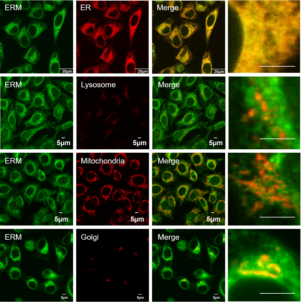
Validation of ER Specificity
Application Data
Comprehensive Proteomic Analysis of Recovered Proteins

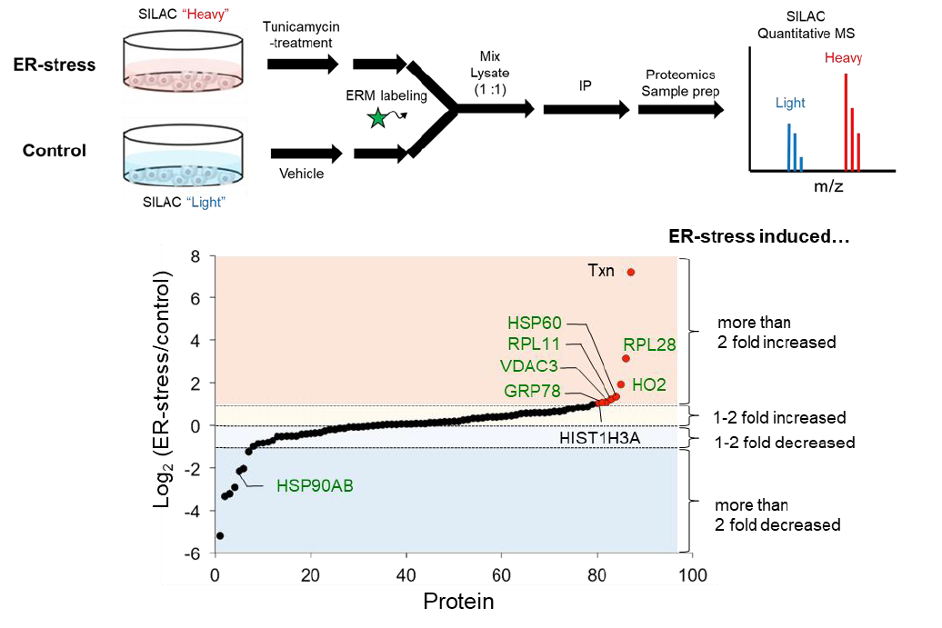
Quantitative Evaluation of ER Protein Dynamics under ER Stress
Reference
- Fujisawa, A., et al., "Chemical Profiling of the Endoplasmic Reticulum Proteome Using Designer Labeling Reagents.", J. Am. Chem. Soc., 140, 17060-17070 (2018). [PMID: 30433779]
Webinar
Product information
[Date : July 16 2026 00:08]
| Detail | Product Name | Product Code | Supplier | Size | Price | ||||||||||||||||||||||||||||||
|---|---|---|---|---|---|---|---|---|---|---|---|---|---|---|---|---|---|---|---|---|---|---|---|---|---|---|---|---|---|---|---|---|---|---|---|
|
ER-Protein Capture Kit DatasheetThis may not be the latest data sheet. |
FDV-0039 | FNAFunakoshi Co.,Ltd. | 1 kit | $600 | |||||||||||||||||||||||||||||||
|
|
|
||||||||||||||||||||||||||||||||||
[Date : July 16 2026 00:08]
ER-Protein Capture Kit
DatasheetThis may not be the latest data sheet.
- Product Code: FDV-0039
- Supplier: FNA
- Size: 1kit
- Price: $600
| Description |
ER-Protein Capture Kit enables to purify ER associated proteins to evaluate various ER roles with easy procedures and no special equipment such as ultracentrifuge machine. |
||
|---|---|---|---|
| Storage | -20°C | CAS | |
| Link | |||
Kit Components Sold Separately
[Date : July 16 2026 00:08]
Detail
Product Name
Product Code
Supplier
Size
Price
ERseeing, Endoplasmic Reticulum Green
DatasheetThis may not be the latest data sheet.
FDV-0038
FNAFunakoshi Co.,Ltd.
10
nmol
$380
Description
ERseeing is irreversible ER labeling dye and suitable for long-term live imaging.
Storage
-20°C
CAS
Link
ERseeing™ | Funakoshi
ERseeing™(Endoplasmic Reticulum Green) in a minute
ER-Protein Capture Kit component B <Anti-Rhodol antibody>
DatasheetThis may not be the latest data sheet.
FDV-0039B
FNAFunakoshi Co.,Ltd.
200
µg
$350
Description
Component B of ER-Protein Capture Kit (#FDV-0039) is sold separately. Anti-Rhodol antibody. Concentration: 1 mg/mL. Formulation: 1 x PBS containing 50% glycerol. Host/Clonality: Rabbit polyclonal. Purification: Protein G purified
Storage
-20°C
CAS
Link
[Date : July 16 2026 00:08]
ERseeing, Endoplasmic Reticulum Green
DatasheetThis may not be the latest data sheet.
- Product Code: FDV-0038
- Supplier: FNA
- Size: 10nmol
-
Price:
$380
Description
ERseeing is irreversible ER labeling dye and suitable for long-term live imaging.
Storage
-20°C
CAS
Link
ERseeing™ | Funakoshi
ERseeing™(Endoplasmic Reticulum Green) in a minute
ER-Protein Capture Kit component B <Anti-Rhodol antibody>
DatasheetThis may not be the latest data sheet.
- Product Code: FDV-0039B
- Supplier: FNA
- Size: 200µg
-
Price:
$350
Description
Component B of ER-Protein Capture Kit (#FDV-0039) is sold separately. Anti-Rhodol antibody. Concentration: 1 mg/mL. Formulation: 1 x PBS containing 50% glycerol. Host/Clonality: Rabbit polyclonal. Purification: Protein G purified
Storage
-20°C
CAS
Link
[Date : July 16 2026 00:08]
| Detail | Product Name | Product Code | Supplier | Size | Price | ||||||||||||||||||||||||||||||
|---|---|---|---|---|---|---|---|---|---|---|---|---|---|---|---|---|---|---|---|---|---|---|---|---|---|---|---|---|---|---|---|---|---|---|---|
|
ERseeing, Endoplasmic Reticulum Green DatasheetThis may not be the latest data sheet. |
FDV-0038 | FNAFunakoshi Co.,Ltd. | 10 nmol | $380 | |||||||||||||||||||||||||||||||
|
|
|
||||||||||||||||||||||||||||||||||
|
ER-Protein Capture Kit component B <Anti-Rhodol antibody> DatasheetThis may not be the latest data sheet. |
FDV-0039B | FNAFunakoshi Co.,Ltd. | 200 µg | $350 | |||||||||||||||||||||||||||||||
|
|
|
||||||||||||||||||||||||||||||||||
[Date : July 16 2026 00:08]
ERseeing, Endoplasmic Reticulum Green
DatasheetThis may not be the latest data sheet.
- Product Code: FDV-0038
- Supplier: FNA
- Size: 10nmol
- Price: $380
| Description |
ERseeing is irreversible ER labeling dye and suitable for long-term live imaging. |
||
|---|---|---|---|
| Storage | -20°C | CAS | |
| Link |
ERseeing™ | Funakoshi |
||
ER-Protein Capture Kit component B <Anti-Rhodol antibody>
DatasheetThis may not be the latest data sheet.
- Product Code: FDV-0039B
- Supplier: FNA
- Size: 200µg
- Price: $350
| Description |
Component B of ER-Protein Capture Kit (#FDV-0039) is sold separately. Anti-Rhodol antibody. Concentration: 1 mg/mL. Formulation: 1 x PBS containing 50% glycerol. Host/Clonality: Rabbit polyclonal. Purification: Protein G purified |
||
|---|---|---|---|
| Storage | -20°C | CAS | |
| Link |
|
||
CONTACT
export@funakoshi.co.jp
- ※Prices on our website are for your reference only. Please inquire your distributor for your prices.
- ※Please note that Product Information or Price may change without notice.