Fatty Acid Oxidation (FAO) activity detection / assay reagent FAOBlue™(Fatty Acid Oxidation Detection Reagent)
Date:August 27 2020Web Page No:81517

Funakoshi Co.,Ltd.
This reagent visualizes fatty acid beta-oxidation (FAO) activity, a common metabolic pathway in the breakdown of fatty acids in living organisms, using blue fluorescence. FAO activity of fatty acid beta-oxidation in living cells, which has been difficult to measure in the past, can be easily measured by the fluorescence imaging method. It can be widely applied to comparative evaluation of β-oxidation activity of fatty acids in different cell types, search for compounds that promote or inhibit β-oxidation activity, and basic research on enzymes involved in β-oxidation.

Image of FAOBlue™ reagent

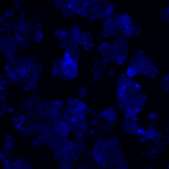
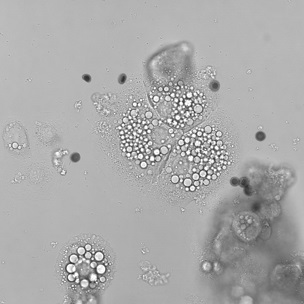
Image of FAOBlue™ in HepG2 (-FAO inhibitor)

Image of FAOBlue™ in HepG2 (+FAO inhibitor)
- Background
- Principle
- Features
- Application
- How to use
- Example of Use
- Original Paper
- Product information
Background
Degradation of fatty acids
Fatty acids (FAs) are basic building blocks for wide variety of lipids, essential components of cells, and are one of primary sources of energy. Especially during starvation due to glucose shortage, a large amount of ATP is produced through the active degradation of fatty acids. There are various types of fatty acids with different carbon chain lengths and degrees of unsaturation and the FAs are mainly degraded in common degradation pathway in mitochondria called fatty acid beta-oxidation (FAO).
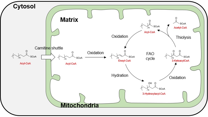
Degradation pathway of fatty acids by β-oxidation
The β-oxidation of fatty acids consists of the following four main enzymatic reaction pathways.
- 1) Oxidation at the β-position of fatty acids
- 2) Hydration at the β-position
- 3) Oxidation at the β-position
- 4) Cleavage into fatty acids with two short carbons and acetyl CoA
The resulting fatty acid with two carbons shorter is further degraded in the same cycle, repeating the degradation of fatty acids that are two carbons shorter each time.
About β-oxidation of fatty acids & FAOBlue™
It has been reported that the FAO activity is highly variable in diseases such as cancer and metabolic dysfunction associated steatohepatitis (MASH), and the development of a method to measure FAO activity has been expected. However, analytical method for FAO activity in living cell is highly limited. FAOBlue™ is the world’s first reagent for directly measuring FAO activity in living cells. FAOBlue™ enables to measure FAO activity with an easy procedure in living cells by fluorescence imaging.
Principle
FAOBlue™ is a fatty acid analog consisting of a nonanoic acid (C9) with a blue fluorescence dye coumarin-derivative at the end of the carbon chain, and further its carboxylic acid is protected with acetoxymethyl ester to improve cell membrane permeability. Although FAOBlue™ contains a coumarin derivative, it shows no-fluorescence excited by 405 nm before metabolization by FAO.

- FAOBlue™ can permeate cell membranes due to the high hydrophobicity of the acetoxymethyl group attached to the carboxy group, and is efficiently taken up into cells.
- Acetoxymethyl ester of FAOBlue™ is hydrolyzed by intracellular esterases and FAOBlue™ is converted to free fatty acid form.
- Free FA type of FAOBlue™ is converted to acyl-CoA by acyl-CoA synthases and further incorporated into mitochondrial FAO pathway.
- Acyl-CoA type of FAOBlue™ is next converted to nonanoic acid (C9), heptanoic acid (C7), pentanoic acid (C5), and propionic acid (C3) sequentially by three FAO cycle. After the 4th FAO cycle, free coumarin dye is released from propionic acid. Coumarin dye emits blue fluorescence upon excitation at 405 nm.
- Since free coumarin dyes diffuse into whole cells, FAO activity can be evaluated quantitatively and time-dependently by measuring the intensity of intracellular blue fluorescence.
※The addition of Etomoxir, a carnitine shuttle inhibitor, strongly suppresses the increase in intracellular fluorescence intensity. This result indicates that FAOBlue™ detects FAO activity primarily in mitochondria.
Features
- FAOBlue™ has little fluorescence excited at 405 nm, and the free coumarin derivative after degradation shows higher fluorescence intensity excited at 405 nm than before degradation.
- Observation is possible within 30 to 120 minutes after addition to the medium.
- β-oxidation activity can be measured without any special manipulation.
- β-oxidation has been observed in several cell types.
- Activity change of β-oxidation due to drug-dependent inhibition or promotion was detected.
- Measurement wavelength: Excitation 405 nm / Fluorescence 460 nm
Note
If a confocal laser microscope is used, excitation with a 405 nm laser is recommend. When this reagent is excited in the range of 330-380 nm, fluorescence in the range of 370-450 nm (maximum fluorescence wavelength of 410 nm) is observed, which is derived regardless of FAO activity. When using an epi-fluorescence microscope, it is recommended to select a filter for excitation light in the range of 390-430 nm for excitation in order to perform FAO activity-specific observation. For details, please refer to example of use: ☞ FAO-dependent changes in absorption and fluorescence spectra
Application
- Evaluation of FAO activity in various cells
- Basic research on genes related with FAO
- Screening of compounds that modulate FAO activity
How to use
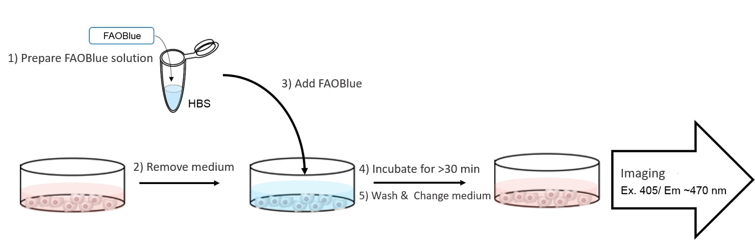
Workflow of FAOBlue™
- Dissolve FAOBlue™ in HEPES-buffered saline (HBS; cf. 20 mM HEPES (pH 7.4), 107 mM NaCl, 6 mM KCl, 1.2 mM MgSO4, 2 mM CaCl2) to a final concentration 5-20 uM*1.
- Remove the cultured medium and wash the cells with HBS twice.
- Add FAOBlue™ containing HBS to cells.
- Incubate cells at 37℃ for at least 30 min*2.
- After changing the culture medium*3, blue fluorescence (Ex. 405 nm / Em. 430-480 nm) is observed by imaging.
- *1: The appropriate concentration varies for each cell type. We recommend examining the concentration for each experiment.
- *2: The optimum incubation time varies for each cell type. We recommend examining it for each experiment.
- *3: Changing the culture medium after staining is not essential. It can be observed without washing.
Note
Since this reagent uses 405 nm wavelength as excitation light, autofluorescence may be observed depending on samples. Negative control (without this reagent) for each experiment is recommended to confirm cell-derived autofluorescence signals. In particular, if a dot-like fluorescent signals are observed during microscopic observation, they may be caused by autofluorescence.
Example of Use
FAO-dependent changes in absorption & fluorescence spectra
FAOBlue™ is converted to a coumarin derivative by FAO, resulting in a long wavelength shift of the absorption maximum from 350 nm to 405 nm. Therefore, under 405 nm excitation, FAOBlue™ doesn’t show fluorescence, and blue fluorescence is observed only when the coumarin derivative is released by FAO.

Absorbance spectra of FAOBlue™ and coumarin derivative

Fluorescence spectra of FAOBlue™ and coumarin derivative
Note
Excitation of FAOBlue™ with around the absorption maximum of 350 nm also causes blue fluorescence (380-450 nm; maximum wavelength 400 nm). If a general DAPI filter for fluorescence microscopy is used, both the unreacted FAOBlue™ and the coumarin derivative after the FAO reaction will be excited. Please be careful in selecting filters.
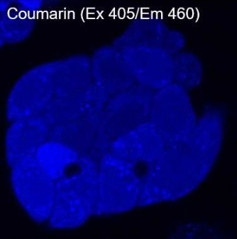
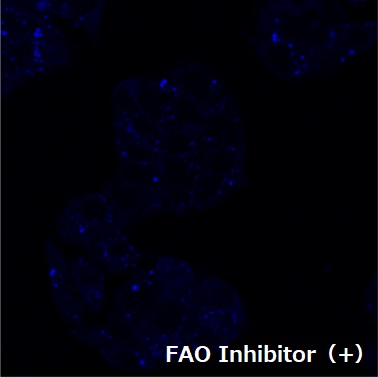
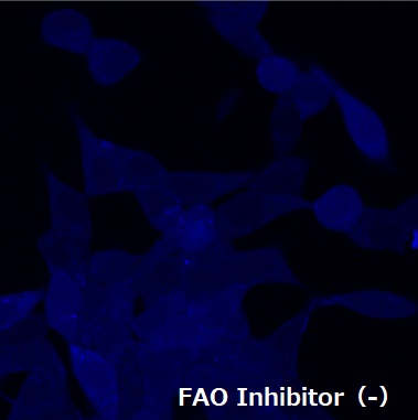
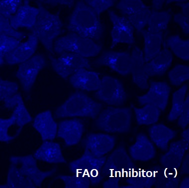
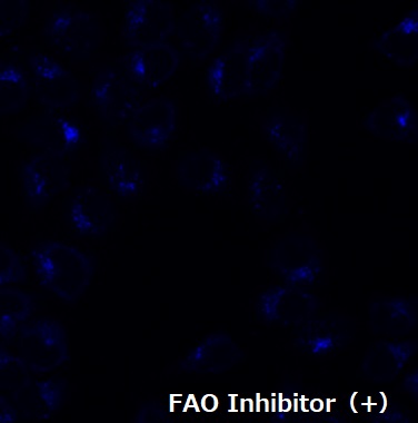
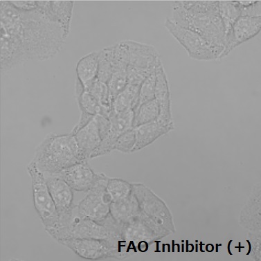
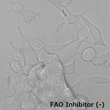
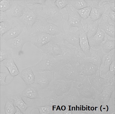
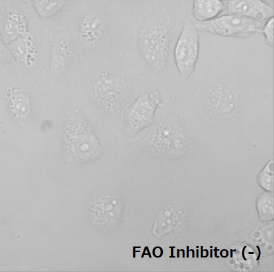
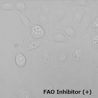
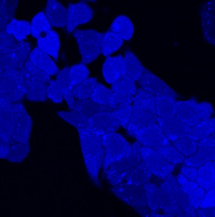
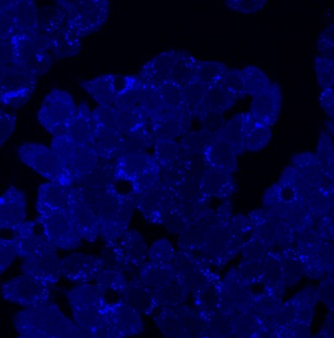
Visualization of FAO activities in various cell lines
Four types of cancer cells were incubated with FAOBlue™ and fluorescence was observed after a certain time of incubation. All cell lines showed blue fluorescence, but pre-treatment of etomoxir clearly decreased fluorescent intensities. These results indicated the blue fluorescence was derived from FAO activity in the cells.
Excitation of FAOBlue™ with around the absorption maximum of 350 nm also causes blue fluorescence (380-450 nm; maximum wavelength 400 nm). If a general DAPI filter for fluorescence microscopy is used, both the unreacted FAOBlue™ and the coumarin derivative after the FAO reaction will be excited. Please be careful in selecting filters.
Visualization of FAO activities in various cell lines
Four types of cancer cells were incubated with FAOBlue™ and fluorescence was observed after a certain time of incubation. All cell lines showed blue fluorescence, but pre-treatment of etomoxir clearly decreased fluorescent intensities. These results indicated the blue fluorescence was derived from FAO activity in the cells.
| Cell Type | HepG2 | LNCap | HeLa | A549 | |||||
|---|---|---|---|---|---|---|---|---|---|
| FAOBlue™ Treatment | 5 μM, 30 min | 20 μM, 120 min | 20 μM, 120 min | 5 μM, 30 min | |||||
| FAO Inbihitor | - | + | - | + | - | + | - | + | |
| Image | Coumarin | 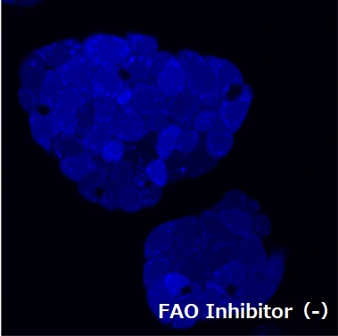 |
 |
 |
 |
 |
 |
 |
 |
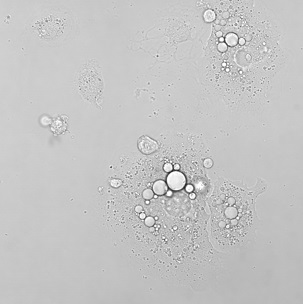
| DIC |  |
 |
 |
 |
 |
 |
 |
 |
|
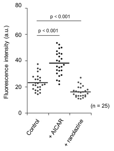
Drug-dependent changes in FAO activity
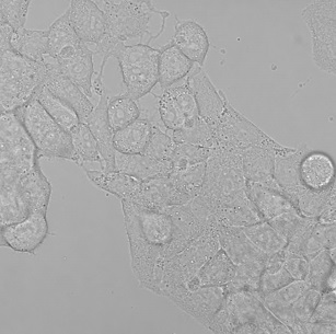
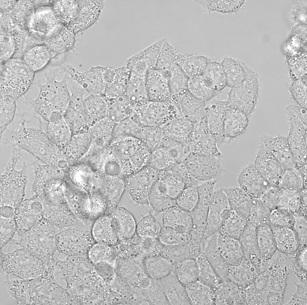
HepG2 cells were pre-incubated with a lipid metabolism enhancer AICAR*(200 μM, 3 hours) or a partial FAO inhibitor Ranolazine (200 μM, 12 hours), and then incubated with FAOBlue™(5 μM) for 30 min. Compared with control cells, pretreatment with AICAR significantly increased blue fluorescence intensity. On the other hand, pretreatment with the partial FAO inhibitor ranolazine clearly decreased blue fluorescence intensity. *AICAR: AMPK (AMP-activated protein kinase) activator, which activates lipid metabolism.
| FAO Activity Modulator | Control | +AICAR | +Ranolazine | ||
|---|---|---|---|---|---|
| Image | Coumarin | 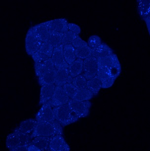 |
 |
 |
|
| DIC | 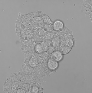 |
 |
 |
||
Quantitative analysis of the drug effects on FAO activity
FAOBlue™ can be used to quantitatively analyze the effects of drugs on FAOs. After HepG2 cells were pre-incubated with ND-630 (acetyl-CoA carboxylase inhibitor), a drug candidate for the treatment of metabolic dysfunction associated steatotic liver disease (MASLD), for 4 hours, the cells were incubated with FAOBlue™(5 μM) for 30 min. Evaluation of blue fluorescence intensity by confocal laser microscopy showed a ND630 concentration-dependent increase in fluorescence intensity, indicating that FAO activity is enhanced by ND-630.
| ND-630 concentration | 0.2 nM | 30 nM | 100 nM | 
|
|
|---|---|---|---|---|---|
| Image | Coumarin | 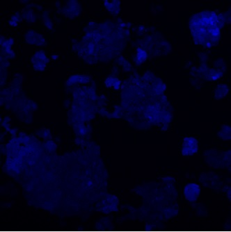 |
 |
 |
|
Analysis of FAO activity using MASH model mouse
Metabolic dysfunction associated steatohepatitis (MASH) is a typical disease which shows low metabolic activity of FAs. After control healthy mice and MASH model mice were orally administered with bezafibrate, a therapeutic agent of MASH, primary hepatocytes were isolated from livers of control mice and MASH model mice. Primary hepatocytes were treated with 5 uM FAOBlue™ for 30 min and fluorescence imaging was performed. Compared with control cells, MASH model mouse-derived hepatocytes showed low FAO activity. On the other hand, Bezafibrate dramatically recovered FAO activity of hepatocytes isolated from MASH model mouse. This reagent is a powerful tool to estimate drug effects and efficiency on FAO activity.
| Control mouse | MASH mouse |
 |
|||
|---|---|---|---|---|---|
| Bezafibrate (-) | Bezafibrate (+) | ||||
| Picture | Coumarin | 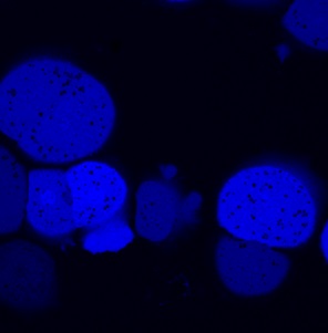 |
 |
 |
|
| DIC |  |
 |
 |
||
High-throughput cell-based detection by fluorescent plate leader
786-O cells were seeded in 96 well and culture for 1 day. Confluent cells were treated with various drugs in serum-free DMEM medium for 20 hours. After wash cells with HBS, cells were treated with final 10 uM of FAOBlue™ in HBS for 2 hours. Without washing cells, fluorescent intensity (Ex 420±5 / Em 460±10) by the fluorescent plate leader with a transparent mode. Fluorescent signals were normalized by the background signal of 10 uM FAOBlue™ without cells. A mitochondrial FAO inhibitor, etomoxir, clearly suppress blue fluorescence signal, indicating that FAOBlue™ detected fluorescence signals dependent on mitochondrial FAO activity. [Drug treatment: 10 uM Etomoxir (mitochondrial FAO inhibitor), 500 uM AICAR (AMPK activator), 5 uM 2-bromopalmitate (2-BP; broad lipid metabolism inhibitor)]

Original Paper
- Uchinomiya, S., et. al., "Fluorescence Detection of Metabolic Activity of Fatty Acid Beta Oxidation Pathway in Living Cells", Chem. Commun. 56(20), 3023-3026 (2020). [PMID: 32048639]
Product information
[Date : July 12 2026 00:07]
| Detail | Product Name | Product Code | Supplier | Size | Price | ||||||||||||||||||||||||||||||
|---|---|---|---|---|---|---|---|---|---|---|---|---|---|---|---|---|---|---|---|---|---|---|---|---|---|---|---|---|---|---|---|---|---|---|---|
|
FAOBlue, Fatty Acid Oxidation Detection Reagent DatasheetThis may not be the latest data sheet. |
FDV-0033 | FNAFunakoshi Co.,Ltd. | 0.2 mg | $350 | |||||||||||||||||||||||||||||||
|
|
|
||||||||||||||||||||||||||||||||||
[Date : July 12 2026 00:07]
FAOBlue, Fatty Acid Oxidation Detection Reagent
DatasheetThis may not be the latest data sheet.
- Product Code: FDV-0033
- Supplier: FNA
- Size: 0.2mg
- Price: $350
| Description |
Cutting-edge fluorescent dye that visualizes fatty acid β-oxidation (FAO) activity, a common pathway for fatty acid degradation, with blue fluorescence. (Ex. 405 nm / Em. 430-480 nm) |
||
|---|---|---|---|
| Storage | -20°C | CAS | |
| Link | |||
You may also like
-25-metabolism_map.jpg)
CONTACT
export@funakoshi.co.jp
- ※Prices on our website are for your reference only. Please inquire your distributor for your prices.
- ※Please note that Product Information or Price may change without notice.