DNA-specific Cell Nucleus Live Imaging Reagent NucleoSeeing™(Live Nucleus Green)
Date:March 12 2019Web Page No:81204

Funakoshi Co.,Ltd.

NucleoSeeing™ is a nuclear staining reagent for live cell imaging that binds specifically to DNA and emits green fluorescence. It exhibits a high S/N values not only in animal cells and tissues, but also in Arabidopsis thaliana leaf cells, making it excellent for observing nuclear dynamics in live cells. NucleoSeeing™ can also be applied for ☞ nucleus-specific pH sensing.
- Cell Nuclear Imaging in Living Cells
- Principle
- Features
- Application
- Example Data
- Nucleus-specific pH sensing
- Original Papers
- Cited Papers
- Video
- Product Information
Cell Nuclear Imaging in Living Cells
Cell nucleus is one of the most important organelle, serving a DNA storehouse and responsible for cell division and epigenetic modulation.
Therefore, monitoring nucleus-dynamics in live cells is considered a very important issue.
Many cell nucleus-specific dyes have been developed. While nucleic acid-responsive blue fluorescent dyes, such as Hoechst series and DAPI have been widely used,
these blue fluorescent dyes require UV excitation, which shows strong photo-toxicity for cells and not applicable to live cell imaging.
Recently, several green or red fluorochromes for nucleus imaging on live cells have been commercially available,
however, they have some disadvantages such as DNA/RNA specificity, or cell toxicity.
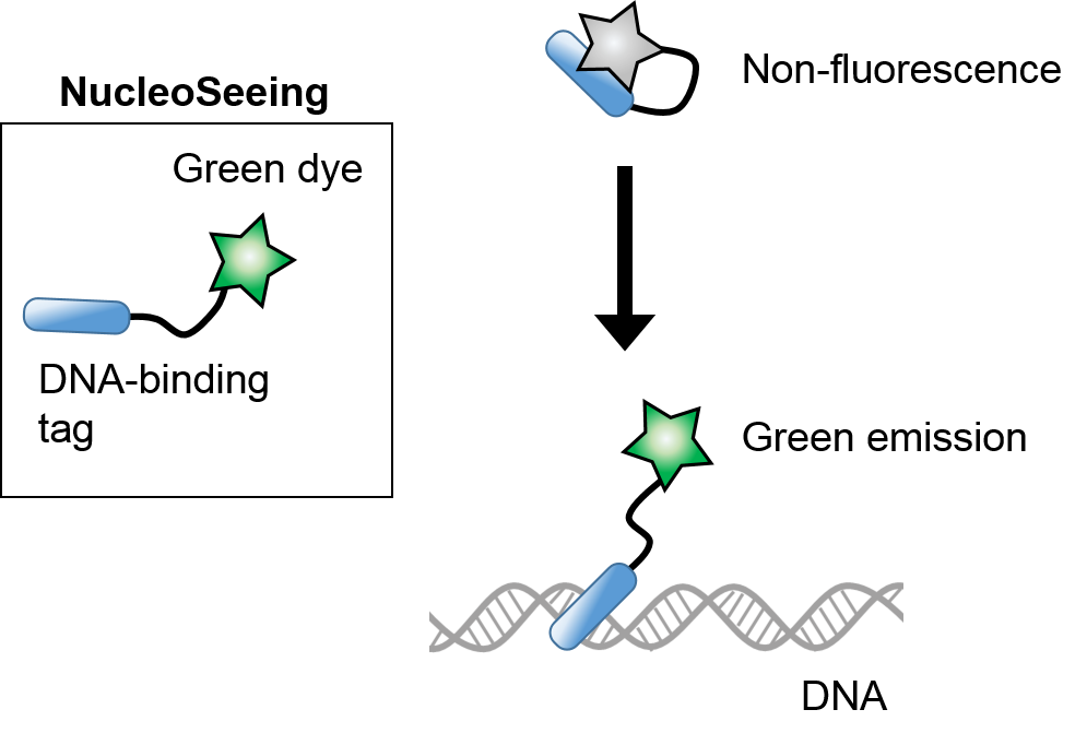
NucleoSeeing™ is a DNA-responsive probe that has a DNA-specific binding tag and emits green fluorescence in the presence of DNA, enabling live cell imaging in cultured cells, cultured tissues, and plant cells such as Arabidopsis thaliana leaf cells.
The product exhibits low cytotoxicity, high DNA-specific S/N value, and excellent cell nuclear staining.
Fixed cells can also be observed, allowing for flexible planning of applications.
Furthermore, it has unique pH-dependent fluorescence properties and can be applied for nucleus-specific pH sensing. With the recent discussion on the importance of nuclear pH, it is expected to be used as a dedicated reagent for nuclear pH measurement.
※ ☞ For more information on nuclear pH measurement, please click here.
Comparison of nuclear staining reagents
| Reagent Name | Optical Properties | Physical Properties | Observation Method | |||||
|---|---|---|---|---|---|---|---|---|
| Wavelength Ex/Em(nm) | Fluorescence | Photo-toxicity* | Nucleus Specificity | Cell Membrane Permeability | Cyto-toxicity | Live Cell | Fixed Cell | |
| NucleoSeeing™ | 488 / 520 | Green | Low | High | Permeable | Low | Good | Good |
| Hoechst | 350 / 461 | Blue | High | High | Permeable | High | Good | Good |
| DAPI | 350 / 461 | Blue | High | High | Impermeable | Unknown | No | Good |
| Company X | 485 / 498 | Green | Low | Middle | Permeable | Unknown | Good | Good |
| Company Y | 646 / 680 | Red | Low | High | Permeable | High | Good | Good |
* Photo-toxicity: cytotoxicity due to light irradiation
Principle
Features
- Emits green fluorescence only upon DNA binding and exhibits a high S/N ratio. Since it is quenched in culture medium, it can be observed with high sensitivity even when added to medium.
※ To increase sensitivity, it is recommended to change the culture medium after staining. - Compared with conventional reagents such as Hoechst, there is almost no cytotoxicity.
- When this reagent is added to culture medium, long-term imaging is possible.
- The effect of FBS is small, and good staining is possible even under 10%FBS conditions.
※ In the presence of FBS, it is necessary to increase the concentration of the reagent. (Please refer ☞ Influence of FBS) - In addition to live-cell imaging, it can also be used for staining with fixed cells and observation of fixed cells after live-cell imaging, and can also be used for immunostaining.
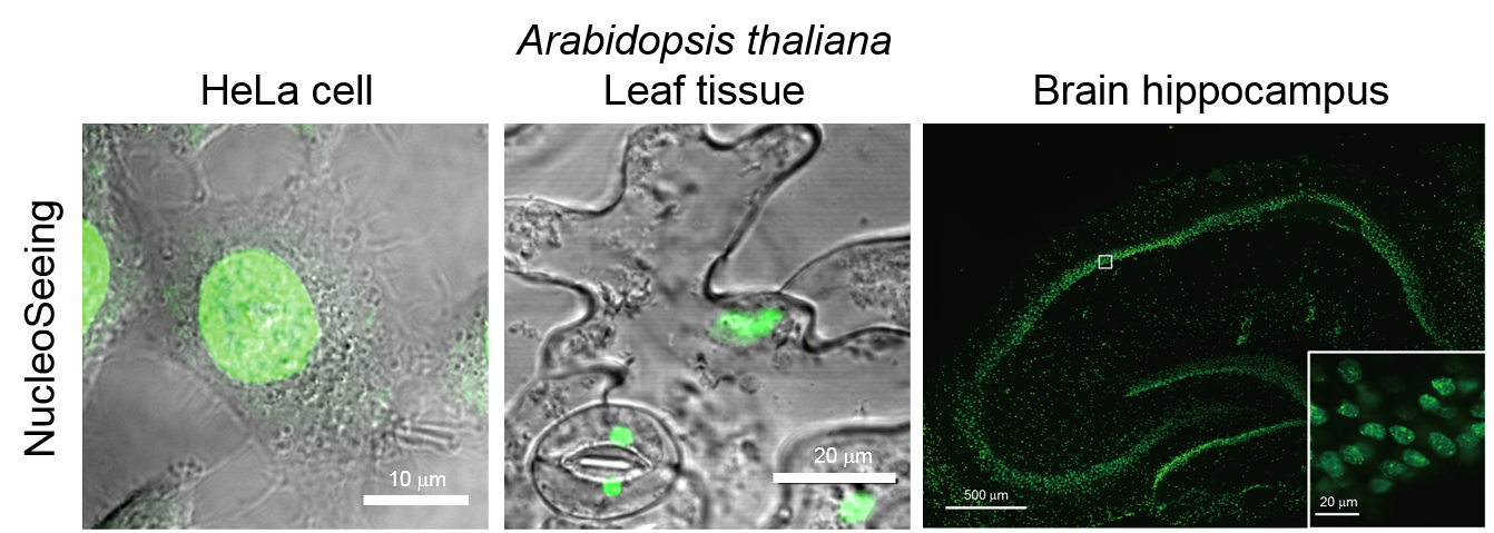
- It has been used in animal-derived cultured cells/tissues and plant cells (Arabidopsis leaf cells), and can stain nuclei with a high S/N ratio in both cases.
- When observing plant cells, it is possible to visualize only nuclei without being affected by autofluorescence (Em: >615 nm) derived from chloroplasts.
- Reversible staining is possible. By changing the medium after staining with this reagent, it is almost completely excreted from the cell in about 12-24 hours after changing the medium.
- Excitation/Emission wavelength: 488 nm/520 nm
- Since the fluorescence intensity changes depending on pH, it can be used as ☞ nuclear pH sensor by taking the fluorescence intensity ratio of 2 wavelengths (Ex 405 nm, Em 520 nm/460 nm) between pH6-8. It emits green fluorescence only upon DNA binding and shows a high S/N ratio.
Application
- Live cell nucleus imaging of cultured cells, cultured tissues
- Live cell nucleus imaging of plant cells
- Cell nucleus staining of fixed cells
- ☞ Cell nucleus-specific pH sensing (pH 6-8)
Example Data
Index
- ☞ DNA-responsive fluorescent properties and nuclear-specific staining
- ☞ Cell toxicity
- ☞ Reversible staining
- ☞ Staining of various cultured cells
- ☞ Long-term time lapse imaging
- ☞ Staining of mouse brain (hippocampus) slice culture tissue
- ☞ Influence of FBS
- ☞ Staining of fixed cells
- ☞ Staining of Arabidopsis thaliana Guard cells
- ☞ Staining of Arabidopsis thaliana epidermal cells
DNA-responsive fluorescent properties and nuclear-specific staining
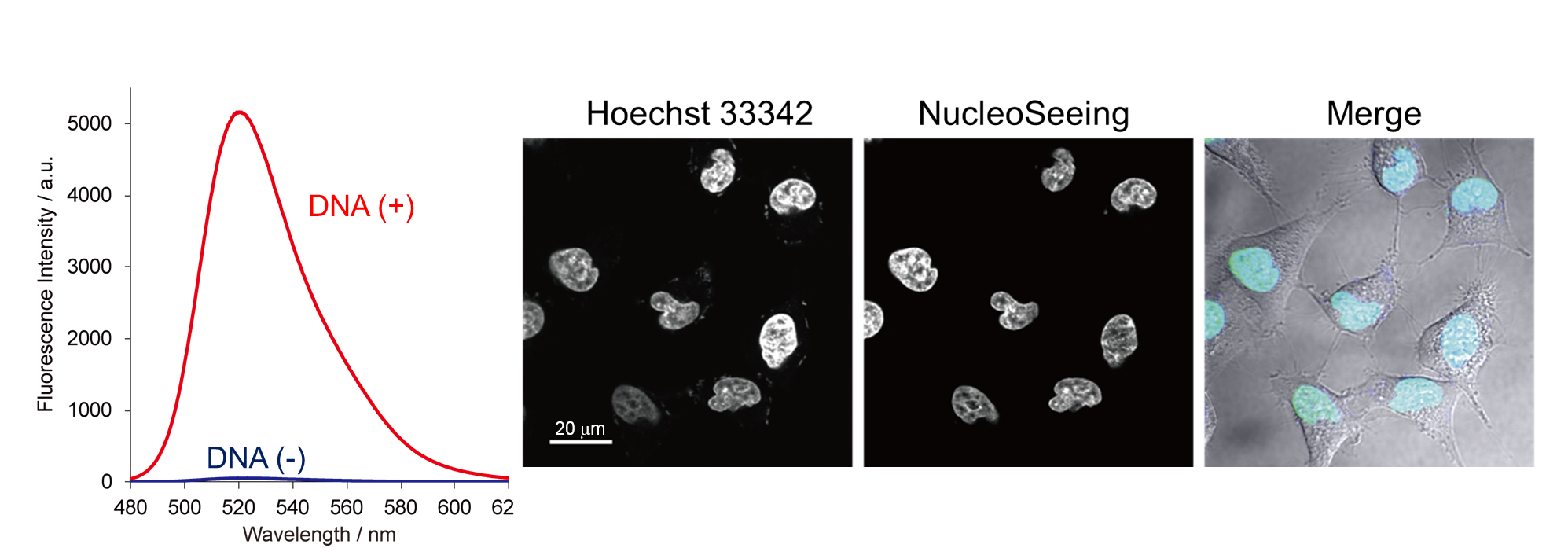
- Left: NucleoSeeing™ shows strong green fluorescent only in the presence of DNA. Excitation 488 nm.
- Right: Co-staining with DNA-specific dye Hoechst33342 (Blue) and NucleoSeeing™(Green) in living cells. They overlap very well.
Cell toxicity
Cell toxicities of Hoechst33342, widely used as blue nuclear staining reagent, and NucleoSeeing™ were tested by MTT assay. While Hoechst causes cell death at 5 μM, NucleoSeeing™ keeps cell viability at least 5 μM.
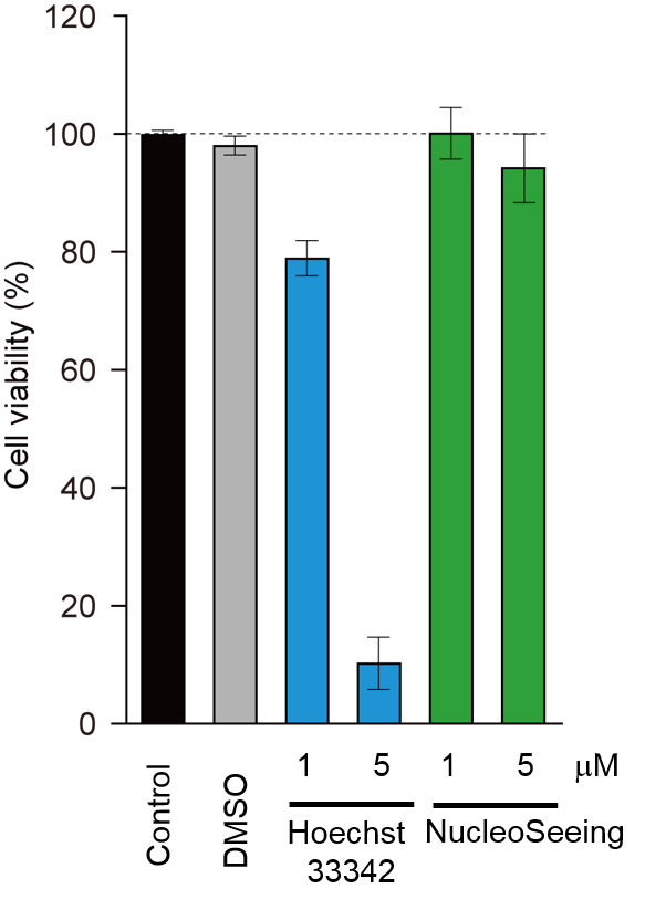
Cytotoxicity of NucleoSeeing™
Reversible staining
To evaluate the cellular retention, cells were treated with 1 μM Hoechst33342 and NucleoSeeing™ for 15 min. The medium was changed to remove excess reagents, and fluorescence intensity was observed for 24 hours. While over 80% of Hoechst33342 signal was still remained, NucleoSeeing™ signals were dramatically reduced within 12 hours. NucleoSeeing™ can be used for reversible nucleus staining.
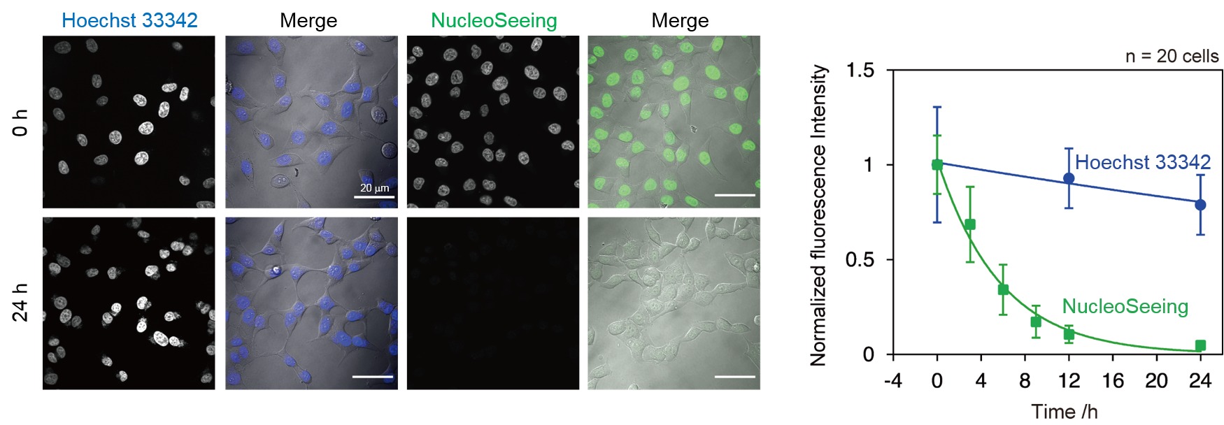
Reversible staining of NucleoSeeing™
Staining of various cultured cells
Various cell lines were treated with 1 μM NucleoSeeing™ for 15 min. After medium change, cells were observed in live cell condition. Nucleus-specific signals were observed in each cell lines.
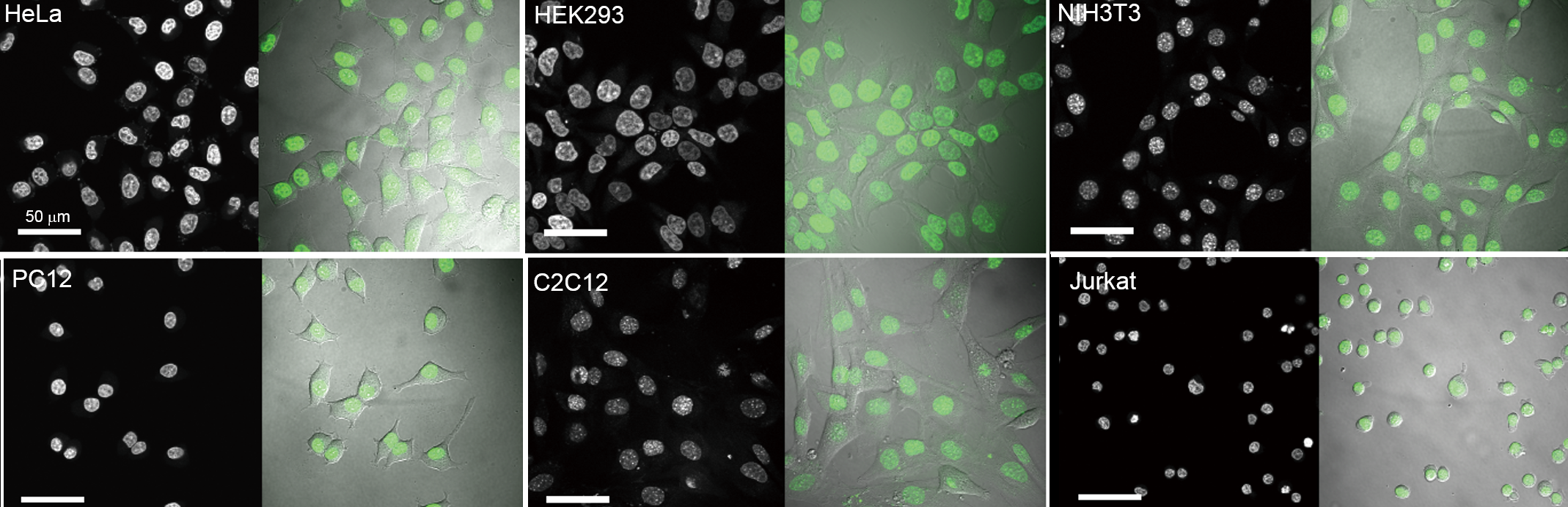
Various cultured cells were stained with NucleoSeeing™
Long-term time lapse imaging
HeLa cells on a glass bottom dish were treated with 0.5 μM NucleoSeeing™ in DMEM (+10% FBS) for
1.5 hours. The cells were observed for 20 hours by confocal microscopy without any washout or medium change (Time lapse condition: Ex 488/Em 500-600 nm, 60x oil lens, 10 min interval).
The process of mitotic cell division and cell proliferation was observed.
※ If you cannot play the video, please see ☞ here.
Staining of mouse brain (hippocampus) slice culture tissue
Cultured mouse hippocampal brain tissue was treated with 20 μM NucleoSeeing™ for 15 min and observed in non-fixed condition.
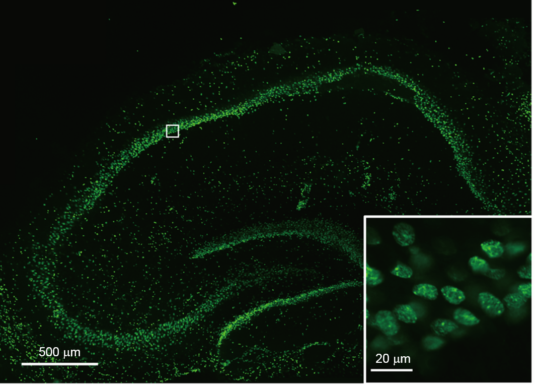
Mouse brain (hippocampus) slice culture tissue was stained with NucleoSeeing™
Influence of FBS
To check any influence of FBS,
cells were treated NucleoSeeing™ in 10% FBS-containing media for 15 min.
Images were acquired by confocal microscopy (Ex. 488/ Em. 500-600 nm) in the same condition (laser power and detection sensitivity).
Compared with 1 μM NucleoSeeing™ in serum-free condition, nucleus fluorescent intensity stained by 1 μM with 10% FBS was partially reduced.
Higher concentration of NucleoSeeing™ in FBS condition recovered fluorescent intensity.
NOTE: NucleoSeeing™ is compatible with FBS-containing medium, however the staining property is lower than FBS-free condition. It is recommended to optimize the reagent concentration.
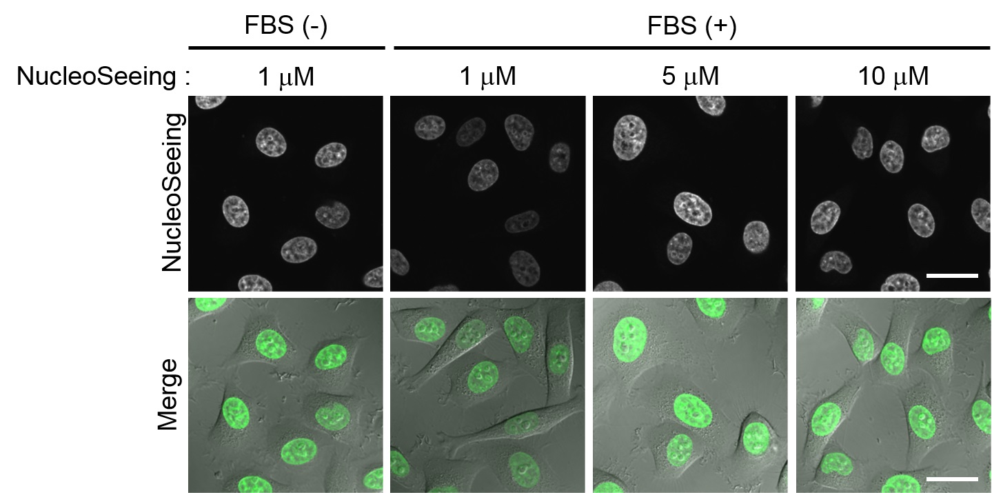
Influence of FBS on NucleoSeeing™ staining
Staining of fixed cells
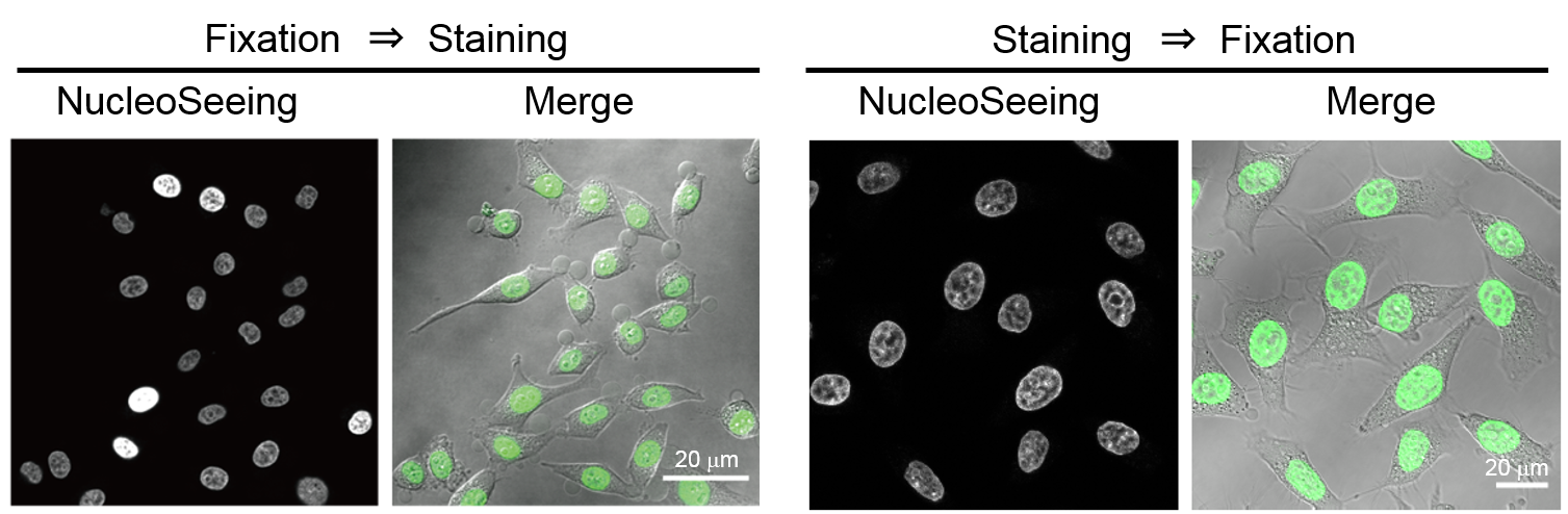
Fixed cells (HeLa cells) were stained with NucleoSeeing™
- Left: PFA-fixed HeLa cells were treated by 1 μM NucleoSeeing™ for 15 min, washed by PBS and observed.
- Right: HeLa cells were treated by 5 μM NucleoSeeing™ for 15 min in live cell condition. After wash the cells, the cells were fixed with 4% PFA and observed.
Staining of Arabidopsis thaliana Guard cells
Leaf slices of Arabidopsis thaliana were treated with 20 μM NucleoSeeing™ for 60 min. After washing, the slices were observed in living condition. When excited at 488 nm, nucleus of guard cells at closed state were observed in green fluorescence (Em. 490-555 nm), and autofluorescence derived from chloroplasts was observed (Em. >615 nm). Using this reagent, cell nucleus can be observed separately from autofluorescence of chloroplasts, and it is expected as a nuclear dynamics imaging reagent for plant cells.
※ After finding nuclear staining in Arabidopsis thaliana Guard cells in ☞ original paper 2, it was reported in ☞ original paper 4 that guard cells are specifically stained when stomata is closed and de-stained when stomata open. Using this phenomenon, a simple and high-throughput evaluation method for stomatal opening and closure using this reagent has been proposed. For details, please refer to ☞ original article 4.

Arabidopsis thaliana Guard cells were stained with NucleoSeeing™
Staining of Arabidopsis thaliana epidermal cells
Arabidopsis thaliana leaves were treated with 20 μM NucleoSeeing™ for 60 min and observed in living condition. Nucleus-specific staining was confirmed in epidermal cells.
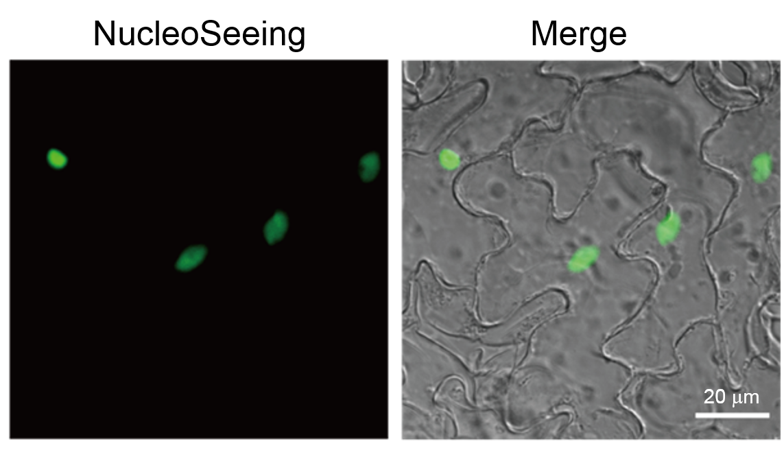
Arabidopsis thaliana epidermal cells were stained with NucleoSeeing™
Nucleus-specific pH sensing
The cell nucleus has a special membrane structure called the nuclear envelope, which consists of two lipid bilayers, an outer membrane and an inner membrane, and is isolated from the cytoplasm. Substances between the nucleus and the cytoplasm are exchanged through nuclear pores, but the environments of the nucleus and the cytoplasm are thought to be different. Recently, it has been suggested that a nuclear membrane-specific proton pump (H+-ATPase) exists in the nuclear envelope, which creates a proton gradient between the nucleus and the cytoplasm. It is expected to measure pH changes in the nucleus in response to physiological phenomena, but a sufficient reagent has not been developed.
☞ The original paper 5 reports that NucleoSeeing™ has pH-dependent fluorescence properties and can be used as a nuclear pH sensing tool. NucleoSeeing™ is usually used as a nuclear staining reagent with excitation light at 480 nm and fluorescence at 520 nm, but when excitation light at 405 nm is used, the fluorescence at around 460 nm and 520 nm changes depending on the pH. By measuring the fluorescence intensity ratio (F520 / F460) at 460 nm and 520 nm at 405 nm, a sigmoid curve can be observed between pH5.5-8.5. By using this principle, pH sensing within the cell nucleus is possible. For details, refer to ☞ the original paper 5.
Principle & Features of Nuclear pH sensing by NucleoSeeing™
(Reference data) pH-dependent fluorescence characteristics of NucleoSeeing™ in vitro
※ Note: The membrane permeability of NucleoSeeing™ is optimized for living cells. After internalization, it is hydrolyzed by esterase and converted to active substance. This data was measured in vitro using chemically synthesized active substance (hydrolysis of NucleoSeeing™) for verification. Please note that this data cannot be reproduced by using NucleoSeeing™ directly in vitro.
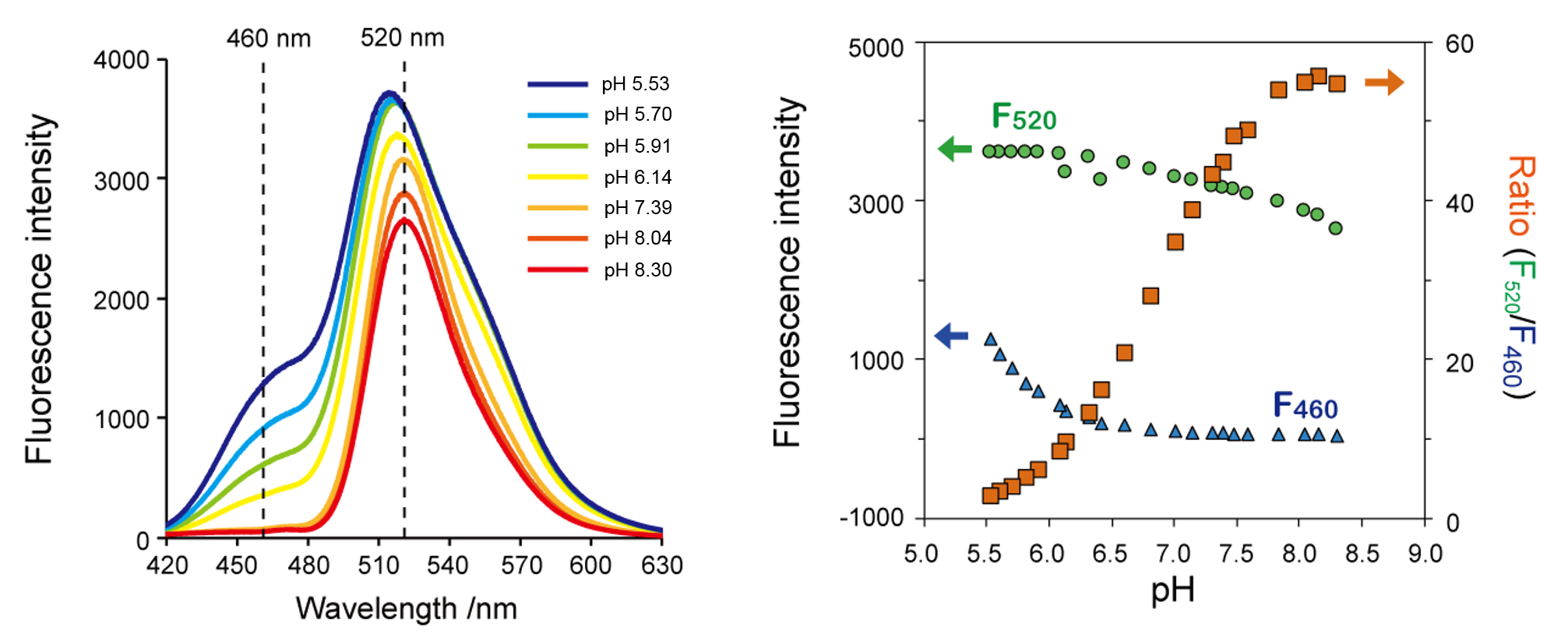
- Left: Fluorescence spectrum and pH dependence of NucleoSeeing™ at 405 nm excitation wavelength. At pH5.5-8.5, the fluorescence intensity around 460 nm and around 520 nm increases with decreasing pH.
- Right: Fluorescence intensity F (excitation wavelength 405 nm) and fluorescence intensity ratio (F520 / F460) at 460 nm and 520 nm at each pH.
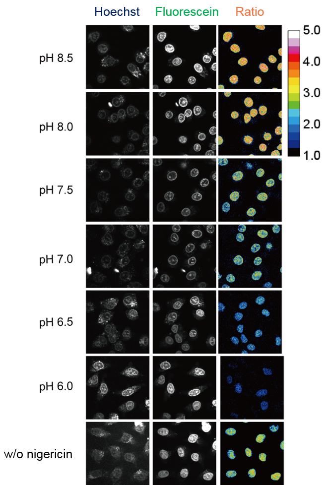
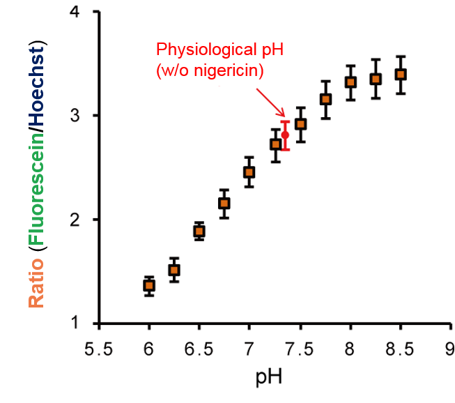
Nuclear pH observation by NucleoSeeing™ in cellulo
After staining cultured cells (HeLa) with NucleoSeeing™, the fluorescence intensity ratio (FFluorescein / FHoechst) at 405 nm of excitation light was observed by confocal microscopy after incubation in pH buffers containing the K+ / H+ ionophore Nigericin*. As in vitro, a pH-dependent response of the fluorescence intensity ratio was observed, and the physiological (non-Nigericin) nuclear pH was estimated to be 7.4.
*Nigericin is a typical K+ / H+ ionophore and is used to match the pH of extracellular solution with that of intracellular pH.
Original Papers
※ This product, NucleoSeeing™ is equal to hoeAc2FL in the papers below.
- Nakamura, A., et al., "Hoechst tagging: a modular strategy to design synthetic fluorescent probes for live-cell nucleus imaging.", Chem. Commun., 50, 6149~6152 (2014). [PMID:24776726]
- Ueda, M., et al., "Noncanonical function of a small-molecular virulence factor coronatine against plant immunity: an in vivo raman imaging approach."ACS Cent. Sci., 3 (5), 462~472 (2017). [PMID:28573209]
- Nakamura, A. and Tsukiji, S., "Ratiometric fluorescence imaging of nuclear pH in living cells using hoechst-tagged fluorescein.", Bioorg. Med. Chem. Lett., 27 (14), 3127~3130 (2017). [PMID:28558968]
- Takaoka, Y., et al., "Hoechst-tagged Fluorescein Diacetate for the Fluorescence Imaging-based Assessment of Stomatal Dynamics in Arabidopsis thaliana.", Sci. Rep., 10, 5333 (2020). (
 )
) - Nakamura, A. and Tsukiji, S., "Ratiometric fluorescence imaging of nuclear pH in living cells using hoechst-tagged fluorescein.", Bioorg. Med. Chem. Lett., 27 (14), 3127~3130 (2017). [PMID:28558968]
Cited Papers
- Okabe, M., et al., "An approach for live imaging of first cleavage in mouse embryos using fluorescent chemical probes for DNA, microtubules, and microfilaments", Reprod. Med. Biol., 22(1), e12551 [PMID:38023339]
Video
Product Information
[Date : July 01 2025 00:09]
| Detail | Product Name | Product Code | Supplier | Size | Price | ||||||||||||||||||||||||||||||
|---|---|---|---|---|---|---|---|---|---|---|---|---|---|---|---|---|---|---|---|---|---|---|---|---|---|---|---|---|---|---|---|---|---|---|---|
|
NucleoSeeing, Live Nucleus Green DatasheetThis may not be the latest data sheet. |
FDV-0029 | FNAFunakoshi Co.,Ltd. | 0.1 mg | $350 | |||||||||||||||||||||||||||||||
|
|
|
||||||||||||||||||||||||||||||||||
[Date : July 01 2025 00:09]
NucleoSeeing, Live Nucleus Green
DatasheetThis may not be the latest data sheet.
- Product Code: FDV-0029
- Supplier: FNA
- Size: 0.1mg
- Price: $350
| Description |
NucleoSeeing is a novel live cell imaging probe which emits green fluorescence by binding to DNA specifically. NucleoSeeing can be used for not only in animal cells or tissues, but also Guard cell of Arabidopsis thaliana with high S/N ratio. Moreover, NucleoSeeing can be used as a pH sensor in nucleus. |
||
|---|---|---|---|
| Storage | -20°C | CAS | |
| Link |
"NucleoSeeing" (Reversible Fluorescent Dye for Nucleus) in a minute! |
||
You may also like

CONTACT
export@funakoshi.co.jp
- ※Prices on our website are for your reference only. Please inquire your distributor for your prices.
- ※Please note that Product Information or Price may change without notice.