HOME>
Products>
Protein / Enzyme>
Protein Labeling Reagents>
LiveReceptor™ AMPAR (Endogenous AMPAR Labeling Reagent)
HOME>
Products>
Cell Culture>
Imaging dye>
LiveReceptor™ AMPAR (Endogenous AMPAR Labeling Reagent)
Endogenous AMPA-Type Glutamate Receptor (AMPAR) Live Cell Imaging Reagents LiveReceptor™ AMPAR (Endogenous AMPAR Labeling Reagent)
Date:June 11 2018Web Page No:81075

Funakoshi Co.,Ltd.

LiveReceptor ™AMPAR is a protein-labeling reagent that specifically labels AMPA type glutamate receptor (AMPA Receptor; AMPAR) with fluorescein in living cells. Since it can label endogenous AMPAR in living cells, it is excellent for analyzing physiological AMPAR behavior by live-cell imaging.
※This product is commercialized by the research result of Professor Hamachi, at Department of Synthetic Chemistry and Biological Chemistry, Graduate School of Engineering, Kyoto University.
Background
Generally, fluorescein direct conjugated ligand has been used for labeling receptors, however, such ligands easiliy detach from ligand binding pocket, and then signal will be faded out. (See below image)
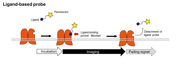
The LiveReceptor™ has a linker of acyl imidazole between fluorescein and ligand. While ligand is binding to receptor, nucleophillic amino acids attacks acyl imidazole and changes its chemical structure. By this method, receptors are directly labeled and keep fluorescence even after the ligand is detached from binding pocket.

Features
- Can label fluorecein to endogenous AMPAR
- Highly specific to AMPAR (GluA2-4 subunit)*. Other ion channel type glutamate receptors will not be labeled.
- Can observe signal for 1 to 4 hours after adding LiveReceptor™ to medium.
- No cell permeability - only AMPAR on cell surface can be labeled.
- It has been used in cultured nerve cells, cultured brain slices, and cell lines overexpressing AMPAR (GluA2, GluA3, or GluA4 subunits).
- Because of its low molecular weight, it has high tissue penetration and has a track record of deep labeling in acute slices of brain tissue.
- Ion channel function is maintained after labeling.
- Low cytotoxicity under recommended concentration (1 μM)
- Since fluorescein is used as a fluorescent dye, it is pH-responsive, and labeled AMPAR after internalization tends to be quenched. It is ideal for observation of cell surface AMPAR.
- It can be used for various applications including live cell imaging. For details, see the Example of Use below.
* GluA1 homomers cannot be visualized because the GluA1 subunit is not labeled.
Application
- Live cell imaging
- Immunocytochemistry with specific antibodies
- Immunoprecipitation with anti-fluorescein antibody
- Immunoblotting with anti-fluorescein antibody
- Drug screening for competitive AMPAR antagonist
Example of Use
Live cell imaging of labelled endogenous
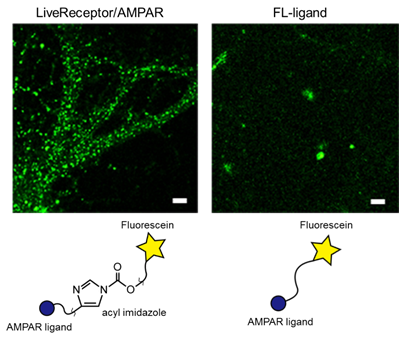
Cultured hippocampal neurons were treated with 1 μM of LiveReceptor™ AMPAR (in left) or Fluorescein-conjugated ligand as negative control (in right) for 1 hour at 17℃ and washed out three times with the basal medium. Dendritic spin-like punctual structures were observed on live cells by specifically LiveReceptor™.
Live cell imaging of labelled endogenous AMPARs in cultured slice tissue
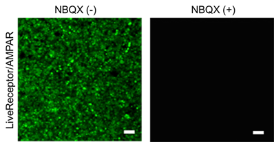
Acutely prepared hippocampal slices were treated with 1 μM of LiveReceptor™ AMPAR for 1 hour at 17℃ in the absence (in left) or presence (in right) of 10 μM NBQX, a potent inhibitor of AMPAR, and washed out three times with the basal medium. Dendritic spin-like punctual structures were observed on live cells and the signal was clearly disappeared by NBQX.
Staining of AMPARs in deep tissue in cultured slice tissue
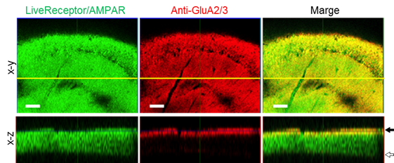
Cultured slice tissue of mouse was stained by LiveReceptor™ AMPAR and fixed. After fixation, stained by anti-GluA2/3 antibody. x-y dimension shows a good concordance between LiveReceptor™ AMPAR and anti-GluA2/3 antibody. However, x-z dimension shows that anti-GluA2/3 antibody stained the surface of slice tissue only. LiveReceptor™ AMPAR penetrates into deep tissue and can label them. (←Black arrowhead : Top of sliced tissue / ⇦White arrowhead : Bottom of sliced tissue)
References
- Fujishima, S., et al., "Ligand-directed acyl imidazole chemistry for labeling of membrane-bound proteins on live cells", J. Am. Chem. Soc., 134(9), 3961~3964 (2012). [PMID: 22352855]
- Miki, T., et al.,"LDAI-based chemical labeling of intact membrane proteins and its pulse-chase analysis under live cell conditions", Chem. Biol., 21(8), 1013~1022 (2014). (
 )
) - Wakayama, S., et al.,"Chemical labelling for visualizing native AMPA receptors in live neurons", Nat. Commun., 8, 14850 (2017). [PMID: 28387242]
Webinar Movie
[Date : July 10 2026 00:08]
| Detail | Product Name | Product Code | Supplier | Size | Price | ||||||||||||||||||||||||||||||
|---|---|---|---|---|---|---|---|---|---|---|---|---|---|---|---|---|---|---|---|---|---|---|---|---|---|---|---|---|---|---|---|---|---|---|---|
|
LiveReceptor AMPAR, Endogenous AMPAR Labeling Reagent DatasheetThis may not be the latest data sheet. |
FDV-0018A | FNAFunakoshi Co.,Ltd. | 10 µg | $600 | |||||||||||||||||||||||||||||||
|
|
|
||||||||||||||||||||||||||||||||||
[Date : July 10 2026 00:08]
LiveReceptor AMPAR, Endogenous AMPAR Labeling Reagent
DatasheetThis may not be the latest data sheet.
- Product Code: FDV-0018A
- Supplier: FNA
- Size: 10µg
- Price: $600
| Description |
LiveReceptor AMPAR is the world-first reagent which can specifically label AMPA-type glutamate receptors (AMPARs) with fluorescein dye on living neurons. |
||
|---|---|---|---|
| Storage | -20°C | CAS | |
| Link | |||
CONTACT
export@funakoshi.co.jp
- ※Prices on our website are for your reference only. Please inquire your distributor for your prices.
- ※Please note that Product Information or Price may change without notice.