
Anti-p63, Rabbit-Poly <Anti-TP63> | GeneTex International Corporation
掲載日情報:2026/06/03 現在Webページ番号:548415

GeneTex International CorporationのAnti-p63, Rabbit-Poly <Anti-TP63>商品情報ページです。
※本製品は研究用です。研究用以外には使用できません。
カートに商品を
追加しました。
追加しました。
価格
[在庫・価格 :2026年07月04日 16時35分現在]
※ 表示されている納期は弊社に在庫が無く、取り寄せた場合の納期目安となります。
| 詳細 | 商品名 |
|
文献数 | ||||||||||||||||||||||||||||||||||||||||||||||||||||||||||||||||||||||||||||||||||
|---|---|---|---|---|---|---|---|---|---|---|---|---|---|---|---|---|---|---|---|---|---|---|---|---|---|---|---|---|---|---|---|---|---|---|---|---|---|---|---|---|---|---|---|---|---|---|---|---|---|---|---|---|---|---|---|---|---|---|---|---|---|---|---|---|---|---|---|---|---|---|---|---|---|---|---|---|---|---|---|---|---|---|---|---|---|
|
Anti-p63, Rabbit-Poly <Anti-TP63> |
|
57 | |||||||||||||||||||||||||||||||||||||||||||||||||||||||||||||||||||||||||||||||||||
|
|||||||||||||||||||||||||||||||||||||||||||||||||||||||||||||||||||||||||||||||||||||


[在庫・価格 :2026年07月04日 16時35分現在]
※ 表示されている納期は弊社に在庫が無く、取り寄せた場合の納期目安となります。
Anti-p63, Rabbit-Poly <Anti-TP63>
文献数: 57
- 商品コード:GTX102425
- メーカー:GNT
- 包装:25μl
- 価格:¥30,000
- 在庫:1個
- 納期:10日程度 ※※ 表示されている納期は弊社に在庫がなく、取り寄せた場合の目安納期となります。
- 法規制等:
| 説明文 | レビューあり。 別名:tumor protein p63,AIS,B(p51A),B(p51B),EEC3,KET,LMS,NBP,OFC8,RHS,SHFM4,TP53CP,TP53L,TP73L,p40,p51,p53CP,p63,p73H,p73L Genbank No: 8626 |
||||||
|---|---|---|---|---|---|---|---|
| 別包装品 | 別包装品あり | ||||||
| 法規制等 | |||||||
| 保存条件 | -20℃ | 法規備考 | |||||
| 抗原種 | Human | 免疫動物 | Rabbit | ||||
| 交差性 | Cat/Dog/Human/Mouse/Rat | 適用 | IC,IF,IHC,IP,PLA,Western Blot | ||||
| 標識 | Unlabeled | 性状 | Purified | ||||
| 吸収処理 | クラス | IgG | |||||
| クロナリティ | Polyclonal | フォーマット | |||||
| 掲載カタログ |
|
||||||
| 製品記事 | 肺癌マーカー関連抗体 使いっきり抗体 |
||||||
| 関連記事 | GeneTex社における抗体の品質管理 |
||||||
カートに商品を
追加しました。
追加しました。
ラインナップ
カートに商品を
追加しました。
追加しました。
画像
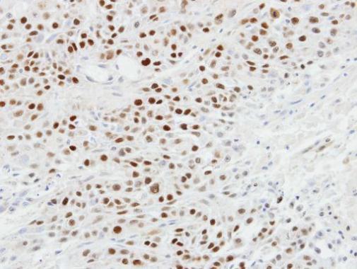
GTX102425 IHC-P Image
Immunohistochemical analysis of paraffin-embedded SCC4 xenograft, using p63(GTX102425) antibody at 1:100 dilution.
Immunohistochemical analysis of paraffin-embedded SCC4 xenograft, using p63(GTX102425) antibody at 1:100 dilution.
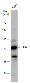
![GTX102425 WB Image<br>p63 antibody [N2C1], Internal detects TP63 protein by western blot analysis.<br>A. 50 ug mouse brain lysate/extract<br>7.5% SDS-PAGE<br>p63 antibody [N2C1], Internal (GTX102425) dilution: 1:500<br>The HRP-conjugated anti-rabbit IgG antibody (GTX213110-01) was used to detect the primary antibody.](/domestic/data/graphics/GNT/graphics/GTX102425_39918_WB_M_brain.jpg)
GTX102425 WB Image
p63 antibody [N2C1], Internal detects TP63 protein by western blot analysis.
A. 50 ug mouse brain lysate/extract
7.5% SDS-PAGE
p63 antibody [N2C1], Internal (GTX102425) dilution: 1:500
The HRP-conjugated anti-rabbit IgG antibody (GTX213110-01) was used to detect the primary antibody.
p63 antibody [N2C1], Internal detects TP63 protein by western blot analysis.
A. 50 ug mouse brain lysate/extract
7.5% SDS-PAGE
p63 antibody [N2C1], Internal (GTX102425) dilution: 1:500
The HRP-conjugated anti-rabbit IgG antibody (GTX213110-01) was used to detect the primary antibody.
![GTX102425 WB Image<br>p63 antibody [N2C1], Internal detects TP63 protein by western blot analysis.<br>A. 50 ug rat brain lysate/extract<br>7.5% SDS-PAGE<br>p63 antibody [N2C1], Internal (GTX102425) dilution: 1:500<br>The HRP-conjugated anti-rabbit IgG antibody (GTX213110-01) was used to detect the primary antibody.](/domestic/data/graphics/GNT/graphics/GTX102425_39918_WB_R_brain.jpg)
GTX102425 WB Image
p63 antibody [N2C1], Internal detects TP63 protein by western blot analysis.
A. 50 ug rat brain lysate/extract
7.5% SDS-PAGE
p63 antibody [N2C1], Internal (GTX102425) dilution: 1:500
The HRP-conjugated anti-rabbit IgG antibody (GTX213110-01) was used to detect the primary antibody.
p63 antibody [N2C1], Internal detects TP63 protein by western blot analysis.
A. 50 ug rat brain lysate/extract
7.5% SDS-PAGE
p63 antibody [N2C1], Internal (GTX102425) dilution: 1:500
The HRP-conjugated anti-rabbit IgG antibody (GTX213110-01) was used to detect the primary antibody.
![GTX102425 ICC/IF Image<br>p63 antibody [N2C1], Internal detects p63 protein at nucleus by immunofluorescent analysis.<br>Sample: A431 cells were fixed in 4% paraformaldehyde at RT for 15 min.<br>Green: p63 protein stained by p63 antibody [N2C1], Internal (GTX102425) diluted at 1:500.<br>Blue: Hoechst 33342 staining.<br>Scale bar = 10 um.](/domestic/data/graphics/GNT/graphics/GTX102425_41472_20151119_IFA.jpg)
GTX102425 ICC/IF Image
p63 antibody [N2C1], Internal detects p63 protein at nucleus by immunofluorescent analysis.
Sample: A431 cells were fixed in 4% paraformaldehyde at RT for 15 min.
Green: p63 protein stained by p63 antibody [N2C1], Internal (GTX102425) diluted at 1:500.
Blue: Hoechst 33342 staining.
Scale bar = 10 um.
![GTX102425 IP Image<br>Immunoprecipitation of p63 protein from A431 whole cell extracts using 5 ug of p63 antibody [N2C1], Internal (GTX102425).<br>Western blot analysis was performed using p63 antibody [N2C1], Internal (GTX102425).<br>EasyBlot anti-Rabbit IgG (GTX221666-01) was used as a secondary reagent.</br></br></br>](/domestic/data/graphics/GNT/graphics/GTX102425_39918_20150910_IP.jpg)
GTX102425 IP Image
Immunoprecipitation of p63 protein from A431 whole cell extracts using 5 ug of p63 antibody [N2C1], Internal (GTX102425).
Western blot analysis was performed using p63 antibody [N2C1], Internal (GTX102425).
EasyBlot anti-Rabbit IgG (GTX221666-01) was used as a secondary reagent.
Immunoprecipitation of p63 protein from A431 whole cell extracts using 5 ug of p63 antibody [N2C1], Internal (GTX102425).
Western blot analysis was performed using p63 antibody [N2C1], Internal (GTX102425).
EasyBlot anti-Rabbit IgG (GTX221666-01) was used as a secondary reagent.
GTX102425 WB Image
p63 antibody detects p63 protein by Western blot analysis. Whole cell extracts (30 ug) was separated by 7.5% SDS-PAGE, and the membrane was blotted with p63 antibody (GTX102425) diluted by 1:500.
p63 antibody detects p63 protein by Western blot analysis. Whole cell extracts (30 ug) was separated by 7.5% SDS-PAGE, and the membrane was blotted with p63 antibody (GTX102425) diluted by 1:500.
![GTX102425 WB Image<br>Non-transfected (?) and transfected (+) A431 whole cell extracts (30 ug) were separated by 7.5% SDS-PAGE, and the membrane was blotted with p63 antibody [N2C1], Internal (GTX102425) diluted at 1:2000. The HRP-conjugated anti-rabbit IgG antibody (GTX213110-01) was used to detect the primary antibody.](/domestic/data/graphics/GNT/graphics/GTX102425_41472_20180105_WB_shRNA_watermark.jpg)
GTX102425 WB Image
Non-transfected (?) and transfected (+) A431 whole cell extracts (30 ug) were separated by 7.5% SDS-PAGE, and the membrane was blotted with p63 antibody [N2C1], Internal (GTX102425) diluted at 1:2000. The HRP-conjugated anti-rabbit IgG antibody (GTX213110-01) was used to detect the primary antibody.
Non-transfected (?) and transfected (+) A431 whole cell extracts (30 ug) were separated by 7.5% SDS-PAGE, and the membrane was blotted with p63 antibody [N2C1], Internal (GTX102425) diluted at 1:2000. The HRP-conjugated anti-rabbit IgG antibody (GTX213110-01) was used to detect the primary antibody.
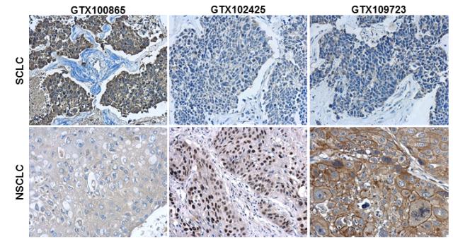
GTX102425 IHC-P Image
Immunohistochemical characterization of Synaptophysin (GTX100865), p63 (GTX102425) and Cytokeratin 7 (GTX109723) in human small cell lung cancer (SCLC) and non-small cell lung cancer (NSCLC) specimens.
Sample: Paraffin-embedded human SCLC (upper panel) and NSCLC (lower panel).
The section was pre-treated using heat mediated antigen retrieval with sodium citrate buffer (pH6) for 15 mins. The section was then incubated with primary antibody at 1:500 overnight at 4 and detected using an HRP conjugated avidin-biotin-peroxidase Complex system. DAB was used as the chromogen and counterstained with haematoxylin.
カートに商品を
追加しました。
追加しました。
商品情報
| 商品説明 | レビューあり |
|---|---|
| 抗原動物 | Human |
| 交差性 | Cat/Dog/Human/Mouse/Rat |
| 免疫動物 | Rabbit |
| 性状 | Purified |
| 適用 | IC, IF, IHC, IP, PLA, Western Blot |
| クラス | IgG |
| 標識 | Unlabeled |
| クロナリティ | Polyclonal |
| 別名 | tumor protein p63, AIS, B(p51A), B(p51B), EEC3, KET, LMS, NBP, OFC8, RHS, SHFM4, TP53CP, TP53L, TP73L, p40, p51, p53CP, p63, p73H, p73L |
| Genbank No | 8626 |
| データシート | データシート |
| メーカーサイト | メーカーサイト |
| 使用文献 | 使用文献 |
| 保存条件 | -20℃ |
カートに商品を
追加しました。
追加しました。
製品情報は掲載時点のものですが、価格表内の価格については随時最新のものに更新されます。お問い合わせいただくタイミングにより製品情報・価格などは変更されている場合があります。
表示価格に、消費税等は含まれていません。一部価格が予告なく変更される場合がありますので、あらかじめご了承下さい。