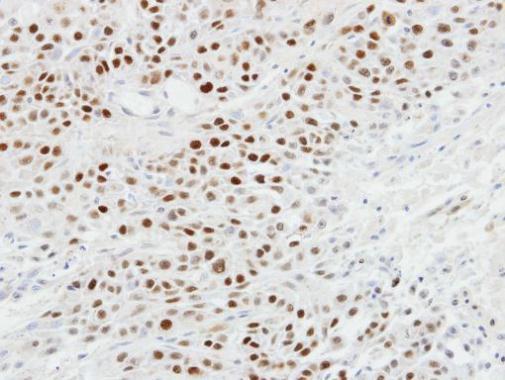
GTX102425 IHC-P Image
Immunohistochemical analysis of paraffin-embedded SCC4 xenograft, using p63(GTX102425) antibody at 1:100 dilution.
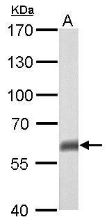
GTX102425 WB Image
p63 antibody [N2C1], Internal detects TP63 protein by western blot analysis.
A. 50 ug mouse brain lysate/extract
7.5% SDS-PAGE
p63 antibody [N2C1], Internal (GTX102425) dilution: 1:500
The HRP-conjugated anti-rabbit IgG antibody (GTX213110-01) was used to detect the primary antibody.
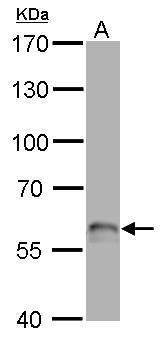
GTX102425 WB Image
p63 antibody [N2C1], Internal detects TP63 protein by western blot analysis.
A. 50 ug rat brain lysate/extract
7.5% SDS-PAGE
p63 antibody [N2C1], Internal (GTX102425) dilution: 1:500
The HRP-conjugated anti-rabbit IgG antibody (GTX213110-01) was used to detect the primary antibody.
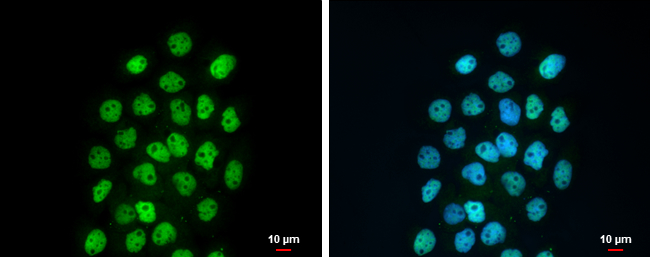
GTX102425 ICC/IF Image
p63 antibody [N2C1], Internal detects p63 protein at nucleus by immunofluorescent analysis.
Sample: A431 cells were fixed in 4% paraformaldehyde at RT for 15 min.
Green: p63 protein stained by p63 antibody [N2C1], Internal (GTX102425) diluted at 1:500.
Blue: Hoechst 33342 staining.
Scale bar = 10 um.
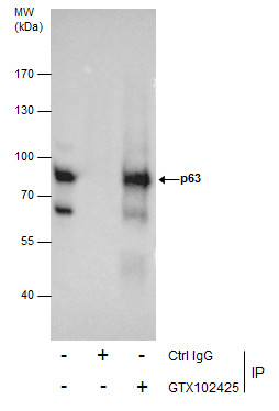
GTX102425 IP Image
Immunoprecipitation of p63 protein from A431 whole cell extracts using 5 ug of p63 antibody [N2C1], Internal (GTX102425).
Western blot analysis was performed using p63 antibody [N2C1], Internal (GTX102425).
EasyBlot anti-Rabbit IgG (GTX221666-01) was used as a secondary reagent.
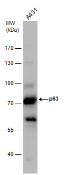
GTX102425 WB Image
p63 antibody detects p63 protein by Western blot analysis. Whole cell extracts (30 ug) was separated by 7.5% SDS-PAGE, and the membrane was blotted with p63 antibody (GTX102425) diluted by 1:500.
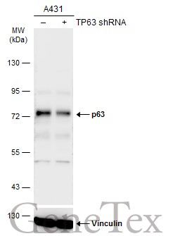
GTX102425 WB Image
Non-transfected (?) and transfected (+) A431 whole cell extracts (30 ug) were separated by 7.5% SDS-PAGE, and the membrane was blotted with p63 antibody [N2C1], Internal (GTX102425) diluted at 1:2000. The HRP-conjugated anti-rabbit IgG antibody (GTX213110-01) was used to detect the primary antibody.
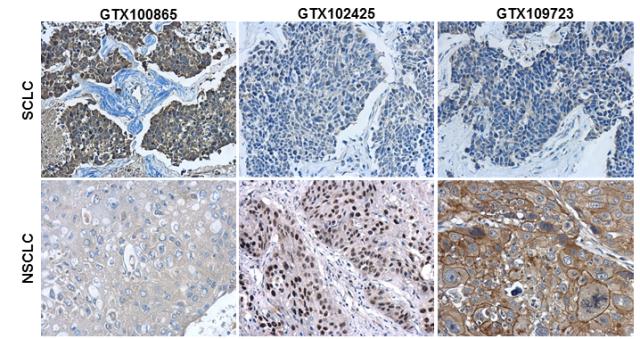
GTX102425 IHC-P Image
Immunohistochemical characterization of Synaptophysin (GTX100865), p63 (GTX102425) and Cytokeratin 7 (GTX109723) in human small cell lung cancer (SCLC) and non-small cell lung cancer (NSCLC) specimens.
Sample: Paraffin-embedded human SCLC (upper panel) and NSCLC (lower panel).
The section was pre-treated using heat mediated antigen retrieval with sodium citrate buffer (pH6) for 15 mins. The section was then incubated with primary antibody at 1:500 overnight at 4Åé and detected using an HRP conjugated avidin-biotin-peroxidase Complex system. DAB was used as the chromogen and counterstained with haematoxylin.