
Anti-HIF-1α, Rabbit-Poly | GeneTex International Corporation
掲載日情報:2026/06/03 現在Webページ番号:540150

GeneTex International CorporationのAnti-HIF-1α, Rabbit-Poly商品情報ページです。
※本製品は研究用です。研究用以外には使用できません。
カートに商品を
追加しました。
追加しました。
価格
[在庫・価格 :2026年07月17日 00時01分現在]
※ 表示されている納期は弊社に在庫が無く、取り寄せた場合の納期目安となります。
| 詳細 | 商品名 |
|
文献数 | ||||||||||||||||||||||||||||||||||||||||||||||||||||||||||||||||||||||||||||||||||
|---|---|---|---|---|---|---|---|---|---|---|---|---|---|---|---|---|---|---|---|---|---|---|---|---|---|---|---|---|---|---|---|---|---|---|---|---|---|---|---|---|---|---|---|---|---|---|---|---|---|---|---|---|---|---|---|---|---|---|---|---|---|---|---|---|---|---|---|---|---|---|---|---|---|---|---|---|---|---|---|---|---|---|---|---|---|
|
Anti-HIF-1α, Rabbit-Poly |
|
177 | |||||||||||||||||||||||||||||||||||||||||||||||||||||||||||||||||||||||||||||||||||
|
|||||||||||||||||||||||||||||||||||||||||||||||||||||||||||||||||||||||||||||||||||||


[在庫・価格 :2026年07月17日 00時01分現在]
※ 表示されている納期は弊社に在庫が無く、取り寄せた場合の納期目安となります。
Anti-HIF-1α, Rabbit-Poly
文献数: 177
- 商品コード:GTX127309
- メーカー:GNT
- 包装:25μl
- 価格:¥30,000
- 在庫:1個
- 納期:10日程度 ※※ 表示されている納期は弊社に在庫がなく、取り寄せた場合の目安納期となります。
- 法規制等:
| 説明文 | レビューあり。KO/KDバリデーション済み抗体。抗原:aa 657~826 別名:hypoxia inducible factor 1 subunit alpha,HIF-1-alpha,HIF-1A,HIF-1alpha,HIF1,HIF1-ALPHA,MOP1,PASD8,bHLHe78 Genbank No: 3091 |
||||||
|---|---|---|---|---|---|---|---|
| 別包装品 | 別包装品あり | ||||||
| 法規制等 | |||||||
| 保存条件 | -20℃ | 法規備考 | |||||
| 抗原種 | Human | 免疫動物 | Rabbit | ||||
| 交差性 | Bovine/Human/Mouse/Rabbit/Rat | 適用 | ChIP,IC,IF,IHC,IP,Western Blot | ||||
| 標識 | Unlabeled | 性状 | Purified | ||||
| 吸収処理 | クラス | IgG | |||||
| クロナリティ | Polyclonal | フォーマット | |||||
| 掲載カタログ |
|
||||||
| 製品記事 | がん転移関連因子抗体 抗HIF-1α抗体(GeneTex社) 低酸素症関連抗体 使いっきり抗体 抗HIF-1α抗体|Anti-HIF-1α Antibody |
||||||
| 関連記事 | GeneTex社における抗体の品質管理 |
||||||
カートに商品を
追加しました。
追加しました。
ラインナップ
カートに商品を
追加しました。
追加しました。
画像
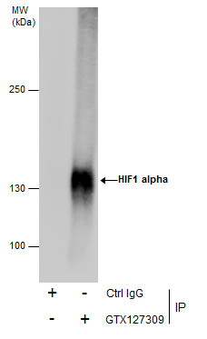
GTX127309 IP Image
Immunoprecipitation of HIF1 alpha protein from HepG2 whole cell extracts treated with 500 uM CoCl2 for 24 hr using 5 ug of HIF1 alpha antibody (GTX127309).
Western blot analysis was performed using HIF1 alpha antibody (GTX127309).
EasyBlot anti-Rabbit IgG (GTX221666-01) was used as a secondary reagent.
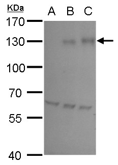
GTX127309 WB Image
HIF1 alpha antibody detects HIF1 alpha protein by western blot analysis.
A. 30 ug HepG2 whole cell lysate/extract (untreated)
B.30 ug HepG2 whole cell lysate/extract ( 200 uM CoCl2 treatment for 24 hr)
C. 30 ug HepG2 whole cell lysate/extract (500 uM CoCl2 treatment for 24 hr)
7.5% SDS-PAGE
HIF1 alpha antibody (GTX127309) dilution: 1:5000
The HRP-conjugated anti-rabbit IgG antibody (GTX213110-01) was used to detect the primary antibody.
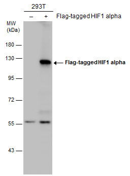
GTX127309 WB Image
Non-transfected (?) and transfected (+) 293T whole cell extracts (30 ug) were separated by 7.5% SDS-PAGE, and the membrane was blotted with HIF1 alpha antibody (GTX127309) diluted at 1:5000. The HRP-conjugated anti-rabbit IgG antibody (GTX213110-01) was used to detect the primary antibody.
Non-transfected (?) and transfected (+) 293T whole cell extracts (30 ug) were separated by 7.5% SDS-PAGE, and the membrane was blotted with HIF1 alpha antibody (GTX127309) diluted at 1:5000. The HRP-conjugated anti-rabbit IgG antibody (GTX213110-01) was used to detect the primary antibody.
GTX127309 WB Image
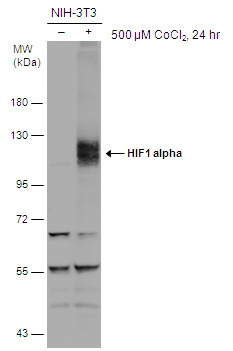
Untreated (?) and treated (+) NIH-3T3 whole cell extracts (30 ug) were separated by 7.5% SDS-PAGE, and the membrane was blotted with HIF1 alpha antibody (GTX127309) diluted at 1:500. The HRP-conjugated anti-rabbit IgG antibody (GTX213110-01) was used to detect the primary antibody.
Untreated (?) and treated (+) NIH-3T3 whole cell extracts (30 ug) were separated by 7.5% SDS-PAGE, and the membrane was blotted with HIF1 alpha antibody (GTX127309) diluted at 1:500. The HRP-conjugated anti-rabbit IgG antibody (GTX213110-01) was used to detect the primary antibody.
GTX127309 ICC/IF Image
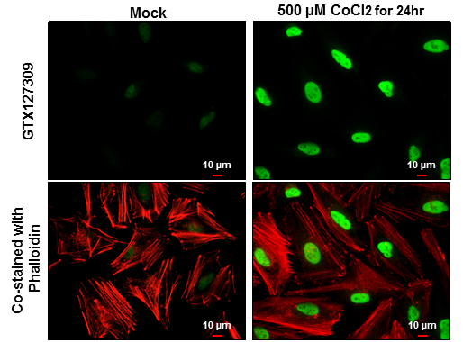
HIF1 alpha antibody detects HIF1 alpha protein at nucleus by immunofluorescent analysis.
Sample: HeLa cells were fixed in 4% paraformaldehyde at RT for 15 min.
Green: HIF1 alpha protein stained by HIF1 alpha antibody (GTX127309) diluted at 1:500.
Red: Phalloidin, a cytoskeleton marker, diluted at 1:100.
Scale bar = 10 um.
GTX127309 ICC/IF Image
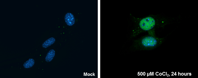
HIF1 alpha antibody detects HIF1 alpha protein at nucleus by immunofluorescent analysis.
Sample: NIH/3T3 cells were fixed in 4% paraformaldehyde at RT for 15 min.
Green: HIF1 alpha protein stained by HIF1 alpha antibody (GTX127309) diluted at 1:200.
Blue: Hoechst 33342 staining.
HIF1 alpha antibody detects HIF1 alpha protein at nucleus by immunofluorescent analysis.
Sample: NIH/3T3 cells were fixed in 4% paraformaldehyde at RT for 15 min.
Green: HIF1 alpha protein stained by HIF1 alpha antibody (GTX127309) diluted at 1:200.
Blue: Hoechst 33342 staining.
GTX127309 WB Image
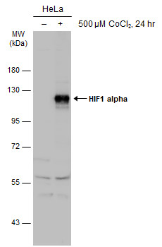
Untreated (?) and treated (+) HeLa whole cell extracts (30 ug) were separated by 7.5% SDS-PAGE, and the membrane was blotted with HIF1 alpha antibody (GTX127309) diluted at 1:1000. The HRP-conjugated anti-rabbit IgG antibody (GTX213110-01) was used to detect the primary antibody.
Untreated (?) and treated (+) HeLa whole cell extracts (30 ug) were separated by 7.5% SDS-PAGE, and the membrane was blotted with HIF1 alpha antibody (GTX127309) diluted at 1:1000. The HRP-conjugated anti-rabbit IgG antibody (GTX213110-01) was used to detect the primary antibody.
GTX127309 WB Image
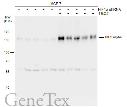
Non-transfected (?) and transfected (+) MCF-7 whole cell extracts (30 ug) were separated by 7.5% SDS-PAGE, and the membrane was blotted with HIF1 alpha antibody (GTX127309) diluted at 1:1000. The HRP-conjugated anti-rabbit IgG antibody (GTX213110-01) was used to detect the primary antibody.
Non-transfected (?) and transfected (+) MCF-7 whole cell extracts (30 ug) were separated by 7.5% SDS-PAGE, and the membrane was blotted with HIF1 alpha antibody (GTX127309) diluted at 1:1000. The HRP-conjugated anti-rabbit IgG antibody (GTX213110-01) was used to detect the primary antibody.
GTX127309 WB Image
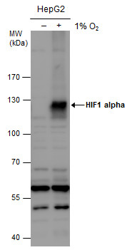
HIF1 alpha antibody detects HIF1 alpha protein by western blot analysis. Un-treated (-) and treated (+, 1% O2 treatment for 24hr) HepG2 whole cell extracts (30 ug) were separated by 7.5% SDS-PAGE, and the membrane was blotted with HIF1 alpha antibody (GTX127309) diluted at 1:1000. The HRP-conjugated anti-rabbit IgG antibody (GTX213110-01) was used to detect the primary antibody.
HIF1 alpha antibody detects HIF1 alpha protein by western blot analysis. Un-treated (-) and treated (+, 1% O2 treatment for 24hr) HepG2 whole cell extracts (30 ug) were separated by 7.5% SDS-PAGE, and the membrane was blotted with HIF1 alpha antibody (GTX127309) diluted at 1:1000. The HRP-conjugated anti-rabbit IgG antibody (GTX213110-01) was used to detect the primary antibody.
GTX127309 ChIP assay Image
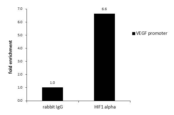
ChIP was performed with HepG2 chromatin extract and 5 ug of either normal rabbit IgG or anti-HIF1 alpha antibody. The precipitated DNA was detected by PCR with primer set targeting to VEGF promoter.
ChIP was performed with HepG2 chromatin extract and 5 ug of either normal rabbit IgG or anti-HIF1 alpha antibody. The precipitated DNA was detected by PCR with primer set targeting to VEGF promoter.
GTX127309 IHC-P Image
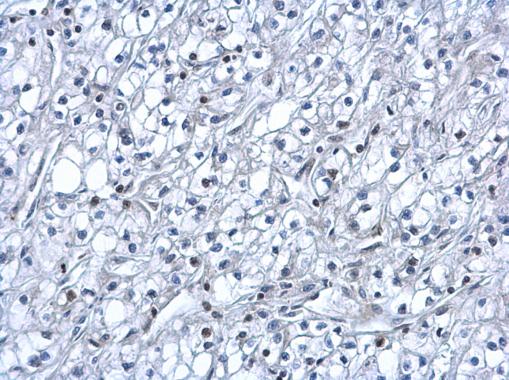
HIF1 alpha antibody detects HIF1 alpha protein at nucleus on human kidney cancer by immunohistochemical analysis.
Sample: Paraffin-embedded human kidney cancer.
HIF1 alpha antibody (GTX127309) dilution: 1:500.
HIF1 alpha antibody detects HIF1 alpha protein at nucleus on human kidney cancer by immunohistochemical analysis.
Sample: Paraffin-embedded human kidney cancer.
HIF1 alpha antibody (GTX127309) dilution: 1:500.
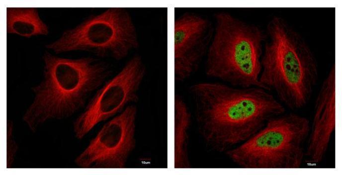
GTX127309 ICC/IF Image
HIF1 alpha antibody detects HIF1 alpha protein at nucleus in CoCl2-treated samples by confocal immunofluorescent analysis.
Samples: Untreated (left panel) and 500uM CoCl2 treated (right panel) HeLa cells for 24 hrs were fixed in 4% paraformaldehyde for 15 min
Green: HIF1alpha protein stained by HIF1 alpha antibody (GTX127309) diluted at 1:1000
Red: Alpha-tubulin, a cytoskeleton marker, stained by Alpha-tubulin antibody (GTX11304) diluted at 1:2500
Blue: Hoechst 33342 staining.
Scale bar = 10 um.
Images captured by Olympus FV10i Confocal Laser Scanning Microscope
カートに商品を
追加しました。
追加しました。
商品情報
| 商品説明 | レビューあり。KO/KDバリデーション済み抗体 |
|---|---|
| 抗原 | aa 657~826 |
| 抗原動物 | Human |
| 交差性 | Bovine/Human/Mouse/Rabbit/Rat |
| 免疫動物 | Rabbit |
| 性状 | Purified |
| 適用 | ChIP, IC, IF, IHC, IP, Western Blot |
| クラス | IgG |
| 標識 | Unlabeled |
| クロナリティ | Polyclonal |
| 別名 | hypoxia inducible factor 1 subunit alpha, HIF-1-alpha, HIF-1A, HIF-1alpha, HIF1, HIF1-ALPHA, MOP1, PASD8, bHLHe78 |
| Genbank No | 3091 |
| データシート | データシート |
| メーカーサイト | メーカーサイト |
| 使用文献 | 使用文献 |
| 保存条件 | -20℃ |
カートに商品を
追加しました。
追加しました。
製品情報は掲載時点のものですが、価格表内の価格については随時最新のものに更新されます。お問い合わせいただくタイミングにより製品情報・価格などは変更されている場合があります。
表示価格に、消費税等は含まれていません。一部価格が予告なく変更される場合がありますので、あらかじめご了承下さい。