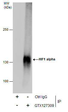
GTX127309 IP Image
Immunoprecipitation of HIF1 alpha protein from HepG2 whole cell extracts treated with 500 uM CoCl2 for 24 hr using 5 ug of HIF1 alpha antibody (GTX127309).
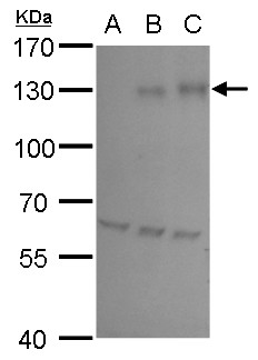
Western blot analysis was performed using HIF1 alpha antibody (GTX127309).
EasyBlot anti-Rabbit IgG (GTX221666-01) was used as a secondary reagent.
GTX127309 WB Image
HIF1 alpha antibody detects HIF1 alpha protein by western blot analysis.
A. 30 ug HepG2 whole cell lysate/extract (untreated)
B.30 ug HepG2 whole cell lysate/extract ( 200 uM CoCl2 treatment for 24 hr)
C. 30 ug HepG2 whole cell lysate/extract (500 uM CoCl2 treatment for 24 hr)
7.5% SDS-PAGE
HIF1 alpha antibody (GTX127309) dilution: 1:5000
The HRP-conjugated anti-rabbit IgG antibody (GTX213110-01) was used to detect the primary antibody.
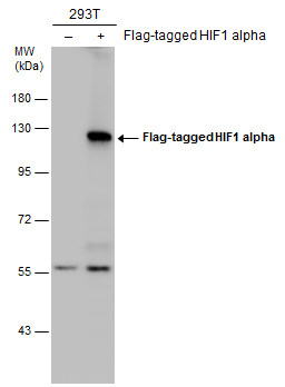
GTX127309 WB Image
Non-transfected (?) and transfected (+) 293T whole cell extracts (30 ug) were separated by 7.5% SDS-PAGE, and the membrane was blotted with HIF1 alpha antibody (GTX127309) diluted at 1:5000. The HRP-conjugated anti-rabbit IgG antibody (GTX213110-01) was used to detect the primary antibody.
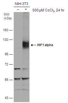
GTX127309 WB Image
Untreated (?) and treated (+) NIH-3T3 whole cell extracts (30 ug) were separated by 7.5% SDS-PAGE, and the membrane was blotted with HIF1 alpha antibody (GTX127309) diluted at 1:500. The HRP-conjugated anti-rabbit IgG antibody (GTX213110-01) was used to detect the primary antibody.
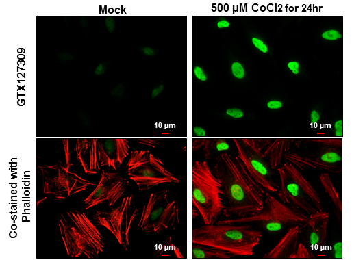
GTX127309 ICC/IF Image
HIF1 alpha antibody detects HIF1 alpha protein at nucleus by immunofluorescent analysis.
Sample: HeLa cells were fixed in 4% paraformaldehyde at RT for 15 min.
Green: HIF1 alpha protein stained by HIF1 alpha antibody (GTX127309) diluted at 1:500.
Red: Phalloidin, a cytoskeleton marker, diluted at 1:100.
Scale bar = 10 um.
GTX127309 ICC/IF Image
HIF1 alpha antibody detects HIF1 alpha protein at nucleus by immunofluorescent analysis.
Sample: NIH/3T3 cells were fixed in 4% paraformaldehyde at RT for 15 min.
Green: HIF1 alpha protein stained by HIF1 alpha antibody (GTX127309) diluted at 1:200.
Blue: Hoechst 33342 staining.
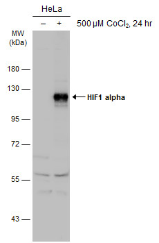
GTX127309 WB Image
Untreated (?) and treated (+) HeLa whole cell extracts (30 ug) were separated by 7.5% SDS-PAGE, and the membrane was blotted with HIF1 alpha antibody (GTX127309) diluted at 1:1000. The HRP-conjugated anti-rabbit IgG antibody (GTX213110-01) was used to detect the primary antibody.
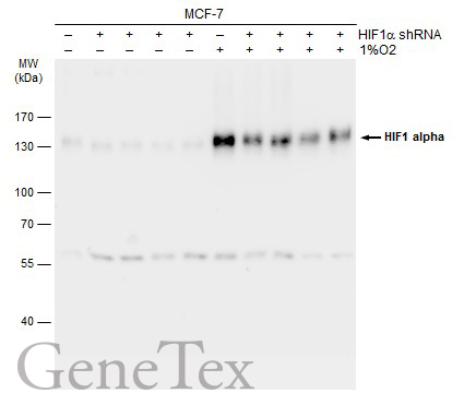
GTX127309 WB Image
Non-transfected (?) and transfected (+) MCF-7 whole cell extracts (30 ug) were separated by 7.5% SDS-PAGE, and the membrane was blotted with HIF1 alpha antibody (GTX127309) diluted at 1:1000. The HRP-conjugated anti-rabbit IgG antibody (GTX213110-01) was used to detect the primary antibody.
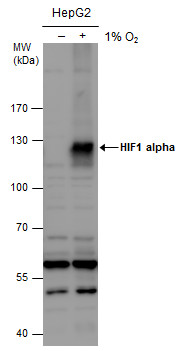
GTX127309 WB Image
HIF1 alpha antibody detects HIF1 alpha protein by western blot analysis. Un-treated (-) and treated (+, 1% O2 treatment for 24hr) HepG2 whole cell extracts (30 ug) were separated by 7.5% SDS-PAGE, and the membrane was blotted with HIF1 alpha antibody (GTX127309) diluted at 1:1000. The HRP-conjugated anti-rabbit IgG antibody (GTX213110-01) was used to detect the primary antibody.
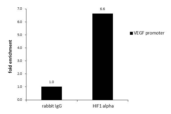
GTX127309 ChIP assay Image
ChIP was performed with HepG2 chromatin extract and 5 ug of either normal rabbit IgG or anti-HIF1 alpha antibody. The precipitated DNA was detected by PCR with primer set targeting to VEGF promoter.
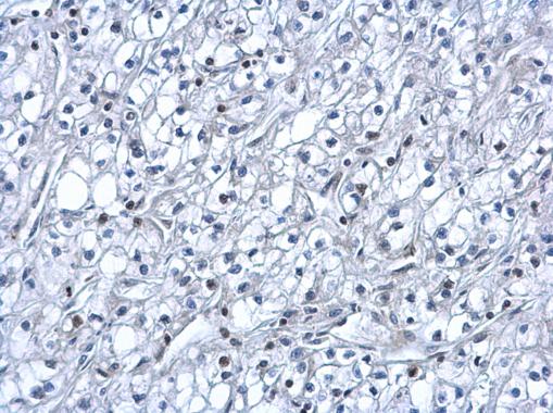
GTX127309 IHC-P Image
HIF1 alpha antibody detects HIF1 alpha protein at nucleus on human kidney cancer by immunohistochemical analysis.
Sample: Paraffin-embedded human kidney cancer.
HIF1 alpha antibody (GTX127309) dilution: 1:500.
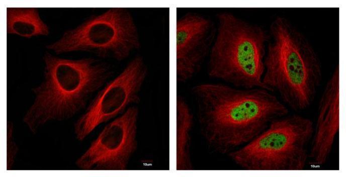
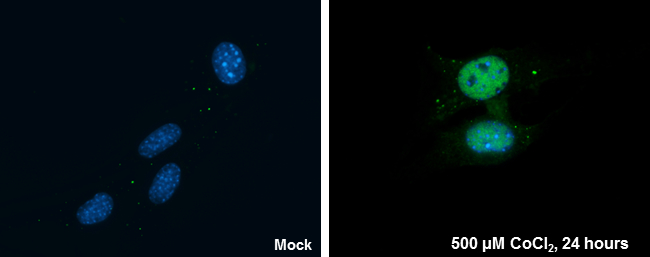
GTX127309 ICC/IF Image
HIF1 alpha antibody detects HIF1 alpha protein at nucleus in CoCl2-treated samples by confocal immunofluorescent analysis.
Samples: Untreated (left panel) and 500uM CoCl2 treated (right panel) HeLa cells for 24 hrs were fixed in 4% paraformaldehyde for 15 min
Green: HIF1alpha protein stained by HIF1 alpha antibody (GTX127309) diluted at 1:1000
Red: Alpha-tubulin, a cytoskeleton marker, stained by Alpha-tubulin antibody (GTX11304) diluted at 1:2500
Blue: Hoechst 33342 staining.
Scale bar = 10 um.
Images captured by Olympus FV10i Confocal Laser Scanning Microscope