
Anti-AIF1/Iba1, Rabbit-Poly | GeneTex International Corporation
掲載日情報:2026/06/03 現在Webページ番号:520141

GeneTex International CorporationのAnti-AIF1/Iba1, Rabbit-Poly商品情報ページです。
※本製品は研究用です。研究用以外には使用できません。
カートに商品を
追加しました。
追加しました。
価格
[在庫・価格 :2026年07月17日 00時01分現在]
※ 表示されている納期は弊社に在庫が無く、取り寄せた場合の納期目安となります。
| 詳細 | 商品名 |
|
文献数 | ||||||||||||||||||||||||||||||||||||||||||||||||||||||||||||||||||||||||||||||||||
|---|---|---|---|---|---|---|---|---|---|---|---|---|---|---|---|---|---|---|---|---|---|---|---|---|---|---|---|---|---|---|---|---|---|---|---|---|---|---|---|---|---|---|---|---|---|---|---|---|---|---|---|---|---|---|---|---|---|---|---|---|---|---|---|---|---|---|---|---|---|---|---|---|---|---|---|---|---|---|---|---|---|---|---|---|---|
|
Anti-AIF1/Iba1, Rabbit-Poly |
|
99 | |||||||||||||||||||||||||||||||||||||||||||||||||||||||||||||||||||||||||||||||||||
|
|||||||||||||||||||||||||||||||||||||||||||||||||||||||||||||||||||||||||||||||||||||


[在庫・価格 :2026年07月17日 00時01分現在]
※ 表示されている納期は弊社に在庫が無く、取り寄せた場合の納期目安となります。
Anti-AIF1/Iba1, Rabbit-Poly
文献数: 99
- 商品コード:GTX100042
- メーカー:GNT
- 包装:100μl
- 価格:¥85,000
- 在庫:1個
- 納期:10日程度 ※※ 表示されている納期は弊社に在庫がなく、取り寄せた場合の目安納期となります。
- 法規制等:
| 説明文 | レビューあり。 別名:allograft inflammatory factor 1,AIF-1,IBA1,IRT-1,IRT1 Genbank No: 199 |
||||||
|---|---|---|---|---|---|---|---|
| 別包装品 | 別包装品あり | ||||||
| 法規制等 | |||||||
| 保存条件 | -20℃ | 法規備考 | |||||
| 抗原種 | Human | 免疫動物 | Rabbit | ||||
| 交差性 | Human/Mouse/Rat | 適用 | FCM,IC,IF,IHC,Western Blot | ||||
| 標識 | Unlabeled | 性状 | Purified | ||||
| 吸収処理 | クラス | IgG | |||||
| クロナリティ | Polyclonal | フォーマット | |||||
| 掲載カタログ |
|
||||||
| 製品記事 | 中枢神経系(CNS)マーカー抗体 抗Iba1抗体(Anti-Iba1 antibody)(GeneTex社) ミクログリアマーカー抗体 |
||||||
| 関連記事 | GeneTex社における抗体の品質管理 |
||||||
カートに商品を
追加しました。
追加しました。
ラインナップ
カートに商品を
追加しました。
追加しました。
画像
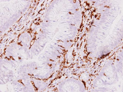
GTX100042 IHC-P Image
Iba1 antibody detects Iba1 protein at cytoplasm on human colon carcinoma stroma by immunohistochemical analysis.
Sample: Paraffin-embedded colon carcinoma stroma.
Iba1 antibody (GTX100042) dilution: 1:500.
Iba1 antibody detects Iba1 protein at cytoplasm on human colon carcinoma stroma by immunohistochemical analysis.
Sample: Paraffin-embedded colon carcinoma stroma.
Iba1 antibody (GTX100042) dilution: 1:500.
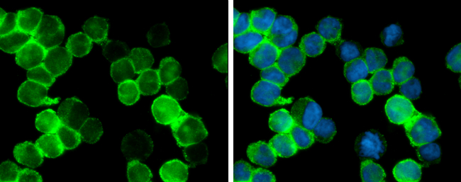
GTX100042 ICC/IF Image
Iba1 antibody detects Iba1 protein at cytoplasm by immunofluorescent analysis.
Sample: THP-1 cells were fixed in 4% paraformaldehyde at RT for 15 min.
Green: Iba1 protein stained by Iba1 antibody (GTX100042) diluted at 1:400.
Blue: Hoechst 33342 staining.
Iba1 antibody detects Iba1 protein at cytoplasm by immunofluorescent analysis.
Sample: THP-1 cells were fixed in 4% paraformaldehyde at RT for 15 min.
Green: Iba1 protein stained by Iba1 antibody (GTX100042) diluted at 1:400.
Blue: Hoechst 33342 staining.
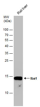
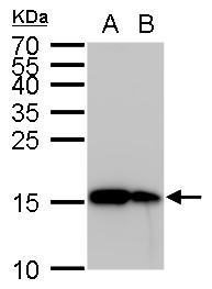
GTX100042 WB Image
Rat tissue extract (50 ug) was separated by 15% SDS-PAGE, and the membrane was blotted with Iba1 antibody (GTX100042) diluted at 1:1000.
Rat tissue extract (50 ug) was separated by 15% SDS-PAGE, and the membrane was blotted with Iba1 antibody (GTX100042) diluted at 1:1000.
GTX100042 WB Image
Iba1 antibody detects Iba1 protein by Western blot analysis.
A. 30 ug THP-1 whole cell lysate/extract
B. 30 ug HL-60 whole cell lysate/extract
15 % SDS-PAGE
Iba1 antibody (GTX100042) dilution: 1:5000
Iba1 antibody detects Iba1 protein by Western blot analysis.
A. 30 ug THP-1 whole cell lysate/extract
B. 30 ug HL-60 whole cell lysate/extract
15 % SDS-PAGE
Iba1 antibody (GTX100042) dilution: 1:5000
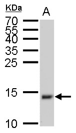
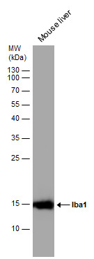
GTX100042 WB Image
Iba1 antibody detects Iba1 protein by Western blot analysis.
A. 50 ug mouse brain lysate/extract
15 % SDS-PAGE
Iba1 antibody (GTX100042) dilution: 1:1000
Iba1 antibody detects Iba1 protein by Western blot analysis.
A. 50 ug mouse brain lysate/extract
15 % SDS-PAGE
Iba1 antibody (GTX100042) dilution: 1:1000
GTX100042 IHC-P Image
Iba1 detects Iba1 protein at cytosol on mouse brain tissue by immunohistochemical analysis.
Sample: Paraffin-embedded mouse brain tissue.
Iba1 (GTX100042) dilution: 1:1000.
Iba1 detects Iba1 protein at cytosol on mouse brain tissue by immunohistochemical analysis.
Sample: Paraffin-embedded mouse brain tissue.
Iba1 (GTX100042) dilution: 1:1000.
GTX100042 IHC-P Image
Iba1 antibody detects Iba1 protein on mouse fore brain by immunohistochemical analysis.
Sample: Paraffin-embedded mouse fore brain.
Iba1 antibody (GTX100042) dilution: 1:500.
Iba1 antibody detects Iba1 protein on mouse fore brain by immunohistochemical analysis.
Sample: Paraffin-embedded mouse fore brain.
Iba1 antibody (GTX100042) dilution: 1:500.
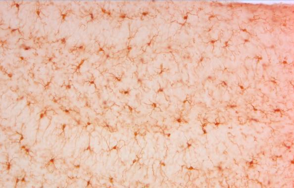
GTX100042 IHC-P Image
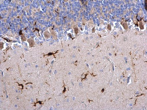
Iba1 antibody detects Iba1 protein on rat hind brain by immunohistochemical analysis.
Sample: Paraffin-embedded rat hind brain.
Iba1 antibody (GTX100042) dilution: 1:500.
Iba1 antibody detects Iba1 protein on rat hind brain by immunohistochemical analysis.
Sample: Paraffin-embedded rat hind brain.
Iba1 antibody (GTX100042) dilution: 1:500.

GTX100042 FACS Image
Iba1 antibody (GTX100042) detects Iba1 by flow cytometry analysis.
Sample: THP-1 cell.
Black: Unlabelled sample was used as a control.
Red: Iba1 antibody (GTX100042) dilution: 1:50.
Iba1 antibody (GTX100042) detects Iba1 by flow cytometry analysis.
Sample: THP-1 cell.
Black: Unlabelled sample was used as a control.
Red: Iba1 antibody (GTX100042) dilution: 1:50.
![GTX100042 IHC-Fr Image<br>Iba1 antibody detects Iba1 protein expression at microglias by immunohistochemical analysis.<br>Sample: Frozen sectioned E13.5 Rat brain.<br>Green: Iba1 protein stained by Iba1 antibody (GTX100042) diluted at 1:250.<br>Red: beta Tubulin 3/ TUJ1, a mature neuron marker, stained by beta Tubulin 3/ TUJ1 antibody [GT11710] (GTX631836) diluted at 1:500.<br>Blue: Fluoroshield with DAPI (GTX30920).<br>](/domestic/data/graphics/GNT/graphics/GTX100042_41556_20161005_IHC-Fr_R.jpg)
GTX100042 IHC-Fr Image
Iba1 antibody detects Iba1 protein expression at microglias by immunohistochemical analysis.
Sample: Frozen sectioned E13.5 Rat brain.
Green: Iba1 protein stained by Iba1 antibody (GTX100042) diluted at 1:250.
Red: beta Tubulin 3/ TUJ1, a mature neuron marker, stained by beta Tubulin 3/ TUJ1 antibody [GT11710] (GTX631836) diluted at 1:500.
Blue: Fluoroshield with DAPI (GTX30920).
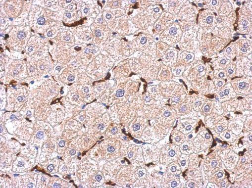
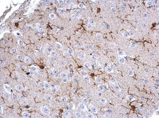
GTX100042 IHC-P Image
Immunohistochemical analysis of paraffin-embedded human hepatoma, using Iba1(GTX100042) antibody at 1:500 dilution.
Immunohistochemical analysis of paraffin-embedded human hepatoma, using Iba1(GTX100042) antibody at 1:500 dilution.
GTX100042 WB Image
Mouse tissue extract (50 ug) was separated by 15% SDS-PAGE, and the membrane was blotted with Iba1 antibody (GTX100042) diluted at 1:1000.
Mouse tissue extract (50 ug) was separated by 15% SDS-PAGE, and the membrane was blotted with Iba1 antibody (GTX100042) diluted at 1:1000.
カートに商品を
追加しました。
追加しました。
商品情報
| 商品説明 | レビューあり |
|---|---|
| 抗原動物 | Human |
| 交差性 | Human/Mouse/Rat |
| 免疫動物 | Rabbit |
| 性状 | Purified |
| 適用 | FCM, IC, IF, IHC, Western Blot |
| クラス | IgG |
| 標識 | Unlabeled |
| クロナリティ | Polyclonal |
| 因子名 | AIF1 |
| 別名 | allograft inflammatory factor 1, AIF-1, IBA1, IRT-1, IRT1 |
| Genbank No | 199 |
| データシート | データシート |
| メーカーサイト | メーカーサイト |
| 使用文献 | 使用文献 |
| 保存条件 | -20℃ |
カートに商品を
追加しました。
追加しました。
製品情報は掲載時点のものですが、価格表内の価格については随時最新のものに更新されます。お問い合わせいただくタイミングにより製品情報・価格などは変更されている場合があります。
表示価格に、消費税等は含まれていません。一部価格が予告なく変更される場合がありますので、あらかじめご了承下さい。