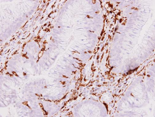
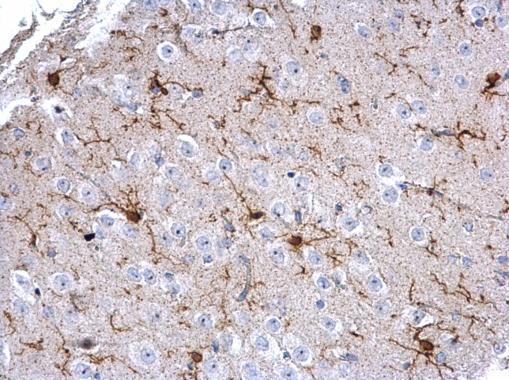
GTX100042 IHC-P Image
Iba1 antibody detects Iba1 protein at cytoplasm on human colon carcinoma stroma by immunohistochemical analysis.
Sample: Paraffin-embedded colon carcinoma stroma.
Iba1 antibody (GTX100042) dilution: 1:500.
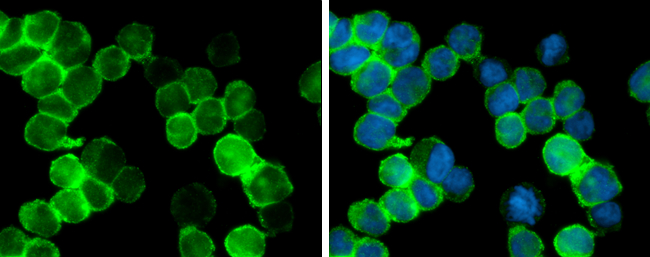
GTX100042 ICC/IF Image
Iba1 antibody detects Iba1 protein at cytoplasm by immunofluorescent analysis.
Sample: THP-1 cells were fixed in 4% paraformaldehyde at RT for 15 min.
Green: Iba1 protein stained by Iba1 antibody (GTX100042) diluted at 1:400.
Blue: Hoechst 33342 staining.
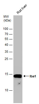
GTX100042 WB Image
Rat tissue extract (50 ug) was separated by 15% SDS-PAGE, and the membrane was blotted with Iba1 antibody (GTX100042) diluted at 1:1000.
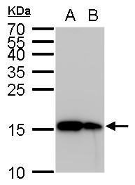
GTX100042 WB Image
Iba1 antibody detects Iba1 protein by Western blot analysis.
A. 30 ug THP-1 whole cell lysate/extract
B. 30 ug HL-60 whole cell lysate/extract
15 % SDS-PAGE
Iba1 antibody (GTX100042) dilution: 1:5000
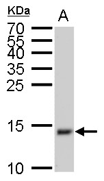
GTX100042 WB Image
Iba1 antibody detects Iba1 protein by Western blot analysis.
A. 50 ug mouse brain lysate/extract
15 % SDS-PAGE
Iba1 antibody (GTX100042) dilution: 1:1000
GTX100042 IHC-P Image
Iba1 detects Iba1 protein at cytosol on mouse brain tissue by immunohistochemical analysis.
Sample: Paraffin-embedded mouse brain tissue.
Iba1 (GTX100042) dilution: 1:1000.
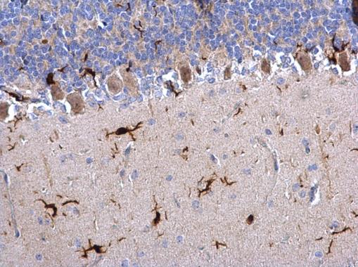
GTX100042 IHC-P Image
Iba1 antibody detects Iba1 protein on mouse fore brain by immunohistochemical analysis.
Sample: Paraffin-embedded mouse fore brain.
Iba1 antibody (GTX100042) dilution: 1:500.
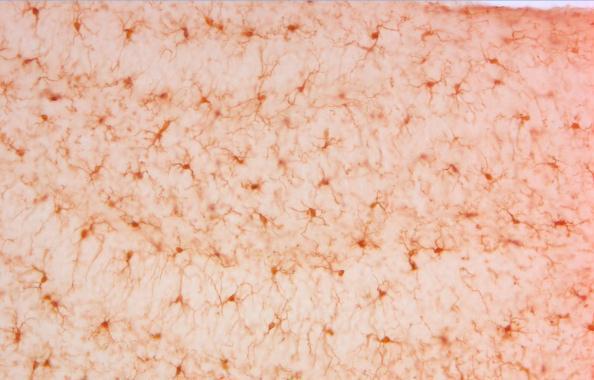
GTX100042 IHC-P Image
Iba1 antibody detects Iba1 protein on rat hind brain by immunohistochemical analysis.
Sample: Paraffin-embedded rat hind brain.
Iba1 antibody (GTX100042) dilution: 1:500.

GTX100042 FACS Image
Iba1 antibody (GTX100042) detects Iba1 by flow cytometry analysis.
Sample: THP-1 cell.
Black: Unlabelled sample was used as a control.
Red: Iba1 antibody (GTX100042) dilution: 1:50.
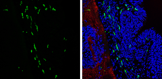
GTX100042 IHC-Fr Image
Iba1 antibody detects Iba1 protein expression at microglias by immunohistochemical analysis.
Sample: Frozen sectioned E13.5 Rat brain.
Green: Iba1 protein stained by Iba1 antibody (GTX100042) diluted at 1:250.
Red: beta Tubulin 3/ TUJ1, a mature neuron marker, stained by beta Tubulin 3/ TUJ1 antibody [GT11710] (GTX631836) diluted at 1:500.
Blue: Fluoroshield with DAPI (GTX30920).
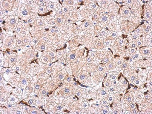
GTX100042 IHC-P Image
Immunohistochemical analysis of paraffin-embedded human hepatoma, using Iba1(GTX100042) antibody at 1:500 dilution.
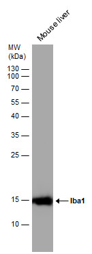
GTX100042 WB Image
Mouse tissue extract (50 ug) was separated by 15% SDS-PAGE, and the membrane was blotted with Iba1 antibody (GTX100042) diluted at 1:1000.