
Anti-LC3B, Rabbit-Poly | GeneTex International Corporation
掲載日情報:2026/06/03 現在Webページ番号:45387

GeneTex International CorporationのAnti-LC3B, Rabbit-Poly商品情報ページです。
※本製品は研究用です。研究用以外には使用できません。
カートに商品を
追加しました。
追加しました。
価格
[在庫・価格 :2026年07月10日 11時15分現在]
※ 表示されている納期は弊社に在庫が無く、取り寄せた場合の納期目安となります。
| 詳細 | 商品名 |
|
文献数 | ||||||||||||||||||||||||||||||||||||||||||||||||||||||||||||||||||||||||||||||||||
|---|---|---|---|---|---|---|---|---|---|---|---|---|---|---|---|---|---|---|---|---|---|---|---|---|---|---|---|---|---|---|---|---|---|---|---|---|---|---|---|---|---|---|---|---|---|---|---|---|---|---|---|---|---|---|---|---|---|---|---|---|---|---|---|---|---|---|---|---|---|---|---|---|---|---|---|---|---|---|---|---|---|---|---|---|---|
|
Anti-LC3B, Rabbit-Poly |
|
99 | |||||||||||||||||||||||||||||||||||||||||||||||||||||||||||||||||||||||||||||||||||
|
|||||||||||||||||||||||||||||||||||||||||||||||||||||||||||||||||||||||||||||||||||||


[在庫・価格 :2026年07月10日 11時15分現在]
※ 表示されている納期は弊社に在庫が無く、取り寄せた場合の納期目安となります。
Anti-LC3B, Rabbit-Poly
文献数: 99
- 商品コード:GTX127375
- メーカー:GNT
- 包装:100μl
- 価格:¥85,000
- 在庫:1個
- 納期:10日程度 ※※ 表示されている納期は弊社に在庫がなく、取り寄せた場合の目安納期となります。
- 法規制等:
| 説明文 | レビューあり。 別名:microtubule associated protein 1 light chain 3 beta,ATG8F,LC3B,MAP1A/1BLC3,MAP1LC3B-a Genbank No: 81631 |
||||||
|---|---|---|---|---|---|---|---|
| 別包装品 | 別包装品あり | ||||||
| 法規制等 | |||||||
| 保存条件 | -20℃ | 法規備考 | |||||
| 抗原種 | Human | 免疫動物 | Rabbit | ||||
| 交差性 | Guinea Pig/Human/Mosquito/Mouse/Pig/Rat | 適用 | Electron Microscopy,FCM,IC,IF,IHC,IP,Western Blot | ||||
| 標識 | Unlabeled | 性状 | Purified | ||||
| 吸収処理 | クラス | IgG | |||||
| クロナリティ | Polyclonal | フォーマット | |||||
| 掲載カタログ |
|
||||||
| 製品記事 | GeneTex社 オルガネラマーカー抗体 アポトーシス・オートファジー・ネクローシス関連抗体 |
||||||
| 関連記事 | GeneTex社における抗体の品質管理 |
||||||
カートに商品を
追加しました。
追加しました。
ラインナップ
カートに商品を
追加しました。
追加しました。
画像
GTX127375 WB Image
LC3B antibody detects LC3B protein by western blot analysis. Various whole cell extracts (30 ug) were separated by 15% SDS-PAGE, and the membrane was blotted with LC3B antibody (GTX127375) diluted at a dilution of 1:1000. The HRP-conjugated anti-rabbit IgG antibody (GTX213110-01) was used to detect the primary antibody.
LC3B antibody detects LC3B protein by western blot analysis. Various whole cell extracts (30 ug) were separated by 15% SDS-PAGE, and the membrane was blotted with LC3B antibody (GTX127375) diluted at a dilution of 1:1000. The HRP-conjugated anti-rabbit IgG antibody (GTX213110-01) was used to detect the primary antibody.
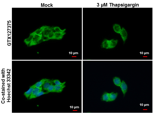
GTX127375 ICC/IF Image
LC3B antibody detects LC3B protein at autophagosome by immunofluorescent analysis.
Samples: Hep G2 cells mock (left) and treated with 3 uM Thapsigargin for 12 hrs (right) were fixed in ice-cold MeOH for 10 min and permeabilized with ice-cold acetone for 1 min.
Green: LC3B protein stained by LC3B antibody (GTX127375) diluted at 1:500.
Blue: Hoechst 33342 staining.
Scale bar = 10 um.
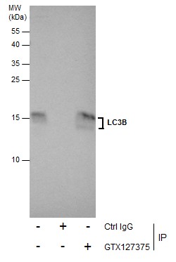
GTX127375 IP Image
Immunoprecipitation of LC3B protein from U87-MG whole cell extracts using 5 ug of LC3B antibody (GTX127375).
Western blot analysis was performed using LC3B antibody (GTX127375).
EasyBlot anti-Rabbit IgG (GTX221666-01) was used as a secondary reagent.
Immunoprecipitation of LC3B protein from U87-MG whole cell extracts using 5 ug of LC3B antibody (GTX127375).
Western blot analysis was performed using LC3B antibody (GTX127375).
EasyBlot anti-Rabbit IgG (GTX221666-01) was used as a secondary reagent.
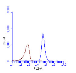
GTX127375 FACS Image
LC3B antibody (GTX127375) detects LC3B protein by flow cytometry analysis.
Sample: HeLa cell fixed in 4% paraformaldehyde at 4oC for 5 min.
Brown: Unlabelled sample was also used as a control.
Blue: LC3B antibody (GTX127375) dilution: 1:100.
Acquisition of >20,000 events were collected using Argon ion laser (488nm) and 525/30 bandpass filter.
GTX127375 ICC/IF Image
LC3B antibody detects LC3B protein at autophagosome by immunofluorescent analysis.
Sample: HeLa cells were fixed in 4% paraformaldehyde at RT for 15 min.
Green: LC3B protein stained by LC3B antibody (GTX127375) diluted at 1:200.
Red: phalloidin, a cytoskeleton marker, diluted at 1:50.
Blue: Hoechst 33342 staining.
Scale bar = 10 um.
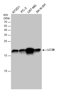
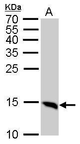
GTX127375 WB Image
LC3B antibody detects MAP1LC3B protein by western blot analysis.
A. 50 ug mouse brain lysate/extract
15% SDS-PAGE
LC3B antibody (GTX127375) dilution: 1:1000
The HRP-conjugated anti-rabbit IgG antibody (GTX213110-01) was used to detect the primary antibody.
LC3B antibody detects MAP1LC3B protein by western blot analysis.
A. 50 ug mouse brain lysate/extract
15% SDS-PAGE
LC3B antibody (GTX127375) dilution: 1:1000
The HRP-conjugated anti-rabbit IgG antibody (GTX213110-01) was used to detect the primary antibody.
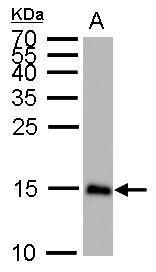
GTX127375 WB Image
LC3B antibody detects MAP1LC3B protein by western blot analysis.
A. 50 ug Rat brain lysate/extract
15% SDS-PAGE
LC3B antibody (GTX127375) dilution: 1:1000
The HRP-conjugated anti-rabbit IgG antibody (GTX213110-01) was used to detect the primary antibody.
LC3B antibody detects MAP1LC3B protein by western blot analysis.
A. 50 ug Rat brain lysate/extract
15% SDS-PAGE
LC3B antibody (GTX127375) dilution: 1:1000
The HRP-conjugated anti-rabbit IgG antibody (GTX213110-01) was used to detect the primary antibody.
GTX127375 WB Image
LC3B antibody detects LC3B protein in Thapsigargin-treated samples by western blot analysis.
Upper panel: LC3B antibody (GTX127375)
Lower panel: Beta-actin antibody (GTX110564)A. 30 ug HepG2 whole cell lysate/extract (untreated)
B. 30 ug HepG2 whole cell lysate/extract (3 uM Thapsigargin treatment for 12 hr)
15% SDS-PAGE
LC3B antibody (GTX127375) dilution: 1:1000
Beta-actin antibody (GTX110564) dilution: 1:20000
The HRP-conjugated anti-rabbit IgG antibody (GTX213110-01) was used to detect the primary antibody.
GTX127375 WB Image
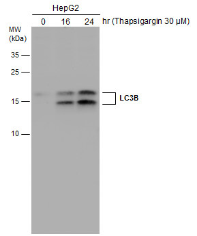
HepG2 cells were untreated or treated with 3 uM thapsigargin for 16 and 24 hrs. Whole cell extracts (30 ug) were separated by 15% SDS-PAGE, and the membrane was blotted with LC3B antibody (GTX127375) diluted at 1:1000. The HRP-conjugated anti-rabbit IgG antibody (GTX213110-01) was used to detect the primary antibody.
HepG2 cells were untreated or treated with 3 uM thapsigargin for 16 and 24 hrs. Whole cell extracts (30 ug) were separated by 15% SDS-PAGE, and the membrane was blotted with LC3B antibody (GTX127375) diluted at 1:1000. The HRP-conjugated anti-rabbit IgG antibody (GTX213110-01) was used to detect the primary antibody.
GTX127375 WB Image
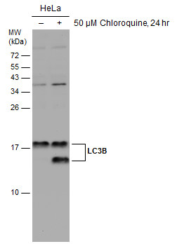
Untreated (?) and treated (+) HeLa whole cell extracts (30 ug) were separated by 15% SDS-PAGE, and the membrane was blotted with LC3B antibody (GTX127375) diluted at 1:2500. The HRP-conjugated anti-rabbit IgG antibody (GTX213110-01) was used to detect the primary antibody.
Untreated (?) and treated (+) HeLa whole cell extracts (30 ug) were separated by 15% SDS-PAGE, and the membrane was blotted with LC3B antibody (GTX127375) diluted at 1:2500. The HRP-conjugated anti-rabbit IgG antibody (GTX213110-01) was used to detect the primary antibody.
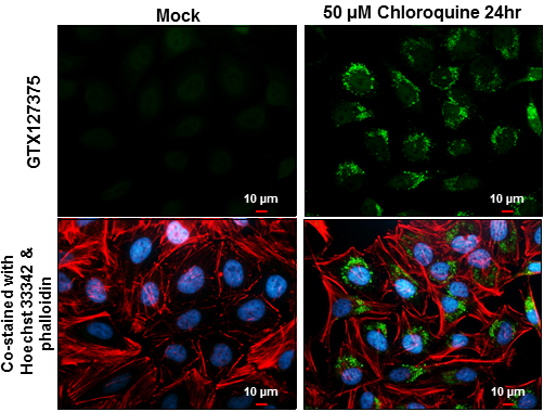
![GTX127375 ICC/IF Image<br>LC3B antibody detects LC3B protein at autophagosome by immunofluorescent analysis.<br>Samples: HeLa cells mock (left) and treated with 50uM Chloroquine for 24 hr (right) were fixed in 4% paraformaldehyde at RT for 15 min.<br>Green: LC3B protein stained by LC3B antibody (GTX127375) diluted at 1:2000.<br>Red: alpha Tubulin, a cytoskeleton marker, stained by alpha Tubulin antibody [GT114] (GTX628802) diluted at 1:1000.<br>Blue: Hoechst 33342 staining.<br>](/domestic/data/graphics/GNT/graphics/GTX127375_41003_20150410_IFA.jpg)
GTX127375 ICC/IF Image
LC3B antibody detects LC3B protein at autophagosome by immunofluorescent analysis.
Samples: HeLa cells mock (left) and treated with 50uM Chloroquine for 24 hr (right) were fixed in 4% paraformaldehyde at RT for 15 min.
Green: LC3B protein stained by LC3B antibody (GTX127375) diluted at 1:2000.
Red: alpha Tubulin, a cytoskeleton marker, stained by alpha Tubulin antibody [GT114] (GTX628802) diluted at 1:1000.
Blue: Hoechst 33342 staining.
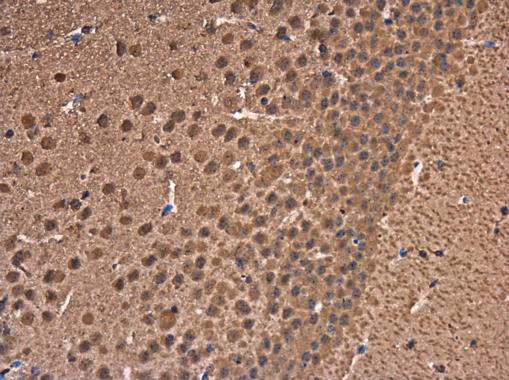
GTX127375 IHC-P Image
LC3B antibody detects LC3B protein at cytoplasm in mouse brain by immunohistochemical analysis.
Sample: Paraffin-embedded mouse brain.
LC3B antibody (GTX127375) diluted at 1:500.
LC3B antibody detects LC3B protein at cytoplasm in mouse brain by immunohistochemical analysis.
Sample: Paraffin-embedded mouse brain.
LC3B antibody (GTX127375) diluted at 1:500.

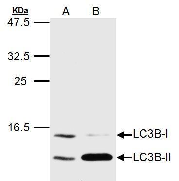
GTX127375 WB Image
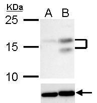
LC3B antibody detects LC3B protein in HCV-infected samples by western blot analysis.
? A. 20 ug Huh7 whole cell lysate/extract (un-infected)
? B. 20 ug Huh7 whole cell lysate/extract (HCV-infected)
? LC3B antibody (GTX127375) dilution: 1:1500
?The HRP-conjugated anti-rabbit IgG antibody (GTX213110-01) was used to detect the primary antibody.
GTX127375 WB Image
LC3B antibody detects MAP1LC3B protein by western blot analysis.
A. 20 ug Huh7 whole cell lysate/extract (untreated)
B. 20 ug Huh7 whole cell lysate/extract (3uM-Thapsigargin treatment for 12hr)
LC3B antibody (GTX127375) dilution: 1:1500
The HRP-conjugated anti-rabbit IgG antibody (GTX213110-01) was used to detect the primary antibody.
カートに商品を
追加しました。
追加しました。
商品情報
| 商品説明 | レビューあり |
|---|---|
| 抗原動物 | Human |
| 交差性 | Guinea Pig/Human/Mosquito/Mouse/Pig/Rat |
| 免疫動物 | Rabbit |
| 性状 | Purified |
| 適用 | Electron Microscopy, FCM, IC, IF, IHC, IP, Western Blot |
| クラス | IgG |
| 標識 | Unlabeled |
| クロナリティ | Polyclonal |
| 別名 | microtubule associated protein 1 light chain 3 beta, ATG8F, LC3B, MAP1A/1BLC3, MAP1LC3B-a |
| Genbank No | 81631 |
| データシート | データシート |
| メーカーサイト | メーカーサイト |
| 使用文献 | 使用文献 |
| 保存条件 | -20℃ |
カートに商品を
追加しました。
追加しました。
製品情報は掲載時点のものですが、価格表内の価格については随時最新のものに更新されます。お問い合わせいただくタイミングにより製品情報・価格などは変更されている場合があります。
表示価格に、消費税等は含まれていません。一部価格が予告なく変更される場合がありますので、あらかじめご了承下さい。