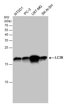
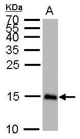
GTX127375 WB Image
LC3B antibody detects LC3B protein by western blot analysis. Various whole cell extracts (30 ug) were separated by 15% SDS-PAGE, and the membrane was blotted with LC3B antibody (GTX127375) diluted at a dilution of 1:1000. The HRP-conjugated anti-rabbit IgG antibody (GTX213110-01) was used to detect the primary antibody.
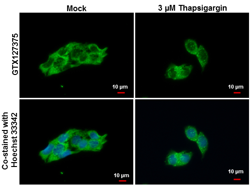
GTX127375 ICC/IF Image
LC3B antibody detects LC3B protein at autophagosome by immunofluorescent analysis.
Samples: Hep G2 cells mock (left) and treated with 3 uM Thapsigargin for 12 hrs (right) were fixed in ice-cold MeOH for 10 min and permeabilized with ice-cold acetone for 1 min.
Green: LC3B protein stained by LC3B antibody (GTX127375) diluted at 1:500.
Blue: Hoechst 33342 staining.
Scale bar = 10 um.
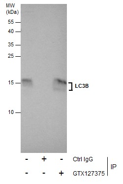
GTX127375 IP Image
Immunoprecipitation of LC3B protein from U87-MG whole cell extracts using 5 ug of LC3B antibody (GTX127375).
Western blot analysis was performed using LC3B antibody (GTX127375).
EasyBlot anti-Rabbit IgG (GTX221666-01) was used as a secondary reagent.
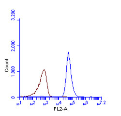
GTX127375 FACS Image
LC3B antibody (GTX127375) detects LC3B protein by flow cytometry analysis.
Sample: HeLa cell fixed in 4% paraformaldehyde at 4oC for 5 min.
Brown: Unlabelled sample was also used as a control.
Blue: LC3B antibody (GTX127375) dilution: 1:100.
Acquisition of >20,000 events were collected using Argon ion laser (488nm) and 525/30 bandpass filter.
GTX127375 ICC/IF Image
LC3B antibody detects LC3B protein at autophagosome by immunofluorescent analysis.
Sample: HeLa cells were fixed in 4% paraformaldehyde at RT for 15 min.
Green: LC3B protein stained by LC3B antibody (GTX127375) diluted at 1:200.
Red: phalloidin, a cytoskeleton marker, diluted at 1:50.
Blue: Hoechst 33342 staining.
Scale bar = 10 um.
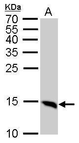
GTX127375 WB Image
LC3B antibody detects MAP1LC3B protein by western blot analysis.
A. 50 ug mouse brain lysate/extract
15% SDS-PAGE
LC3B antibody (GTX127375) dilution: 1:1000
The HRP-conjugated anti-rabbit IgG antibody (GTX213110-01) was used to detect the primary antibody.
GTX127375 WB Image
LC3B antibody detects MAP1LC3B protein by western blot analysis.
A. 50 ug Rat brain lysate/extract
15% SDS-PAGE
LC3B antibody (GTX127375) dilution: 1:1000
The HRP-conjugated anti-rabbit IgG antibody (GTX213110-01) was used to detect the primary antibody.
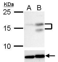
GTX127375 WB Image
LC3B antibody detects LC3B protein in Thapsigargin-treated samples by western blot analysis.
Upper panel: LC3B antibody (GTX127375)
Lower panel: Beta-actin antibody (GTX110564)A. 30 ug HepG2 whole cell lysate/extract (untreated)
B. 30 ug HepG2 whole cell lysate/extract (3 uM Thapsigargin treatment for 12 hr)
15% SDS-PAGE
LC3B antibody (GTX127375) dilution: 1:1000
Beta-actin antibody (GTX110564) dilution: 1:20000
The HRP-conjugated anti-rabbit IgG antibody (GTX213110-01) was used to detect the primary antibody.
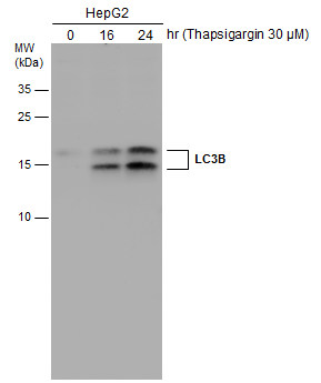
GTX127375 WB Image
HepG2 cells were untreated or treated with 3 uM thapsigargin for 16 and 24 hrs. Whole cell extracts (30 ug) were separated by 15% SDS-PAGE, and the membrane was blotted with LC3B antibody (GTX127375) diluted at 1:1000. The HRP-conjugated anti-rabbit IgG antibody (GTX213110-01) was used to detect the primary antibody.
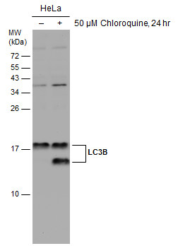
GTX127375 WB Image
Untreated (?) and treated (+) HeLa whole cell extracts (30 ug) were separated by 15% SDS-PAGE, and the membrane was blotted with LC3B antibody (GTX127375) diluted at 1:2500. The HRP-conjugated anti-rabbit IgG antibody (GTX213110-01) was used to detect the primary antibody.
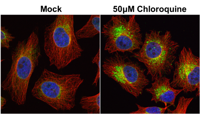
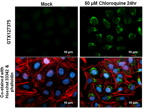
GTX127375 ICC/IF Image
LC3B antibody detects LC3B protein at autophagosome by immunofluorescent analysis.
Samples: HeLa cells mock (left) and treated with 50uM Chloroquine for 24 hr (right) were fixed in 4% paraformaldehyde at RT for 15 min.
Green: LC3B protein stained by LC3B antibody (GTX127375) diluted at 1:2000.
Red: alpha Tubulin, a cytoskeleton marker, stained by alpha Tubulin antibody [GT114] (GTX628802) diluted at 1:1000.
Blue: Hoechst 33342 staining.
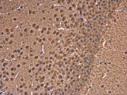
GTX127375 IHC-P Image
LC3B antibody detects LC3B protein at cytoplasm in mouse brain by immunohistochemical analysis.
Sample: Paraffin-embedded mouse brain.
LC3B antibody (GTX127375) diluted at 1:500.

GTX127375 WB Image
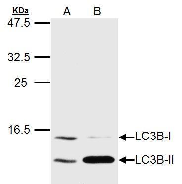
LC3B antibody detects LC3B protein in HCV-infected samples by western blot analysis.
? A. 20 ug Huh7 whole cell lysate/extract (un-infected)
? B. 20 ug Huh7 whole cell lysate/extract (HCV-infected)
? LC3B antibody (GTX127375) dilution: 1:1500
?The HRP-conjugated anti-rabbit IgG antibody (GTX213110-01) was used to detect the primary antibody.
GTX127375 WB Image
LC3B antibody detects MAP1LC3B protein by western blot analysis.
A. 20 ug Huh7 whole cell lysate/extract (untreated)
B. 20 ug Huh7 whole cell lysate/extract (3uM-Thapsigargin treatment for 12hr)
LC3B antibody (GTX127375) dilution: 1:1500
The HRP-conjugated anti-rabbit IgG antibody (GTX213110-01) was used to detect the primary antibody.