
Anti-PD-L1, Rabbit-Poly <Anti-CD274> | GeneTex International Corporation
掲載日情報:2026/06/03 現在Webページ番号:37012

GeneTex International CorporationのAnti-PD-L1, Rabbit-Poly <Anti-CD274>商品情報ページです。
※本製品は研究用です。研究用以外には使用できません。
カートに商品を
追加しました。
追加しました。
価格
[在庫・価格 :2026年07月10日 11時55分現在]
※ 表示されている納期は弊社に在庫が無く、取り寄せた場合の納期目安となります。
| 詳細 | 商品名 |
|
文献数 | ||||||||||||||||||||||||||||||||||||||||||||||||||||||||||||||||||||||||||||||||||
|---|---|---|---|---|---|---|---|---|---|---|---|---|---|---|---|---|---|---|---|---|---|---|---|---|---|---|---|---|---|---|---|---|---|---|---|---|---|---|---|---|---|---|---|---|---|---|---|---|---|---|---|---|---|---|---|---|---|---|---|---|---|---|---|---|---|---|---|---|---|---|---|---|---|---|---|---|---|---|---|---|---|---|---|---|---|
|
Anti-PD-L1, Rabbit-Poly <Anti-CD274> |
|
40 | |||||||||||||||||||||||||||||||||||||||||||||||||||||||||||||||||||||||||||||||||||
|
|||||||||||||||||||||||||||||||||||||||||||||||||||||||||||||||||||||||||||||||||||||


[在庫・価格 :2026年07月10日 11時55分現在]
※ 表示されている納期は弊社に在庫が無く、取り寄せた場合の納期目安となります。
Anti-PD-L1, Rabbit-Poly <Anti-CD274>
文献数: 40
- 商品コード:GTX104763
- メーカー:GNT
- 包装:100μl
- 価格:¥85,000
- 在庫:1個
- 納期:10日程度 ※※ 表示されている納期は弊社に在庫がなく、取り寄せた場合の目安納期となります。
- 法規制等:
| 説明文 | レビューあり。KO/KDバリデーション済み抗体。 別名:CD274 molecule,B7-H,B7H1,PD-L1,PDCD1L1,PDCD1LG1,PDL1 Genbank No: 29126 |
||||||
|---|---|---|---|---|---|---|---|
| 別包装品 | 別包装品あり | ||||||
| 法規制等 | |||||||
| 保存条件 | -20℃ | 法規備考 | |||||
| 抗原種 | Human | 免疫動物 | Rabbit | ||||
| 交差性 | Human | 適用 | FCM,IC,IF,IHC,Western Blot | ||||
| 標識 | Unlabeled | 性状 | Purified | ||||
| 吸収処理 | クラス | IgG | |||||
| クロナリティ | Polyclonal | フォーマット | |||||
| 掲載カタログ |
ニュース2019年8月15日号 p.7 ニュース2018年7月1日号 p.7
|
||||||
| 製品記事 | 免疫チェックポイント関連抗体 GeneTex社 抗腫瘍マーカー抗体 -肺がん- GeneTex社 がん研究用抗体特集 |
||||||
| 関連記事 | GeneTex社における抗体の品質管理 |
||||||
カートに商品を
追加しました。
追加しました。
ラインナップ
カートに商品を
追加しました。
追加しました。
画像
GTX104763 WB Image
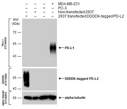
Various whole cell extracts were separated by 10% SDS-PAGE, and the membranes were blotted with PD-L1 antibody (GTX104763) diluted at 1:600 and with DDDDK tag antibody (GTX115043) diluted at 1:3000 to detect DDDDK-tagged PD-L2. The HRP-conjugated anti-rabbit IgG antibody (GTX213110-01) was used to detect the primary antibody.
Various whole cell extracts were separated by 10% SDS-PAGE, and the membranes were blotted with PD-L1 antibody (GTX104763) diluted at 1:600 and with DDDDK tag antibody (GTX115043) diluted at 1:3000 to detect DDDDK-tagged PD-L2. The HRP-conjugated anti-rabbit IgG antibody (GTX213110-01) was used to detect the primary antibody.
GTX104763 WB Image
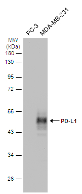
Various whole cell extracts (30 ug) were separated by 10% SDS-PAGE, and the membrane was blotted with PD-L1 antibody (GTX104763) diluted at 1:2000. The HRP-conjugated anti-rabbit IgG antibody (GTX213110-01) was used to detect the primary antibody.
Various whole cell extracts (30 ug) were separated by 10% SDS-PAGE, and the membrane was blotted with PD-L1 antibody (GTX104763) diluted at 1:2000. The HRP-conjugated anti-rabbit IgG antibody (GTX213110-01) was used to detect the primary antibody.
GTX104763 WB Image
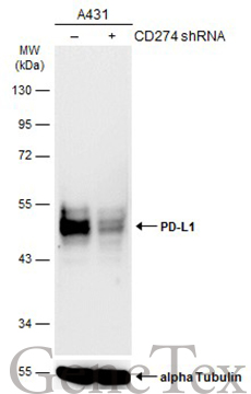
Non-transfected (?) and transfected (+) A431 whole cell extracts (30 ug) were separated by 10% SDS-PAGE, and the membrane was blotted with PD-L1 antibody (GTX104763) diluted at 1:1000. The HRP-conjugated anti-rabbit IgG antibody (GTX213110-01) was used to detect the primary antibody.
Non-transfected (?) and transfected (+) A431 whole cell extracts (30 ug) were separated by 10% SDS-PAGE, and the membrane was blotted with PD-L1 antibody (GTX104763) diluted at 1:1000. The HRP-conjugated anti-rabbit IgG antibody (GTX213110-01) was used to detect the primary antibody.
GTX104763 WB Image
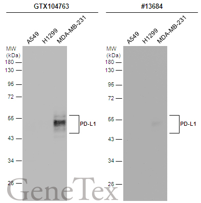
Various whole cell extracts (30 ug) were separated by 12% SDS-PAGE, and the membranes were blotted with PD-L1 antibody (GTX104763) diluted at 1:2000 and competitor's antibody (CST#13684) diluted by 1:500. The HRP-conjugated anti-rabbit IgG antibody (GTX213110-01) was used to detect the primary antibody.
Various whole cell extracts (30 ug) were separated by 12% SDS-PAGE, and the membranes were blotted with PD-L1 antibody (GTX104763) diluted at 1:2000 and competitor's antibody (CST#13684) diluted by 1:500. The HRP-conjugated anti-rabbit IgG antibody (GTX213110-01) was used to detect the primary antibody.
GTX104763 ICC/IF Image
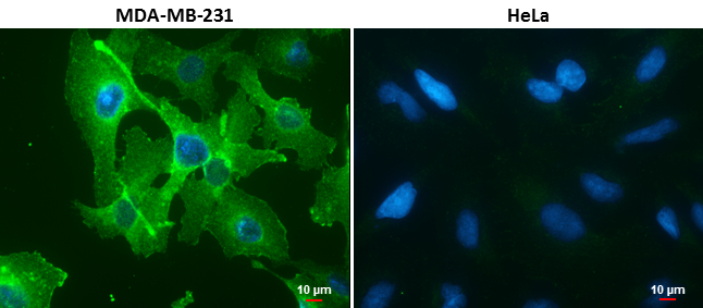
PD-L1 antibody detects PD-L1 protein at cell membrane in MDA-MB-231 cells by immunofluorescent analysis.
Sample: MDA-MB-231 (left) and HeLa (right) cells.
Green: PD-L1 protein stained by PD-L1 antibody (GTX104763) diluted at 1:1000.
Blue: Hoechst 33342 staining.
Scale bar = 10 um.
PD-L1 antibody detects PD-L1 protein at cell membrane in MDA-MB-231 cells by immunofluorescent analysis.
Sample: MDA-MB-231 (left) and HeLa (right) cells.
Green: PD-L1 protein stained by PD-L1 antibody (GTX104763) diluted at 1:1000.
Blue: Hoechst 33342 staining.
Scale bar = 10 um.
GTX104763 IHC-P Image
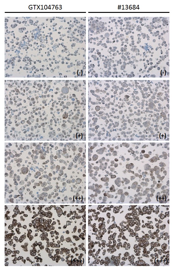
PD-L1 antibody detects PD-L1 protein at cell membrane in PD-L1 protein-expressing cell lines by immunohistochemical analysis. Antibodies: PD-L1 antibody (GTX104763) diluted at 1:1000, and competitor's antibody diluted at 1:50. Samples: Negative (-), low positive (+), intermediate positive (++) and strong positive (+++) cell line cores assessed using Quantitative Digital Pathology.
PD-L1 antibody detects PD-L1 protein at cell membrane in PD-L1 protein-expressing cell lines by immunohistochemical analysis. Antibodies: PD-L1 antibody (GTX104763) diluted at 1:1000, and competitor's antibody diluted at 1:50. Samples: Negative (-), low positive (+), intermediate positive (++) and strong positive (+++) cell line cores assessed using Quantitative Digital Pathology.
GTX104763 IHC-P Image
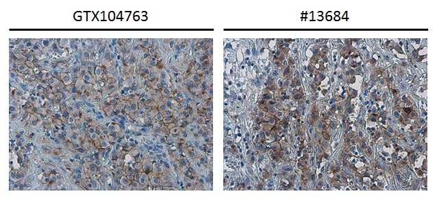
PD-L1 antibody detects PD-L1 protein at cell membrane in human ovarian carcinoma by immunohistochemical analysis. Antibodies: PD-L1 antibody (GTX104763) diluted at 1:1000, and competitor's antibody diluted at 1:50.
PD-L1 antibody detects PD-L1 protein at cell membrane in human ovarian carcinoma by immunohistochemical analysis. Antibodies: PD-L1 antibody (GTX104763) diluted at 1:1000, and competitor's antibody diluted at 1:50.
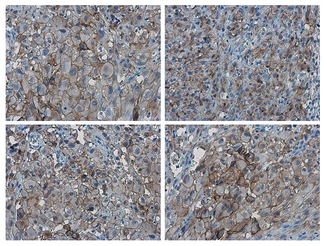
GTX104763 IHC-P Image
PD-L1 antibody detects PD-L1 proteinat cell membrane in human ovarian carcinoma by immunohistochemical analysis.
Sample: Paraffin-embedded human ovarian carcinoma.
PD-L1 antibody (GTX104763) diluted at 1:1000.
PD-L1 antibody detects PD-L1 proteinat cell membrane in human ovarian carcinoma by immunohistochemical analysis.
Sample: Paraffin-embedded human ovarian carcinoma.
PD-L1 antibody (GTX104763) diluted at 1:1000.
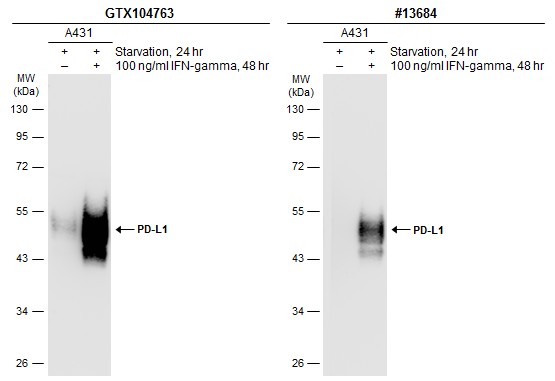
GTX104763 WB Image
Untreated (?) and treated (+) A431 whole cell extracts (30 ug) were separated by 10% SDS-PAGE, and the membranes were blotted with PD-L1 antibody (GTX104763) diluted at 1:1200 and competitor's antibody (CST#13684) diluted at 1:500. The HRP-conjugated anti-rabbit IgG antibody (GTX213110-01) was used to detect the primary antibody.
Untreated (?) and treated (+) A431 whole cell extracts (30 ug) were separated by 10% SDS-PAGE, and the membranes were blotted with PD-L1 antibody (GTX104763) diluted at 1:1200 and competitor's antibody (CST#13684) diluted at 1:500. The HRP-conjugated anti-rabbit IgG antibody (GTX213110-01) was used to detect the primary antibody.
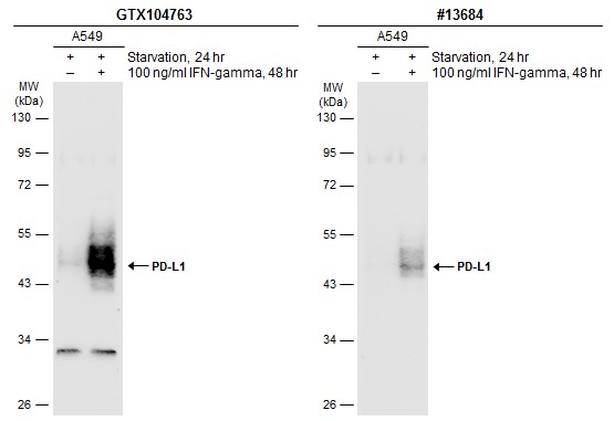
GTX104763 WB Image
Untreated (?) and treated (+) A549 whole cell extracts (30 ug) were separated by 10% SDS-PAGE, and the membranes were blotted with PD-L1 antibody (GTX104763) diluted at 1:600 and competitor's antibody (CST#13684) diluted at 1:500. The HRP-conjugated anti-rabbit IgG antibody (GTX213110-01) was used to detect the primary antibody.
Untreated (?) and treated (+) A549 whole cell extracts (30 ug) were separated by 10% SDS-PAGE, and the membranes were blotted with PD-L1 antibody (GTX104763) diluted at 1:600 and competitor's antibody (CST#13684) diluted at 1:500. The HRP-conjugated anti-rabbit IgG antibody (GTX213110-01) was used to detect the primary antibody.
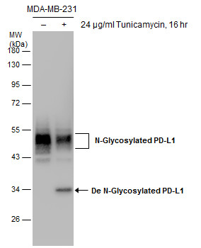
GTX104763 WB Image
Untreated (?) and treated (+) MDA-MB-231 whole cell extracts (30 ug) were separated by 10% SDS-PAGE, and the membrane was blotted with PD-L1 antibody (GTX104763) diluted at 1:1000.
Untreated (?) and treated (+) MDA-MB-231 whole cell extracts (30 ug) were separated by 10% SDS-PAGE, and the membrane was blotted with PD-L1 antibody (GTX104763) diluted at 1:1000.
カートに商品を
追加しました。
追加しました。
商品情報
| 商品説明 | レビューあり。KO/KDバリデーション済み抗体 |
|---|---|
| 抗原動物 | Human |
| 交差性 | Human |
| 免疫動物 | Rabbit |
| 性状 | Purified |
| 適用 | FCM, IC, IF, IHC, Western Blot |
| クラス | IgG |
| 標識 | Unlabeled |
| クロナリティ | Polyclonal |
| 別名 | CD274 molecule, B7-H, B7H1, PD-L1, PDCD1L1, PDCD1LG1, PDL1 |
| Genbank No | 29126 |
| データシート | データシート |
| メーカーサイト | メーカーサイト |
| 使用文献 | 使用文献 |
| 保存条件 | -20℃ |
カートに商品を
追加しました。
追加しました。
製品情報は掲載時点のものですが、価格表内の価格については随時最新のものに更新されます。お問い合わせいただくタイミングにより製品情報・価格などは変更されている場合があります。
表示価格に、消費税等は含まれていません。一部価格が予告なく変更される場合がありますので、あらかじめご了承下さい。