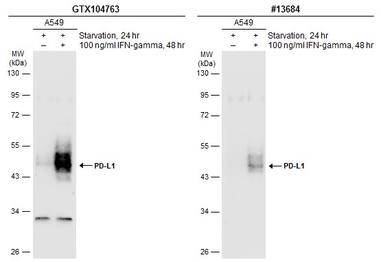
GTX104763 WB Image
Untreated (?) and treated (+) A549 whole cell extracts (30 ug) were separated by 10% SDS-PAGE, and the membranes were blotted with PD-L1 antibody (GTX104763) diluted at 1:600 and competitor's antibody (CST#13684) diluted at 1:500. The HRP-conjugated anti-rabbit IgG antibody (GTX213110-01) was used to detect the primary antibody.
GTX104763 WB Image
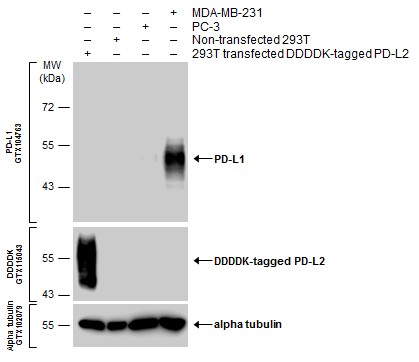
Various whole cell extracts were separated by 10% SDS-PAGE, and the membranes were blotted with PD-L1 antibody (GTX104763) diluted at 1:600 and with DDDDK tag antibody (GTX115043) diluted at 1:3000 to detect DDDDK-tagged PD-L2. The HRP-conjugated anti-rabbit IgG antibody (GTX213110-01) was used to detect the primary antibody.
GTX104763 WB Image
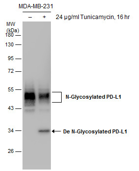
Untreated (?) and treated (+) MDA-MB-231 whole cell extracts (30 ug) were separated by 10% SDS-PAGE, and the membrane was blotted with PD-L1 antibody (GTX104763) diluted at 1:1000.
GTX104763 WB Image
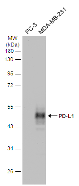
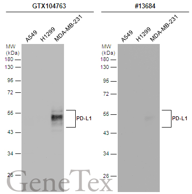
Various whole cell extracts (30 ug) were separated by 10% SDS-PAGE, and the membrane was blotted with PD-L1 antibody (GTX104763) diluted at 1:2000. The HRP-conjugated anti-rabbit IgG antibody (GTX213110-01) was used to detect the primary antibody.
GTX104763 WB Image
Non-transfected (?) and transfected (+) A431 whole cell extracts (30 ug) were separated by 10% SDS-PAGE, and the membrane was blotted with PD-L1 antibody (GTX104763) diluted at 1:1000. The HRP-conjugated anti-rabbit IgG antibody (GTX213110-01) was used to detect the primary antibody.
GTX104763 WB Image
Various whole cell extracts (30 ug) were separated by 12% SDS-PAGE, and the membranes were blotted with PD-L1 antibody (GTX104763) diluted at 1:2000 and competitor's antibody (CST#13684) diluted by 1:500. The HRP-conjugated anti-rabbit IgG antibody (GTX213110-01) was used to detect the primary antibody.
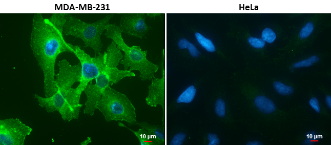
GTX104763 ICC/IF Image
PD-L1 antibody detects PD-L1 protein at cell membrane in MDA-MB-231 cells by immunofluorescent analysis.
Sample: MDA-MB-231 (left) and HeLa (right) cells.
Green: PD-L1 protein stained by PD-L1 antibody (GTX104763) diluted at 1:1000.
Blue: Hoechst 33342 staining.
Scale bar = 10 um.
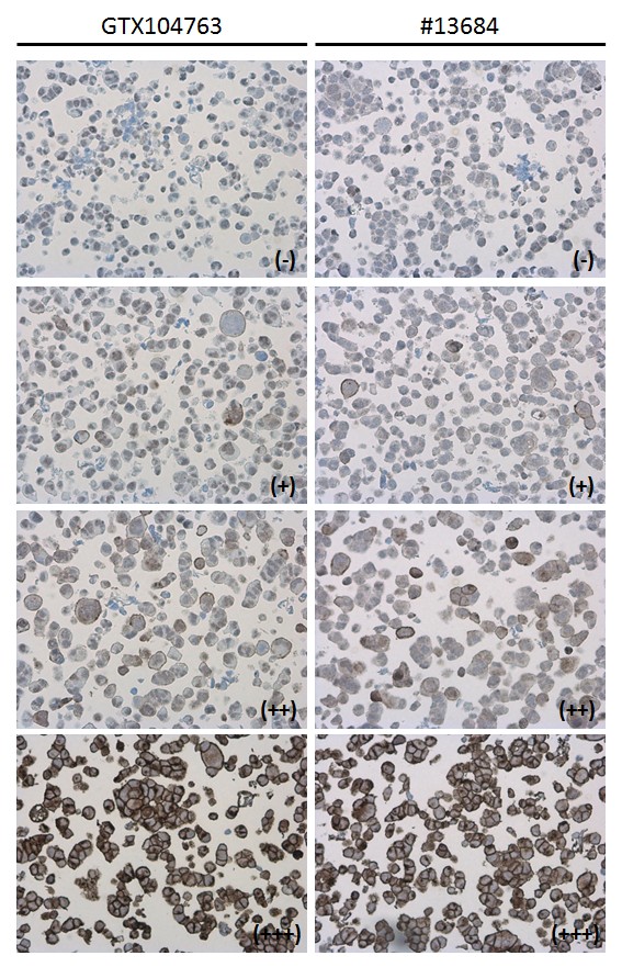
GTX104763 IHC-P Image
PD-L1 antibody detects PD-L1 protein at cell membrane in PD-L1 protein-expressing cell lines by immunohistochemical analysis. Antibodies: PD-L1 antibody (GTX104763) diluted at 1:1000, and competitor's antibody diluted at 1:50. Samples: Negative (-), low positive (+), intermediate positive (++) and strong positive (+++) cell line cores assessed using Quantitative Digital Pathology.
GTX104763 IHC-P Image
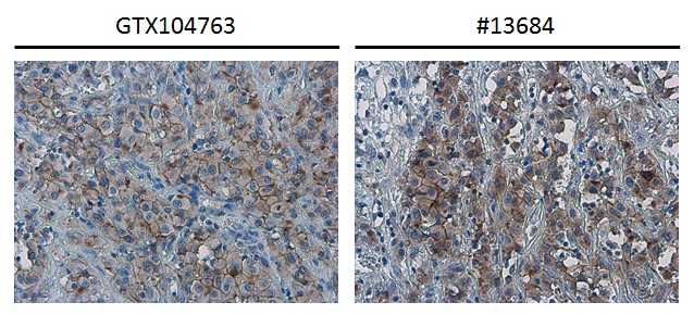
PD-L1 antibody detects PD-L1 protein at cell membrane in human ovarian carcinoma by immunohistochemical analysis. Antibodies: PD-L1 antibody (GTX104763) diluted at 1:1000, and competitor's antibody diluted at 1:50.
GTX104763 IHC-P Image
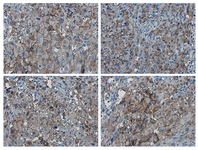
PD-L1 antibody detects PD-L1 proteinat cell membrane in human ovarian carcinoma by immunohistochemical analysis.
Sample: Paraffin-embedded human ovarian carcinoma.
PD-L1 antibody (GTX104763) diluted at 1:1000.
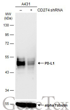
GTX104763 WB Image
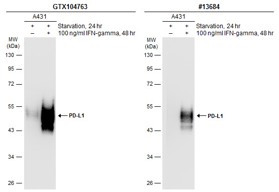
Untreated (?) and treated (+) A431 whole cell extracts (30 ug) were separated by 10% SDS-PAGE, and the membranes were blotted with PD-L1 antibody (GTX104763) diluted at 1:1200 and competitor's antibody (CST#13684) diluted at 1:500. The HRP-conjugated anti-rabbit IgG antibody (GTX213110-01) was used to detect the primary antibody.