
Anti-E-Cadherin, Rabbit-Poly | GeneTex International Corporation
掲載日情報:2026/06/03 現在Webページ番号:35107

GeneTex International CorporationのAnti-E-Cadherin, Rabbit-Poly商品情報ページです。
※本製品は研究用です。研究用以外には使用できません。
カートに商品を
追加しました。
追加しました。
価格
[在庫・価格 :2026年07月17日 00時01分現在]
※ 表示されている納期は弊社に在庫が無く、取り寄せた場合の納期目安となります。
| 詳細 | 商品名 |
|
文献数 | ||||||||||||||||||||||||||||||||||||||||||||||||||||||||||||||||||||||||||||||||||
|---|---|---|---|---|---|---|---|---|---|---|---|---|---|---|---|---|---|---|---|---|---|---|---|---|---|---|---|---|---|---|---|---|---|---|---|---|---|---|---|---|---|---|---|---|---|---|---|---|---|---|---|---|---|---|---|---|---|---|---|---|---|---|---|---|---|---|---|---|---|---|---|---|---|---|---|---|---|---|---|---|---|---|---|---|---|
|
Anti-E-Cadherin, Rabbit-Poly |
|
111 | |||||||||||||||||||||||||||||||||||||||||||||||||||||||||||||||||||||||||||||||||||

|
|||||||||||||||||||||||||||||||||||||||||||||||||||||||||||||||||||||||||||||||||||||


[在庫・価格 :2026年07月17日 00時01分現在]
※ 表示されている納期は弊社に在庫が無く、取り寄せた場合の納期目安となります。
Anti-E-Cadherin, Rabbit-Poly
文献数: 111
- 商品コード:GTX100443
- メーカー:GNT
- 包装:100μl
- 価格:¥85,000
- 在庫:1個
- 納期:10日程度 ※※ 表示されている納期は弊社に在庫がなく、取り寄せた場合の目安納期となります。
- 法規制等:
| 説明文 | レビューあり。KO/KDバリデーション済み抗体。抗原:aa 462~732 別名:cadherin 1,Arc-1,BCDS1,CD324,CDHE,ECAD,LCAM,UVO Genbank No: 999 |
||||||
|---|---|---|---|---|---|---|---|
| 別包装品 | 別包装品あり | ||||||
| 法規制等 | |||||||
| 保存条件 | -20℃ | 法規備考 | |||||
| 抗原種 | Human | 免疫動物 | Rabbit | ||||
| 交差性 | Dog/Human/Mouse/Rat/Zebrafish | 適用 | IC,IF,IHC,IP,PLA,Western Blot | ||||
| 標識 | Unlabeled | 性状 | Purified | ||||
| 吸収処理 | クラス | IgG | |||||
| クロナリティ | Polyclonal | フォーマット | |||||
| 掲載カタログ |
|
||||||
| 製品記事 | 抗SSEA-5抗体 ゼブラフィッシュ(zebrafish)研究用製品特集 GeneTex社:抗E-カドヘリン抗体 獣医学研究用抗体 VetSignalシリーズ |
||||||
| 関連記事 | GeneTex社における抗体の品質管理 |
||||||
カートに商品を
追加しました。
追加しました。
ラインナップ
カートに商品を
追加しました。
追加しました。
画像
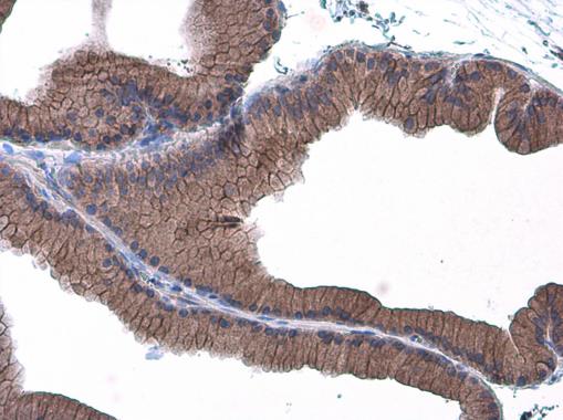
GTX100443 IHC-P Image
E-Cadherin antibody detects E-Cadherin protein at cell membrane in rat prostate by immunohistochemical analysis.
Sample: Paraffin-embedded rat prostate.
E-Cadherin antibody (GTX100443) diluted at 1:500.
E-Cadherin antibody detects E-Cadherin protein at cell membrane in rat prostate by immunohistochemical analysis.
Sample: Paraffin-embedded rat prostate.
E-Cadherin antibody (GTX100443) diluted at 1:500.
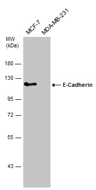
GTX100443 WB Image
Various whole cell extracts (30 ug) were separated by 7.5% SDS-PAGE, and the membrane was blotted with E-Cadherin antibody (GTX100443) diluted at 1:2000.
Various whole cell extracts (30 ug) were separated by 7.5% SDS-PAGE, and the membrane was blotted with E-Cadherin antibody (GTX100443) diluted at 1:2000.
![GTX100443 IP Image<br>E-cadherin antibody immunoprecipitates E-cadherin protein in IP experiments.<br>IP samples: MCF-7 whole cell extract<br>A. Control with 3 ug of preimmune Rabbit IgG<br>B. Immunoprecipitation of E-cadherin protein by 3 ug E-cadherin antibody (GTX100443)<br>5 % SDS-PAGE<br>The immunoprecipitated E-cadherin protein was detected by E-cadherin antibody (GTX100443) diluted at 1:500.<br>[EasyBlot anti-rabbit IgG (GTX221666-01) was used as a secondary reagent]<br></br></br>](/domestic/data/graphics/GNT/graphics/GTX100443_41388_IP.jpg)
GTX100443 IP Image
E-cadherin antibody immunoprecipitates E-cadherin protein in IP experiments.
IP samples: MCF-7 whole cell extract
A. Control with 3 ug of preimmune Rabbit IgG
B. Immunoprecipitation of E-cadherin protein by 3 ug E-cadherin antibody (GTX100443)
5 % SDS-PAGE
The immunoprecipitated E-cadherin protein was detected by E-cadherin antibody (GTX100443) diluted at 1:500.
[EasyBlot anti-rabbit IgG (GTX221666-01) was used as a secondary reagent]
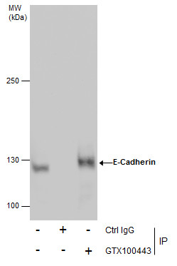
GTX100443 IP Image
Immunoprecipitation of E-Cadherin protein from MCF-7 whole cell extracts using 5 ug of E-Cadherin antibody (GTX100443).
Western blot analysis was performed using E-Cadherin antibody (GTX100443).
EasyBlot anti-Rabbit IgG (GTX221666-01) was used as a secondary reagent.
Immunoprecipitation of E-Cadherin protein from MCF-7 whole cell extracts using 5 ug of E-Cadherin antibody (GTX100443).
Western blot analysis was performed using E-Cadherin antibody (GTX100443).
EasyBlot anti-Rabbit IgG (GTX221666-01) was used as a secondary reagent.
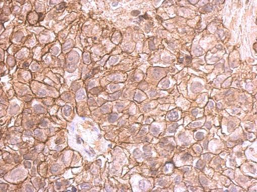
GTX100443 IHC-P Image
E-cadherin antibody detects E-cadherin protein at membrane on human breast cancer by immunohistochemical analysis.
Sample: Paraffin-embedded breast cancer.
E-cadherin antibody (GTX100443) dilution: 1:500.
E-cadherin antibody detects E-cadherin protein at membrane on human breast cancer by immunohistochemical analysis.
Sample: Paraffin-embedded breast cancer.
E-cadherin antibody (GTX100443) dilution: 1:500.
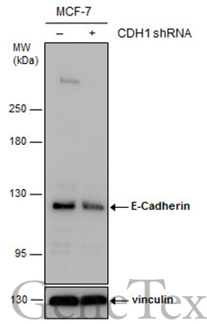
GTX100443 WB Image
Non-transfected (?) and transfected (+) MCF-7 whole cell extracts (30 ug) were separated by 5% SDS-PAGE, and the membrane was blotted with E-Cadherin antibody (GTX100443) diluted at 1:7000. The HRP-conjugated anti-rabbit IgG antibody (GTX213110-01) was used to detect the primary antibody.
Non-transfected (?) and transfected (+) MCF-7 whole cell extracts (30 ug) were separated by 5% SDS-PAGE, and the membrane was blotted with E-Cadherin antibody (GTX100443) diluted at 1:7000. The HRP-conjugated anti-rabbit IgG antibody (GTX213110-01) was used to detect the primary antibody.
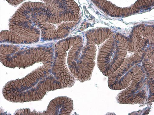
GTX100443 IHC-P Image
E-Cadherin antibody detects E-Cadherin protein at cell membrane in rat prostate by immunohistochemical analysis.
Sample: Paraffin-embedded rat prostate.
E-Cadherin antibody (GTX100443) diluted at 1:500.
E-Cadherin antibody detects E-Cadherin protein at cell membrane in rat prostate by immunohistochemical analysis.
Sample: Paraffin-embedded rat prostate.
E-Cadherin antibody (GTX100443) diluted at 1:500.
GTX100443 IHC-P Image
E-Cadherin antibody detects E-Cadherin protein at cell membrane in mouse pancreas by immunohistochemical analysis.
Sample: Paraffin-embedded mouse pancreas.
E-Cadherin antibody (GTX100443) diluted at 1:400.
E-Cadherin antibody detects E-Cadherin protein at cell membrane in mouse pancreas by immunohistochemical analysis.
Sample: Paraffin-embedded mouse pancreas.
E-Cadherin antibody (GTX100443) diluted at 1:400.
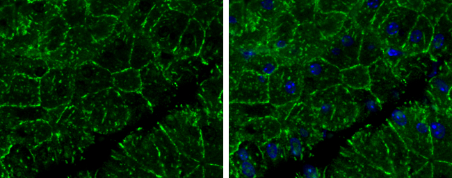
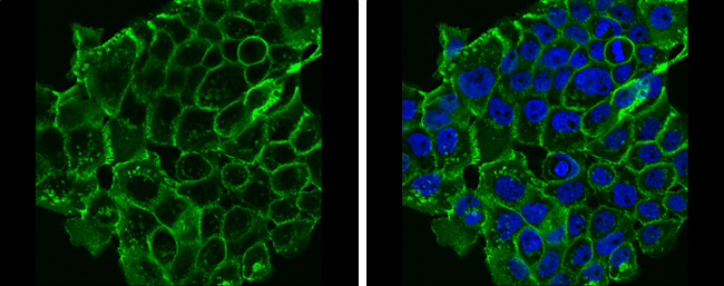
GTX100443 ICC/IF Image
E-Cadherin antibody detects E-Cadherin protein at cell membrane by immunofluorescent analysis.
Sample: HCT 116 cells were fixed in 4% paraformaldehyde at RT for 15 min.
Green: E-Cadherin protein stained by E-Cadherin antibody (GTX100443) diluted at 1:500.
Blue: Hoechst 33342 staining.
Scale bar = 10 um.

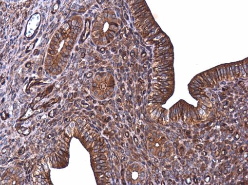
GTX100443 IHC-P Image
Immunohistochemical analysis of paraffin-embedded human ulcerative colitis tissue using E-Cadherin antibody (GTX100443).
Immunohistochemical analysis of paraffin-embedded human ulcerative colitis tissue using E-Cadherin antibody (GTX100443).
GTX100443 ICC/IF Image
E-Cadherin antibody detects E-Cadherin protein at cell membrane by immunofluorescent analysis.
Sample: A431 cells were fixed in 4% paraformaldehyde at RT for 15 min.
Green: E-Cadherin protein stained by E-Cadherin antibody (GTX100443) diluted at 1:500.
Blue: Hoechst 33342 staining.
E-Cadherin antibody detects E-Cadherin protein at cell membrane by immunofluorescent analysis.
Sample: A431 cells were fixed in 4% paraformaldehyde at RT for 15 min.
Green: E-Cadherin protein stained by E-Cadherin antibody (GTX100443) diluted at 1:500.
Blue: Hoechst 33342 staining.
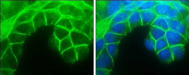
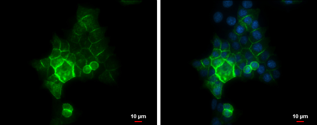
GTX100443 ICC/IF Image
E-Cadherin antibody detects E-Cadherin protein at cell membrane by immunofluorescent analysis.
Sample: MCF7 cells were fixed in 4% paraformaldehyde at RT for 15 min.
Green: E-Cadherin protein stained by E-Cadherin antibody (GTX100443) diluted at 1:500.
Blue: Hoechst 33342 staining.
E-Cadherin antibody detects E-Cadherin protein at cell membrane by immunofluorescent analysis.
Sample: MCF7 cells were fixed in 4% paraformaldehyde at RT for 15 min.
Green: E-Cadherin protein stained by E-Cadherin antibody (GTX100443) diluted at 1:500.
Blue: Hoechst 33342 staining.
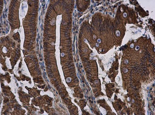
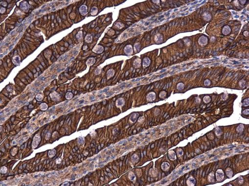
GTX100443 IHC-P Image
E-Cadherin antibody detects E-Cadherin protein at cell membrane and cytoplasm in rat duodenum by immunohistochemical analysis.
Sample: Paraffin-embedded rat duodenum.
E-Cadherin antibody (GTX100443) diluted at 1:500.
E-Cadherin antibody detects E-Cadherin protein at cell membrane and cytoplasm in rat duodenum by immunohistochemical analysis.
Sample: Paraffin-embedded rat duodenum.
E-Cadherin antibody (GTX100443) diluted at 1:500.
GTX100443 IHC-P Image
E-Cadherin antibody detects E-Cadherin protein at cell membrane in mouse cervix by immunohistochemical analysis.
Sample: Paraffin-embedded mouse cervix.
E-Cadherin antibody (GTX100443) diluted at 1:500.
E-Cadherin antibody detects E-Cadherin protein at cell membrane in mouse cervix by immunohistochemical analysis.
Sample: Paraffin-embedded mouse cervix.
E-Cadherin antibody (GTX100443) diluted at 1:500.
GTX100443 IHC-P Image
E-Cadherin antibody detects E-Cadherin protein at cell membrane in rat intestine by immunohistochemical analysis.
Sample: Paraffin-embedded rat intestine.
E-Cadherin antibody (GTX100443) diluted at 1:500.
E-Cadherin antibody detects E-Cadherin protein at cell membrane in rat intestine by immunohistochemical analysis.
Sample: Paraffin-embedded rat intestine.
E-Cadherin antibody (GTX100443) diluted at 1:500.
カートに商品を
追加しました。
追加しました。
商品情報
| 商品説明 | レビューあり。KO/KDバリデーション済み抗体 |
|---|---|
| 抗原 | aa 462~732 |
| 抗原動物 | Human |
| 交差性 | Dog/Human/Mouse/Rat/Zebrafish |
| 免疫動物 | Rabbit |
| 性状 | Purified |
| 適用 | IC, IF, IHC, IP, PLA, Western Blot |
| クラス | IgG |
| 標識 | Unlabeled |
| クロナリティ | Polyclonal |
| 別名 | cadherin 1, Arc-1, BCDS1, CD324, CDHE, ECAD, LCAM, UVO |
| Genbank No | 999 |
| データシート | データシート |
| メーカーサイト | メーカーサイト |
| 使用文献 | 使用文献 |
| 保存条件 | -20℃ |
カートに商品を
追加しました。
追加しました。
製品情報は掲載時点のものですが、価格表内の価格については随時最新のものに更新されます。お問い合わせいただくタイミングにより製品情報・価格などは変更されている場合があります。
表示価格に、消費税等は含まれていません。一部価格が予告なく変更される場合がありますので、あらかじめご了承下さい。