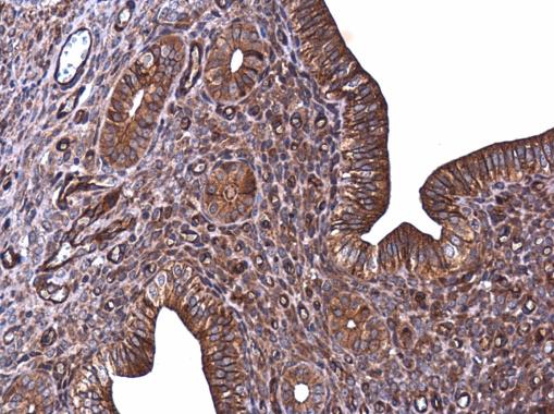
GTX100443 IHC-P Image
E-Cadherin antibody detects E-Cadherin protein at cell membrane in rat prostate by immunohistochemical analysis.
Sample: Paraffin-embedded rat prostate.
E-Cadherin antibody (GTX100443) diluted at 1:500.
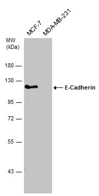
GTX100443 WB Image
Various whole cell extracts (30 ug) were separated by 7.5% SDS-PAGE, and the membrane was blotted with E-Cadherin antibody (GTX100443) diluted at 1:2000.
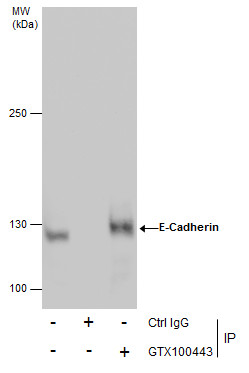
GTX100443 IP Image
E-cadherin antibody immunoprecipitates E-cadherin protein in IP experiments.
IP samples: MCF-7 whole cell extract
A. Control with 3 ug of preimmune Rabbit IgG
B. Immunoprecipitation of E-cadherin protein by 3 ug E-cadherin antibody (GTX100443)
5 % SDS-PAGE
The immunoprecipitated E-cadherin protein was detected by E-cadherin antibody (GTX100443) diluted at 1:500.
[EasyBlot anti-rabbit IgG (GTX221666-01) was used as a secondary reagent]
GTX100443 IP Image
Immunoprecipitation of E-Cadherin protein from MCF-7 whole cell extracts using 5 ug of E-Cadherin antibody (GTX100443).
Western blot analysis was performed using E-Cadherin antibody (GTX100443).
EasyBlot anti-Rabbit IgG (GTX221666-01) was used as a secondary reagent.
GTX100443 IHC-P Image
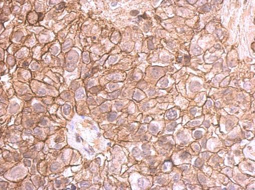
E-cadherin antibody detects E-cadherin protein at membrane on human breast cancer by immunohistochemical analysis.
Sample: Paraffin-embedded breast cancer.
E-cadherin antibody (GTX100443) dilution: 1:500.
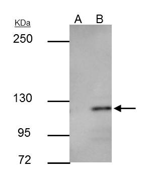
GTX100443 WB Image
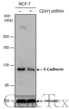
Non-transfected (?) and transfected (+) MCF-7 whole cell extracts (30 ug) were separated by 5% SDS-PAGE, and the membrane was blotted with E-Cadherin antibody (GTX100443) diluted at 1:7000. The HRP-conjugated anti-rabbit IgG antibody (GTX213110-01) was used to detect the primary antibody.
GTX100443 IHC-P Image
E-Cadherin antibody detects E-Cadherin protein at cell membrane in rat prostate by immunohistochemical analysis.
Sample: Paraffin-embedded rat prostate.
E-Cadherin antibody (GTX100443) diluted at 1:500.
GTX100443 IHC-P Image
E-Cadherin antibody detects E-Cadherin protein at cell membrane in mouse pancreas by immunohistochemical analysis.
Sample: Paraffin-embedded mouse pancreas.
E-Cadherin antibody (GTX100443) diluted at 1:400.
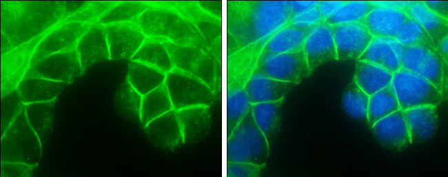
GTX100443 ICC/IF Image
E-Cadherin antibody detects E-Cadherin protein at cell membrane by immunofluorescent analysis.
Sample: HCT 116 cells were fixed in 4% paraformaldehyde at RT for 15 min.
Green: E-Cadherin protein stained by E-Cadherin antibody (GTX100443) diluted at 1:500.
Blue: Hoechst 33342 staining.
Scale bar = 10 um.

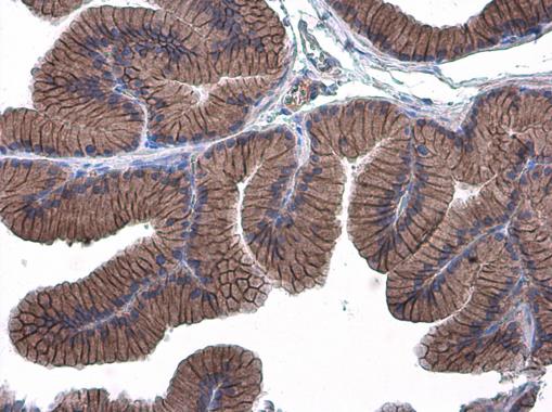
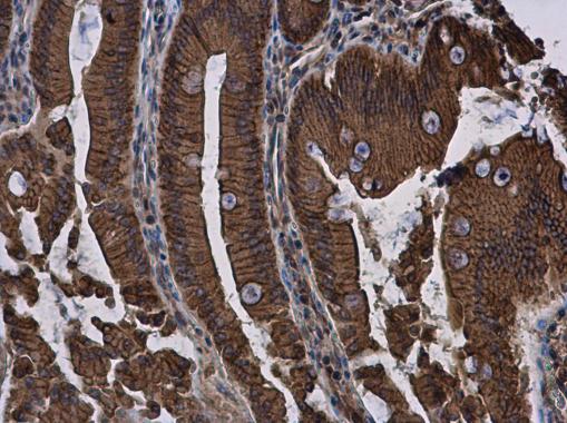
GTX100443 IHC-P Image
Immunohistochemical analysis of paraffin-embedded human ulcerative colitis tissue using E-Cadherin antibody (GTX100443).
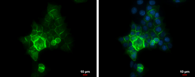
GTX100443 ICC/IF Image
E-Cadherin antibody detects E-Cadherin protein at cell membrane by immunofluorescent analysis.
Sample: A431 cells were fixed in 4% paraformaldehyde at RT for 15 min.
Green: E-Cadherin protein stained by E-Cadherin antibody (GTX100443) diluted at 1:500.
Blue: Hoechst 33342 staining.
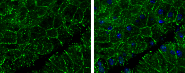
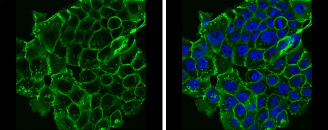
GTX100443 ICC/IF Image
E-Cadherin antibody detects E-Cadherin protein at cell membrane by immunofluorescent analysis.
Sample: MCF7 cells were fixed in 4% paraformaldehyde at RT for 15 min.
Green: E-Cadherin protein stained by E-Cadherin antibody (GTX100443) diluted at 1:500.
Blue: Hoechst 33342 staining.
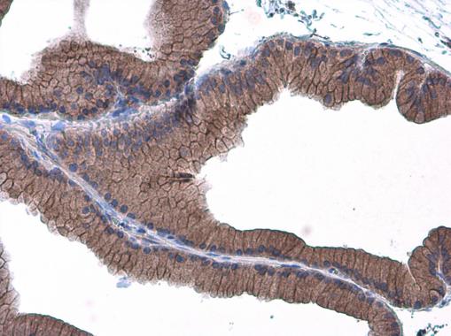
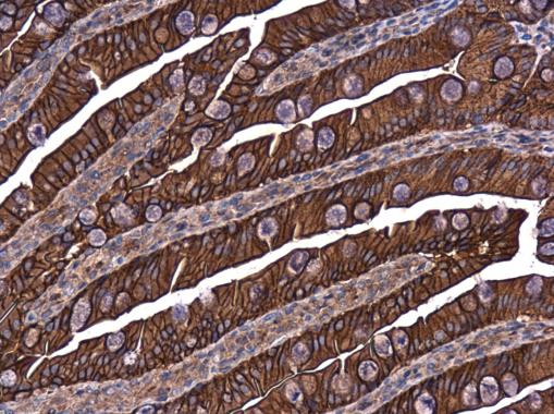
GTX100443 IHC-P Image
E-Cadherin antibody detects E-Cadherin protein at cell membrane and cytoplasm in rat duodenum by immunohistochemical analysis.
Sample: Paraffin-embedded rat duodenum.
E-Cadherin antibody (GTX100443) diluted at 1:500.
GTX100443 IHC-P Image
E-Cadherin antibody detects E-Cadherin protein at cell membrane in mouse cervix by immunohistochemical analysis.
Sample: Paraffin-embedded mouse cervix.
E-Cadherin antibody (GTX100443) diluted at 1:500.
GTX100443 IHC-P Image
E-Cadherin antibody detects E-Cadherin protein at cell membrane in rat intestine by immunohistochemical analysis.
Sample: Paraffin-embedded rat intestine.
E-Cadherin antibody (GTX100443) diluted at 1:500.