
抗EGF Receptor抗体(Anti-EGF Receptor, Human, Goat-Poly antibody)
掲載日情報:2021/01/28 現在Webページ番号:26841

EGF Receptorに対する抗体(Anti-EGF Receptor, Human, Goat-Poly )です。
※ 本製品は研究用です。研究用以外には使用できません。
追加しました。
- 価格
- Product Details
- Applications and Data
- Data Examples
- References
- Related Research Areas
- Related Product & Information
- Citations
価格
[在庫・価格 :2024年05月18日 00時00分現在]
| 詳細 | 商品名 |
|
文献数 | ||||||||||||||||||||||||||||||||||||||||||||||||||||||||||||||||||||||||||||||||||
|---|---|---|---|---|---|---|---|---|---|---|---|---|---|---|---|---|---|---|---|---|---|---|---|---|---|---|---|---|---|---|---|---|---|---|---|---|---|---|---|---|---|---|---|---|---|---|---|---|---|---|---|---|---|---|---|---|---|---|---|---|---|---|---|---|---|---|---|---|---|---|---|---|---|---|---|---|---|---|---|---|---|---|---|---|---|
|
Anti-EGF Receptor, Human, Goat-Poly |
|
13 | |||||||||||||||||||||||||||||||||||||||||||||||||||||||||||||||||||||||||||||||||||
|
|||||||||||||||||||||||||||||||||||||||||||||||||||||||||||||||||||||||||||||||||||||
|
Anti-Human EGFR Affinity Purified Polyclonal Ab |
|
0 | |||||||||||||||||||||||||||||||||||||||||||||||||||||||||||||||||||||||||||||||||||
|
|||||||||||||||||||||||||||||||||||||||||||||||||||||||||||||||||||||||||||||||||||||
[在庫・価格 :2024年05月18日 00時00分現在]
Anti-EGF Receptor, Human, Goat-Poly
文献数: 13
- 商品コード:AF231
- メーカー:RSD
- 包装:100μg
- 価格:¥106,000
- 在庫:無(未発注)
- 納期:10日程度 ※※ 表示されている納期は弊社に在庫がなく、取り寄せた場合の目安納期となります。
- 法規制等:
| 説明文 |
マッチドペア:Human EGF R/ErbB1 サンドイッチELISAの補足用抗体として利用可能,検出用抗体として#BAF231,スタンダードとして#1095-ER-002を用いる。 別名:avian erythroblastic leukemia viral (v-erb-b) oncogene homolog Genbank No: 1956 Protein Accession No: CAA25240 |
||||||
|---|---|---|---|---|---|---|---|
| 別包装品 | 別包装品あり | ||||||
| 法規制等 | |||||||
| 保存条件 | -20℃ | 法規備考 | |||||
| 抗原種 | Human | 免疫動物 | Goat | ||||
| 交差性 | Human | 適用 | ELISA,FCM,IC,IHC,IP,Simple Western,Western Blot | ||||
| 標識 | Unlabeled | 性状 | Antigen Affinity Purified | ||||
| 吸収処理 | クラス | IgG | |||||
| クロナリティ | Polyclonal | フォーマット | |||||
| 掲載カタログ |
|
||||||
| 製品記事 |
免疫染色システム ImmPRESS® Reagent Anti-Goat IgG |
||||||
| 関連記事 |
R&D Systems(R&Dシステムズ)社 ELISA用ペア抗体を使用したELISA 構築ガイド |
||||||
Anti-Human EGFR Affinity Purified Polyclonal Ab
文献数: 0
- 商品コード:AF231-SP
- メーカー:RSD
- 包装:25μg
- 価格:¥30,000
- 在庫:無(未発注)
- 納期:2~3週間 ※※ 表示されている納期は弊社に在庫がなく、取り寄せた場合の目安納期となります。
- 法規制等:
| 説明文 |
※受注発注品。形状:溶液または凍結乾燥 別名:avian erythroblastic leukemia viral (v-erb-b) oncogene homolog Genbank No: 1956 Protein Accession No: CAA25240 |
||||||
|---|---|---|---|---|---|---|---|
| 別包装品 | 別包装品あり | ||||||
| 法規制等 | |||||||
| 保存条件 | -20℃ | 法規備考 | |||||
| 抗原種 | 免疫動物 | Goat | |||||
| 交差性 | Human | 適用 | ELISA,FCM,IC,IHC,IP,Simple Western,Western Blot | ||||
| 標識 | Unlabeled | 性状 | Antigen Affinity Purified | ||||
| 吸収処理 | クラス | IgG | |||||
| クロナリティ | Polyclonal | フォーマット | |||||
| 掲載カタログ |
|
||||||
| 製品記事 |
免疫染色システム ImmPRESS® Reagent Anti-Goat IgG |
||||||
| 関連記事 |
R&D Systems(R&Dシステムズ)社 ELISA用ペア抗体を使用したELISA 構築ガイド |
||||||
追加しました。
Product Details
| Species Reactivity | Human |
|---|---|
| Label | Unconjugated |
| Immunogen | Mouse myeloma cell line NS0-derived recombinant human EGFRLeu25-Ser645Accession # CAA25240 |
| Source | Polyclonal Goat IgG |
| Purification | Antigen Affinity-purified |
| Specificity | Detects human EGFR in ELISAs and Western blots. In sandwich ELISAs, approximately 3% cross-reactivity with recombinant mouse EGFR is observed and less than 0.1% cross-reactivity with recombinant human (rh) ErbB2 and rhErbB3 is observed. |
追加しました。
Applications and Data
| Recommended Concentration | Sample | |
| Western Blot | 1 µg/mL | See below |
| Flow Cytometry | 0.25 µg/106 cells | See below |
| Immunohistochemistry | 1-15 µg/mL | See below |
| Immunoprecipitation | 1 µg/mL | A431 human epithelial carcinoma cell line, see our available Western blot detection antibodies |
| CyTOF-ready | Ready to be labeled using established conjugation methods. No BSA or other carrier proteins that could interfere with conjugation. | |
| Immunocytochemistry | 1-15 µg/mL | See below |
| Human EGFR Sandwich Immunoassay | Reagent | |
| ELISA Capture (Matched Antibody Pair) | 0.2-0.8 µg/mL | Human EGFR Antibody (Catalog #AF231 ) |
| ELISA Detection (Matched Antibody Pair) | 0.1-0.4 µg/mL | Human EGFR Biotinylated Antibody (Catalog #BAF231 ) |
| ELISA Standard | Recombinant Human EGFR Protein (Catalog #1095-ER ) | |
| Please Note: Optimal dilutions should be determined by each laboratory for each application.General Protocolsare available in the Technical Information section on our website. | ||
追加しました。
Data Examples
| Western Blot |
![Western Blot EGFR Antibody [Unconjugated]](http://resources.rndsystems.com/images/datasheets/antibody/EGF_R_AF231_Western_Blot_19925.jpg) click image to view larger | Detection of Human EGFR by Western Blot. Western blot shows lysates of HeLa human cervical epithelial carcinoma cell line and MDA‑MB‑231 human breast cancer cell line. PVDF membrane was probed with 1 µg/mL of Goat Anti-Human EGFR Antigen Affinity-purified Polyclonal Antibody (Catalog # AF231) followed by HRP-conjugated Anti-Goat IgG Secondary Antibody (Catalog # HAF017). A specific band was detected for EGFR at approximately 175 kDa (as indicated). This experiment was conducted under reducing conditions and using Immunoblot Buffer Group 1. |
| Flow Cytometry |
![Flow Cytometry EGFR Antibody [Unconjugated]](http://resources.rndsystems.com/images/datasheets/antibody/EGF_R_AF231_Flow_Cytometry_20401.jpg) click image to view larger | Detection of EGFR in A431 Human Cell Line by Flow Cytometry. A431 human epithelial carcinoma cell line was stained with Goat Anti-Human EGFR Antigen Affinity-purified Polyclonal Antibody (Catalog # AF231, filled histogram) or isotype control antibody (Catalog # AB-108-C, open histogram), followed by Phycoerythrin-conjugated Anti-Goat IgG Secondary Antibody (Catalog # F0107). View our protocol for Staining Membrane-associated Proteins. |
| Immunocytochemistry |
![Immunocytochemistry EGFR Antibody [Unconjugated]](http://resources.rndsystems.com/images/datasheets/antibody/EGF_R_AF231_Immunocytochemistry__Immunofluorescence_21143.jpg) click image to view larger | EGFR in A431 Human Cell Line. EGFR was detected in immersion fixed A431 human epithelial carcinoma cell line using Goat Anti-Human EGFR Antigen Affinity-purified Polyclonal Antibody (Catalog # AF231) at 1 µg/mL for 3 hours at room temperature. Cells were stained using the NorthernLights™ 557-conjugated Anti-Goat IgG Secondary Antibody (red; Catalog # NL001) and counterstained with DAPI (blue). Specific staining was localized to plasma membrane. View our protocol for Fluorescent ICC Staining of Cells on Coverslips. |
| Immunohistochemistry |
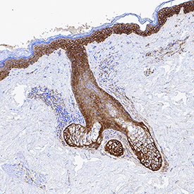
![Immunohistochemistry EGFR Antibody [Unconjugated]](http://resources.rndsystems.com/images/datasheets/antibody/EGF_R_AF231_Immunohistochemistry_20892.jpg) click image to view larger | EGFR in Human Skin. EGFR was detected in immersion fixed frozen sections of human skin using Goat Anti-Human EGFR Antigen Affinity-purified Polyclonal Antibody (Catalog # AF231) at 1 µg/mL for 1 hour at room temperature followed by incubation with the Anti-Goat IgG VisUCyte™ HRP Polymer Antibody (Catalog # VC004). Tissue was stained using DAB (brown) and counterstained with hematoxylin (blue). Specific staining was localized to plasma membrane. View our protocol for IHC Staining with VisUCyte HRP Polymer Detection Reagents. |
| Reconstitution | Reconstitute at 0.2 mg/mL in sterile PBS. | Reconstitution Buffer Available |
| Shipping | The product is shipped at ambient temperature. Upon receipt, store it immediately at the temperature recommended below. *Small pack size (SP) is shipped with polar packs. Upon receipt, store it immediately at -20 to -70 °C | |
| Stability & Storage | Use a manual defrost freezer and avoid repeated freeze-thaw cycles. 12 months from date of receipt, -20 to -70 °C as supplied. | |
| 1 month, 2 to 8 °C under sterile conditions after reconstitution. | ||
| 6 months, -20 to -70 °C under sterile conditions after reconstitution. | ||
| Background: EGFR | The epidermal growth factor receptor (EGFR) subfamily of receptor tyrosine kinases comprises four members: EGFR (also known as HER1, ErbB1 or ErbB), ErbB2 (Neu, HER2), ErbB3 (HER3), and ErbB4 (HER4). All family members are type I transmembrane glycoproteins that have an extracellular domain which contains two cysteine-rich domains separated by a spacer region that is involved in ligand binding, and a cytoplasmic domain which has a membrane-proximal tyrosine kinase domain and a C-terminal tail with multiple tyrosine autophosphorylation sites. The human EGFR gene encodes a 1210 amino acid (aa) residue precursor with a 24 aa putative signal peptide, a 621 aa extracellular domain, a 23 aa transmembrane domain, and a 542 aa cytoplasmic domain. EGFR has been shown to bind a subset of the EGF family ligands, including EGF, amphiregulin, TGF-alpha, betacellulin, epiregulin, heparin-binding EGF and neuregulin-2 alpha in the absence of a co-receptor. Ligand binding induces EGFR homodimerization as well as heterodimerization with ErbB2, resulting in kinase activation, tyrosine phosphorylation and cell signaling. EGFR can also be recruited to form heterodimers with the ligand-activated ErbB3 or ErbB4. EGFR signaling has been shown to regulate multiple biological functions including cell proliferation, differentiation, motility and apoptosis. In addition, EGFR signaling has also been shown to play a role in carcinogenesis (1 - 3). |
|
追加しました。
References
| Daly, R.J. (1999) Growth Factors, 16:255. | |
| Schlessinger, J. (2000) Cell. 103:211. | |
| Maihle, N.J. et al. (2002) Cancer Treat. Res. 107:247. | |
| Long Name: | Epidermal Growth Factor Receptor |
| Entrez Gene IDs: | 1956 (Human); 13649 (Mouse) |
| Alternate Names: | avian erythroblastic leukemia viral (v-erb-b) oncogene homolog; cell growth inhibiting protein 40; cell proliferation-inducing protein 61; EC 2.7.10; EC 2.7.10.1; EGF R; EGFR; epidermal growth factor receptor (avian erythroblastic leukemia viral (v-erb-b)oncogene homolog); epidermal growth factor receptor; ErbB; ErbB1; ERBB1PIG61; HER1; HER-1; mENA; Proto-oncogene c-ErbB-1; Receptor tyrosine-protein kinase erbB-1 |
追加しました。
Related Research Areas
| Adult Neurogenesis | ||
| Cancer Biomarkers | ||
| Cytokine and Growth Factor Receptors on VSMC | ||
| EGF Family | ||
| Receptor Tyrosine Kinases (RTKs) | ||
| Receptor Tyrosine Kinases (RTKs) in the Akt Pathway | ||
| Receptors in the Jak/STAT Pathway | ||
| ||
| Western Blot | |
|---|---|
 | Detection of Human EGFR by Western Blot. Western blot shows lysates of HeLa human cervical epithelial carcinoma cell line and MDA‑MB‑231 human breast cancer cell line. PVDF membrane was probed with 1 µg/mL of Goat Anti-Human EGFR Antigen Affinity-purified Polyclonal Antibody (Catalog # AF231) followed by HRP-conjugated Anti-Goat IgG Secondary Antibody (Catalog # HAF017). A specific band was detected for EGFR at approximately 175 kDa (as indicated). This experiment was conducted under reducing conditions and using Immunoblot Buffer Group 1. |
| Flow Cytometry | |
 | Detection of EGFR in A431 Human Cell Line by Flow Cytometry. A431 human epithelial carcinoma cell line was stained with Goat Anti-Human EGFR Antigen Affinity-purified Polyclonal Antibody (Catalog # AF231, filled histogram) or isotype control antibody (Catalog # AB-108-C, open histogram), followed by Phycoerythrin-conjugated Anti-Goat IgG Secondary Antibody (Catalog # F0107). View our protocol for Staining Membrane-associated Proteins. |
| Immunocytochemistry | |
 | EGFR in A431 Human Cell Line. EGFR was detected in immersion fixed A431 human epithelial carcinoma cell line using Goat Anti-Human EGFR Antigen Affinity-purified Polyclonal Antibody (Catalog # AF231) at 1 µg/mL for 3 hours at room temperature. Cells were stained using the NorthernLights™ 557-conjugated Anti-Goat IgG Secondary Antibody (red; Catalog # NL001) and counterstained with DAPI (blue). Specific staining was localized to plasma membrane. View our protocol for Fluorescent ICC Staining of Cells on Coverslips. |
| Immunohistochemistry | |
 | EGFR in Human Skin. EGFR was detected in immersion fixed frozen sections of human skin using Goat Anti-Human EGFR Antigen Affinity-purified Polyclonal Antibody (Catalog # AF231) at 1 µg/mL for 1 hour at room temperature followed by incubation with the Anti-Goat IgG VisUCyte™ HRP Polymer Antibody (Catalog # VC004). Tissue was stained using DAB (brown) and counterstained with hematoxylin (blue). Specific staining was localized to plasma membrane. View our protocol for IHC Staining with VisUCyte HRP Polymer Detection Reagents. |
追加しました。
Related Product & Information
| Background | EGFR |
|---|---|
| background_content | Background: EGFR The epidermal growth factor receptor (EGFR) subfamily of receptor tyrosine kinases comprises four members: EGFR (also known as HER1, ErbB1 or ErbB), ErbB2 (Neu, HER2), ErbB3 (HER3), and ErbB4 (HER4). All family members are type I transmembrane glycoproteins that have an extracellular domain which contains two cysteine-rich domains separated by a spacer region that is involved in ligand binding, and a cytoplasmic domain which has a membrane-proximal tyrosine kinase domain and a C-terminal tail with multiple tyrosine autophosphorylation sites. The human EGFR gene encodes a 1210 amino acid (aa) residue precursor with a 24 aa putative signal peptide, a 621 aa extracellular domain, a 23 aa transmembrane domain, and a 542 aa cytoplasmic domain. EGFR has been shown to bind a subset of the EGF family ligands, including EGF, amphiregulin, TGF-alpha, betacellulin, epiregulin, heparin-binding EGF and neuregulin-2 alpha in the absence of a co-receptor. Ligand binding induces EGFR homodimerization as well as heterodimerization with ErbB2, resulting in kinase activation, tyrosine phosphorylation and cell signaling. EGFR can also be recruited to form heterodimers with the ligand-activated ErbB3 or ErbB4. EGFR signaling has been shown to regulate multiple biological functions including cell proliferation, differentiation, motility and apoptosis. In addition, EGFR signaling has also been shown to play a role in carcinogenesis (1 - 3). |
追加しました。
Citations
- Improved efficiency of in situ protein analysis by proximity ligation using UnFold probes
Authors: A Klaesson, K Grannas, T Ebai, J Heldin, B Koos, M Leino, D Raykova, J Oelrich, L Arngården, O Söderberg, U Landegren
Sci Rep, 2018;8(1):5400.
Species: Human
Sample Type: Whole Tissue
Application: IHC-P - Soluble fms-like tyrosine kinase 1 promotes angiotensin II sensitivity in preeclampsia
Authors: Suzanne D Burke
J Clin Invest, 2016;0(0):.
Species: Human
Sample Type: Whole Tissue
Application: IHC - Paraffin embedded - Ability of the Met kinase inhibitor crizotinib and new generation EGFR inhibitors to overcome resistance to EGFR inhibitors.
Authors: Nanjo, Shigeki, Yamada, Tadaaki, Nishihara, Hiroshi, Takeuchi, Shinji, Sano, Takako, Nakagawa, Takayuki, Ishikawa, Daisuke, Zhao, Lu, Ebi, Hiromich, Yasumoto, Kazuo, Matsumoto, Kunio, Yano, Seiji
PLoS ONE, 2013;8(12):e84700.
Species: Human
Sample Type: Cell Lysates
Application: WB - Human epidermal growth factor receptor (HER-1:HER-3) Fc-mediated heterodimer has broad antiproliferative activity in vitro and in human tumor xenografts.
Authors: Sarup J, Jin P, Turin L, Bai X, Beryt M, Brdlik C, Higaki JN, Jorgensen B, Lau FW, Lindley P, Liu J, Ni I, Rozzelle J, Kumari R, Watson SA, Zhang J, Shepard HM
Mol. Cancer Ther., 2008;7(10):3223-36.
Species: Human
Sample Type: Cell Lysates
Application: ELISA Development - Development and validation of sandwich ELISA microarrays with minimal assay interference.
Authors: Gonzalez RM, Seurynck-Servoss SL, Crowley SA
J. Proteome Res., 2008;7(6):2406-14.
Species: Human
Sample Type: Serum
Application: ELISA Microarray Development - FGFR2-amplified gastric cancer cell lines require FGFR2 and Erbb3 signaling for growth and survival.
Authors: Kunii K, Davis L, Gorenstein J, Hatch H, Yashiro M, Di Bacco A, Elbi C, Lutterbach B
Cancer Res., 2008;68(7):2340-8.
Species: Human
Sample Type: Cell Lysates
Application: IP - Expression of growth factors and growth factor receptor in non-healing and healing ischaemic ulceration.
Authors: Murphy MO, Ghosh J, Fulford P, Khwaja N, Halka AT, Carter A, Turner NJ, Walker MG
Eur J Vasc Endovasc Surg, 2006;31(5):516-22.
Species: Human
Sample Type: Whole Tissue
Application: IHC
追加しました。
製品情報は掲載時点のものですが、価格表内の価格については随時最新のものに更新されます。お問い合わせいただくタイミングにより製品情報・価格などは変更されている場合があります。
表示価格に、消費税等は含まれていません。一部価格が予告なく変更される場合がありますので、あらかじめご了承下さい。