Membrane Lipid Order Imaging Dye LipiORDER™ | Funakoshi
Date:December 03 2020Web Page No:95007

Funakoshi Co.,Ltd.
LipiORDER™ is a novel solvatochromic dye for membrane lipid order imaging
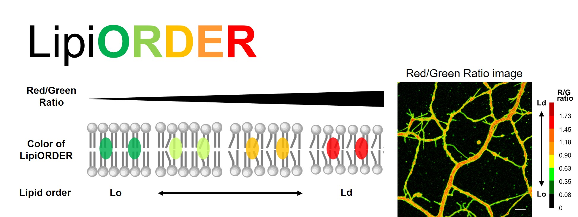
Graphical image of LipiORDER™ and Typical ratio imaging of cultured neurons
Fluorescent change of LipiORDER™ depending on lipid order of membrane (Left) and an example of ratiometric imaging on dendrites of cultured neurons.
Membrane lipid order is a biophysical parameter that defines a membrane organization and is often described by the degree of lipid packing. For example, phospholipids only containing saturated lipids create high packing and thick lipid bilayer, called liquid-order (Lo) phase. On the other hand, phospholipids containing unsaturated lipids, which have bent structure, form low packing and thin membrane structure, called liquid-disorder (Ld) phase. In the model membrane mixing saturated lipids and unsaturated lipids, Lo and Ld are clearly separated and create individual domains. While the model membrane composition can be discussed membrane lipid order (Lo/Ld) easily, actual cells have numerous types of lipids and form very complicated membrane lipid orders. Furthermore, the lipid order is also influenced by various factors in cellular membranes, including sterol lipids such as cholesterol and membrane proteins, etc. Lipid raft, a continuous interest topic in biology, which serves as functional microdomains on cellular membranes, is one of the specialized Lo domains, with highly accumulated saturated lipids such as sphingomyelin, cholesterol, functional membrane proteins and lipidated proteins. Membrane lipid order has been considered as a fundamental factor in providing physical properties of cellular membranes, such as membrane fluidity, membrane tension and membrane curvature. Observation of cellular lipid order may lead to an understanding of the various function of cellular membranes.
To measure membrane lipid order, some solvatochromic dyes which change fluorescence intensity and color in response to their solvent polarity are applied. These solvatochromic dye fluorescent properties change depending on membrane lipid order. Among them, Laurdan is the most well-known dye for membrane lipid order imaging. However, conventional dyes have some limitations. For example, Laurdan requires UV light excitation and exhibits low photostability. So Laurdan is not suitable for live-cell imaging. Dyes which can be excited by longer wavelength with more photostability and chemically stable in cells are desirable traits for cellular imaging of membrane lipid order. LipiORDER™ is a novel solvatochromic dye for membrane lipid order imaging originally developed by Dr. Yosuke Niko, Kochi University, and Dr. Andrey S. Klymchenko, University of Strasbourg (original compound name PK in Ref.1). LipiORDER™ is excited at around 400 nm wavelength, which is compatible with live-cell imaging and changes its emission fluorescent color from green to red depending on membrane lipid order. LipiORDER™ also has high photostability and chemical stability on the cell membranes. LipiORDER™ is a convincing tool to monitor cellular membrane lipid order imaging on live-cells.
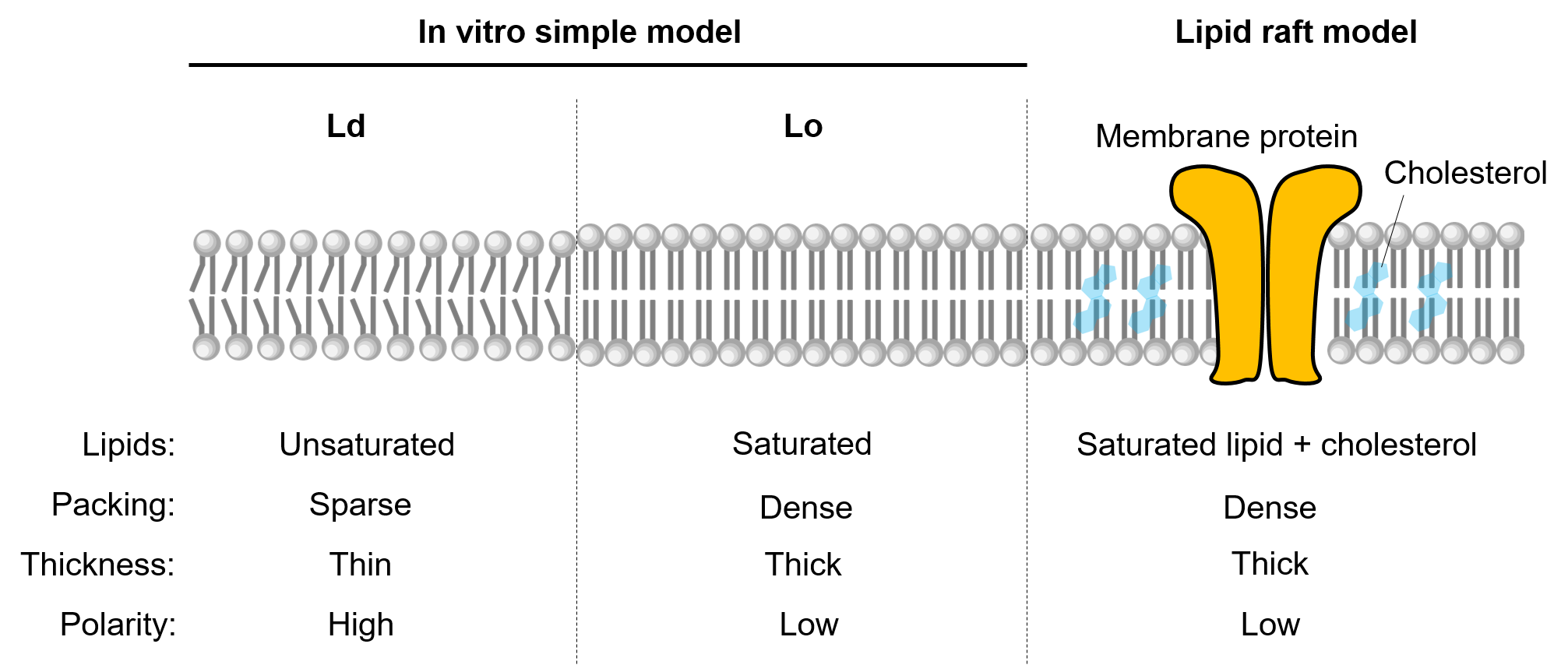
Image of phase state of lipid membrane
- Principle and Reference data
- Specification
- Application Data
- Reference
- Product Information
- You may also like
Principle and Reference data
Sensing of lipid order by using LipiORDER™ is based on the following two unique properties.
1) LipiORDER™ is a pyren-based solvatochromic fluorescent dye which changes fluorescent property in response to their solvent environment (Figure P1). In low polaric solvents such as toluene, LipiORDER™ shows green fluorescence. On the other hand, in highly polaric solvents such as DMSO and methanol, this dye changes color to orange or red.
2) LipiORDER™ is a highly hydrophobic compound and quickly accumulates in the various biological membranes. Combining the two features above, LipiORDER™ can sense the local environment in a lipid bilayer. Generally, Lo is a high packing lipid bilayer and shows lower polarity, whereas Ld is a sparse packing lipid bilayer and shows high polarity. Based on polarity of lipid bilayer derived from lipid order, LipiORDER™ will change fluorescent color, from green on Lo membrane to red on Ld membrane (Figure P2).
Ratiometric fluorescent value (FR/FG ) is correlated to lipid order (Lo and Ld).
Actually, in sphingomyeline/cholesterol (SM/Chol) liposome, one of the model Lo, LipiORDER™ emits green fluorescence and in 1,2-dioleoyl-sn-glucero-3-phosphocholine (DOPC) liposome, a model Ld, shows red fluorescence. In DOPC/Chol, an intermediate model, the reagent show yellow to orange. The ratiometric values (F575/F510) clearly depend on lipid order, SM/Chol (Lo) is low and DOPC (Ld) is high (Figure P3).
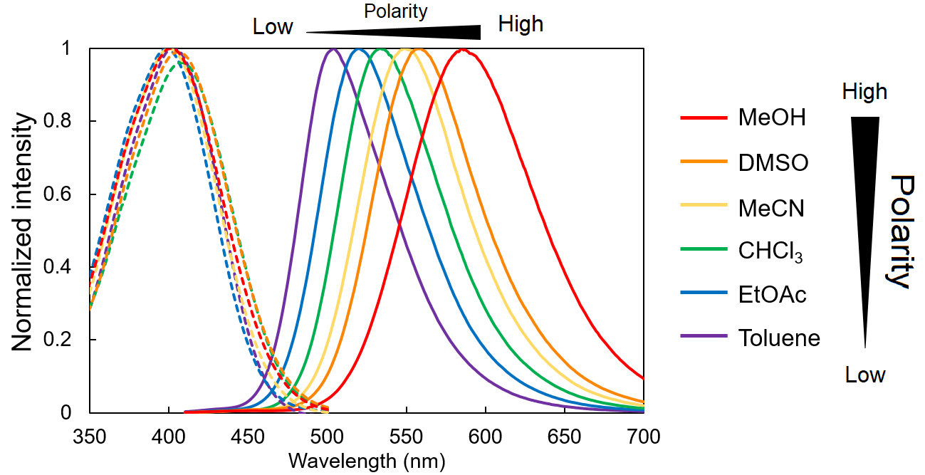
Figure P1 Absorption and fluorescent spectrum of LipiORDER™ in various solvent
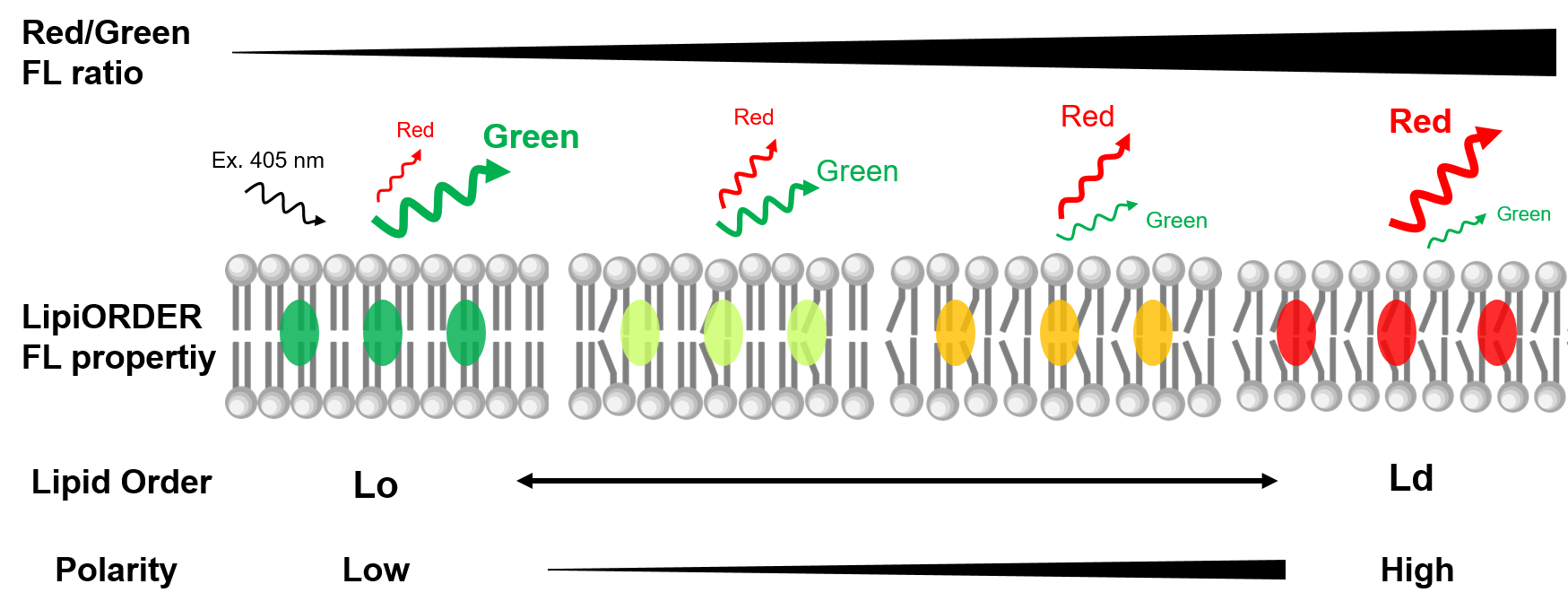
Figure P2 Graphical overview of lipid order-dependent fluorescent change of LipiORDER™
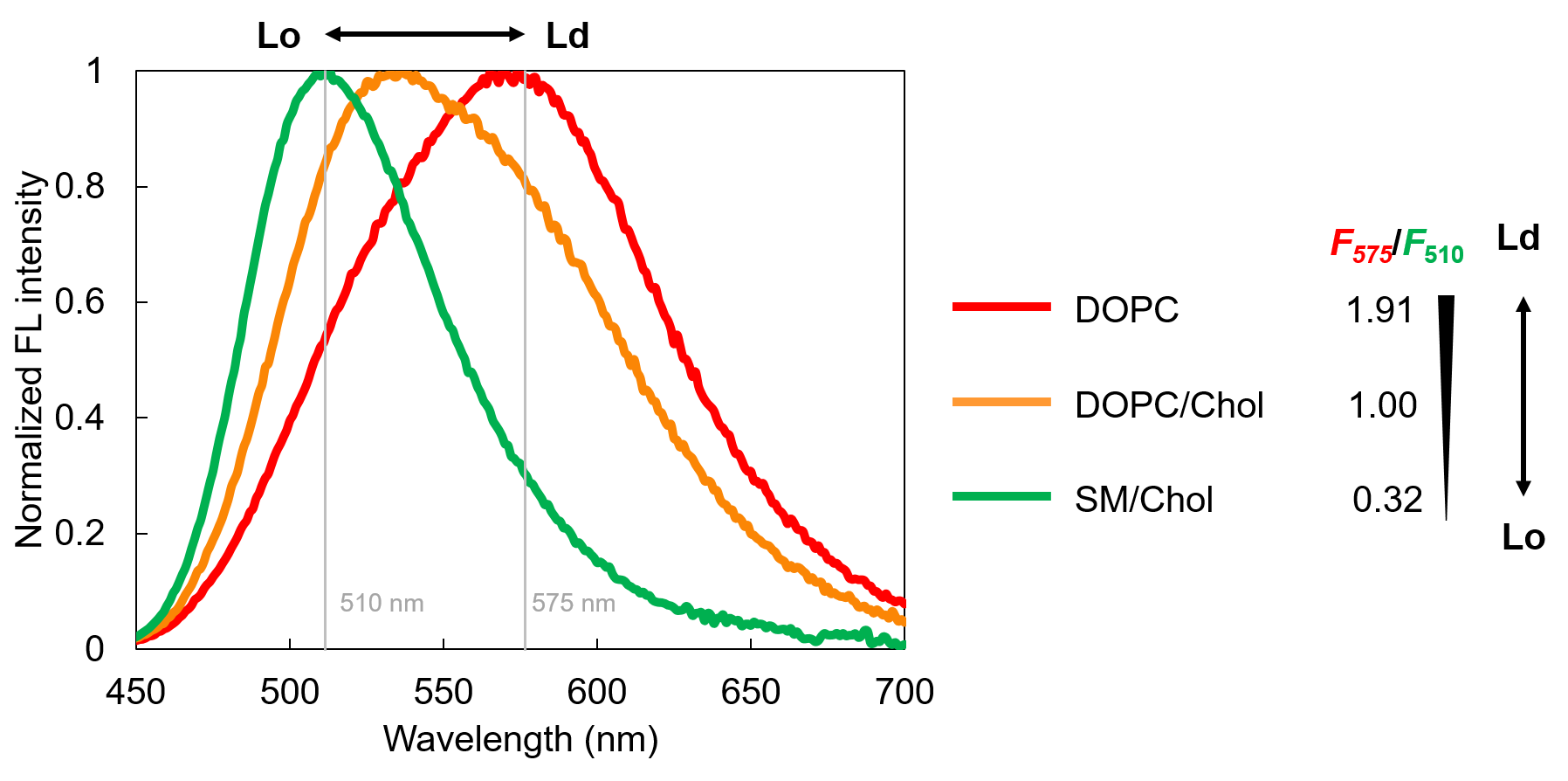
Figure P3 Fluorescent spectrum of LipiORDER™ in model liposomes
Specification
- Formulation: C23H21NO
- Molecular weight: 327.4 g/mol
- Solubility: Soluble in DMSO
- Fluorescent characteristics: Ex. 405 nm/Em. 450-650 nm (dependent on solvents)
Application Data
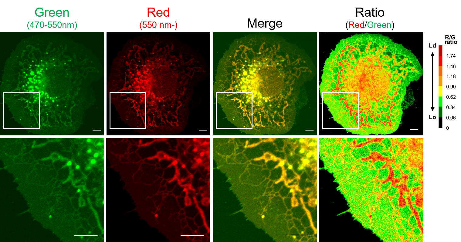
Ratiometric imaging of COS7 cells
COS7 cells were treated with 300 nM LipiORDER™ in HBSS for 10 min and observed by confocal laser microscopy (Ex. 405 nm, Em 470-550 nm for Green channel and >550 nm for Red channel). Ratiometric analysis was performed with ImageJ using green and red channel data and lipid order was shown by green-to-red pseudocolor (Lo■■■■■■■Ld). Plasma membrane and intramembranes are shown Lo and Ld, respectively.
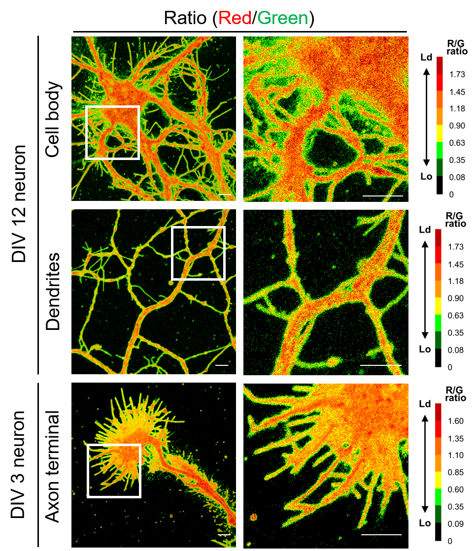
Ratiometric imaging of neuronal cells
Primary cultured hippocampal neurons (DIV 3 or DIV 12) from E17.5 mice were stained with 300 nM LipiORDER™ in HBSS for 10 min and observed by confocal laser microscopy (Ex. 405 nm, Em. 470-550 nm for Green channel and >550 nm for Red channel). Ratiometric analysis was performed with ImageJ using green and red channel data and lipid order was shown by green-to-red pseudocolor (Lo■■■■■■■Ld).
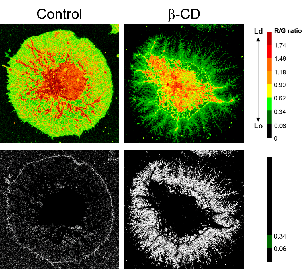
Drug-induced cellular lipid order changes
COS7 cells were treated with 15 mM beta-cyclodextrin (beta-CD), a membrane-disrupting chemical via removing endogenous cholesterol, for 4 hours. After beta-CD treatment, cells were washed and stained with 300 nM LipiORDER™ in HBSS for 10 min. The cells were observed by confocal laser microscopy (Ex. 405 nm, Em. 470-550 nm for Green channel and >550 nm for Red channel). Ratiometric analysis was performed with ImageJ using green and red channel data and lipid order is shown by green-to-red pseudocolor (Lo■■■■■■■Ld).
The cell structure was dramatically changed by beta-CD and at the same time, the distribution of Lo phase (■) clearly changed.
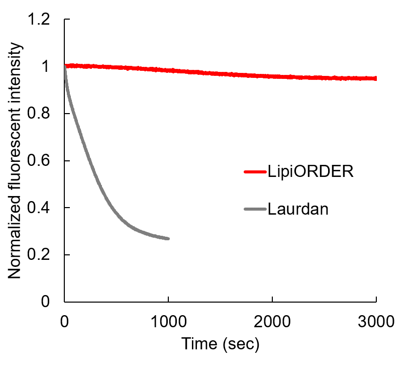
Photostability of LipiORDER
LipiORDER™ and Laurdan, a conventional membrane lipid order imaging dye in lipid vesicles composed of 0.2 mM DOPC in 20 mM HEPES (pH 7.4) were irradiated with Xe lamp. LipiORDER™ and Laurdan were excited at 405 nm and 360 nm, respectively and fluorescent intensity was measured. Laurdan was quickly photodegraded, whereas LipiORDER™ maintains fluorescent intensity for at least 1 hour. LipiORDER™ is highly stable compared to Laurdan.
Reference
1. Valanciunaite et al., Anal. Chem., 92, 6512-6520 (2020) Polarity Mapping of Cells and Embryos by Improved Fluorescent Solvatochromic Pyrene Probe.
Product Information
[Date : July 27 2026 00:07]
| Detail | Product Name | Product Code | Supplier | Size | Price | ||||||||||||||||||||||||||||||
|---|---|---|---|---|---|---|---|---|---|---|---|---|---|---|---|---|---|---|---|---|---|---|---|---|---|---|---|---|---|---|---|---|---|---|---|
|
LipiORDER, Membrane Lipid Order Imaging Dye DatasheetThis may not be the latest data sheet. |
FDV-0041 | FNAFunakoshi Co.,Ltd. | 0.1 mg | $380 | |||||||||||||||||||||||||||||||
|
|
|
||||||||||||||||||||||||||||||||||
[Date : July 27 2026 00:07]
LipiORDER, Membrane Lipid Order Imaging Dye
DatasheetThis may not be the latest data sheet.
- Product Code: FDV-0041
- Supplier: FNA
- Size: 0.1mg
- Price: $380
| Description |
LipiORDER™ is a novel solvatochromic dye for lipid order imaging for various biological membranes. This new dye enables to observe lipid order of membranes such as cell membrane and intracellular membrane, as ratiometric fluorescent value. |
||
|---|---|---|---|
| Storage | -20°C | CAS | |
| Link |
|
||
You may also like
-25-metabolism_map.jpg)
CONTACT
export@funakoshi.co.jp
- ※Prices on our website are for your reference only. Please inquire your distributor for your prices.
- ※Please note that Product Information or Price may change without notice.