Monoclonal Antibody for Scirrhous Stomach Cancer Anti-MUC1 Antibody (014E)
Date:November 22 2016Web Page No:80739

Funakoshi Co.,Ltd.
This antibody can detect the tumor marker MUC1 (Mucin-1; also PEM, PEMT and Episialin) in various tumor tissues by immunohistochemistry. In particular, it can specifically detect scirrhous gastric cancer, which has been considered difficult to detect. ※ This antibody has been commercialized under a license from Kagoshima University.
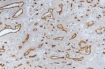
IHC of human gastric carcinoma tissue
- Sample: Human stomach cancer
- Detection reagent: ImmPRESSReagent, Anti-Mouse IgG (Vector Laboratories #MP-7402-15)
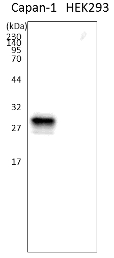
WB of cell lysate (5 μg)
- Left: Capan-1 (Human pancreas cell line: MUC1 positive)
- Right: HEK293 (MUC1 negative)
MUC1 & MUC1 Antibody(014E)
MUC1 (Mucin1), a mucin-type glycoprotein, is expressed in various cancers and is thought to be involved in cancer cell invasion and metastasis.
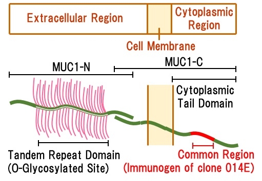
MUC1 is expressed from a single gene as a heterodimer of the extracellular region (MUC1-N) and the extracellular to transmembrane to cytoplasmic region (MUC1-C). MUC1-N contains a tandem repeat sequence (O-glycosylated site), and glycosylation is performed on this region. In some cases, MUC1-N is cleaved. On the other hand, MUC1-C has many splicing variants.
Detection of gastric cancer including scirrhous
IHC with antibodies (e.g. MUC1 antibody, keratin antibody), HE stain and PAS stain with diastase are used to detect stomach cancer.
However, these detection methods may miss cancer cells due to no staining of scirrhous gastric carcinoma (signet ring cell carcinoma: sig, poorly differentiated adenocarcinoma non-solid type: por2), unclear images and non-specific staining. The clone 014E antibody can specifically detect even scirrhous gastric cancer, which has been considered difficult to detect 1, 5.
Detection of pancreatic cancer
In pancreatic cancer detection, some clones of MUC1 antibody could only detect pancreatic ductal adenocarcinoma (PDAC) and could not detect intraductal papillary mucinous tumor (IPMN-gastric type and IPMN-intestinal type). This is because cleavage or glycosylation status of MUC1-N varies in these tumors, and antibodies with the extracellular region as antigen could not detect them.
Since clone 014E antibody is raised against cytoplasmic region, it can detect MUC1-N regardless of MUC1' structure of the extracellular region 4, 5. Also, a correlation between the MUC1 detection results by clone 014E and methylation of promoter region of MUC1 gene has been reported 4, 5.
参考文献
- Yonezawa, S., et al.,"A novel anti-MUC1 antibody against the MUC1 cytoplasmic tail domain: use in sensitive identification of poorly differentiated cells in adenocarcinoma of the stomach", Gastric Cancer, 15(4), 370~381 (2012) [PMID: 22237656].
- Yokoyama, S., et al., "The application of methylation specific electrophoresis (MSE) to DNA methylation analysis of the 5' CpG island of mucin in cancer cells", BMC Cancer, 12, 67 (2012). [PMID: 22329852].
- Kitamoto, S., et al., "MUC1 enhances hypoxia-driven angiogenesis through the regulation of multiple proangiogenic factors", Oncogene, 32(39), 4614~4621 (2013). [PMID: 23108411].
- Yokoyama, S., et al., "Diagnosis of pancreatic neoplasms using a novel method of DNA methylation analysis of mucin expression in pancreatic juice", PLoS ONE, 9(4):e93760 (2014). [PMID: 24714692].
- Japan Patent 5660486, Patent US8951526 B2
In pancreatic cancer detection, some clones of MUC1 antibody could only detect pancreatic ductal adenocarcinoma (PDAC) and could not detect intraductal papillary mucinous tumor (IPMN-gastric type and IPMN-intestinal type). This is because cleavage or glycosylation status of MUC1-N varies in these tumors, and antibodies with the extracellular region as antigen could not detect them.
Since clone 014E antibody is raised against cytoplasmic region, it can detect MUC1-N regardless of MUC1' structure of the extracellular region 4, 5. Also, a correlation between the MUC1 detection results by clone 014E and methylation of promoter region of MUC1 gene has been reported 4, 5.
- Yonezawa, S., et al.,"A novel anti-MUC1 antibody against the MUC1 cytoplasmic tail domain: use in sensitive identification of poorly differentiated cells in adenocarcinoma of the stomach", Gastric Cancer, 15(4), 370~381 (2012) [PMID: 22237656].
- Yokoyama, S., et al., "The application of methylation specific electrophoresis (MSE) to DNA methylation analysis of the 5' CpG island of mucin in cancer cells", BMC Cancer, 12, 67 (2012). [PMID: 22329852].
- Kitamoto, S., et al., "MUC1 enhances hypoxia-driven angiogenesis through the regulation of multiple proangiogenic factors", Oncogene, 32(39), 4614~4621 (2013). [PMID: 23108411].
- Yokoyama, S., et al., "Diagnosis of pancreatic neoplasms using a novel method of DNA methylation analysis of mucin expression in pancreatic juice", PLoS ONE, 9(4):e93760 (2014). [PMID: 24714692].
- Japan Patent 5660486, Patent US8951526 B2
Features
- The majority of splicing variants can be detected regardless of the structure of the extracellular region of MUC1.
- Isotype : IgG1κ
- Immunogen : Synthetic Peptide, common region in Cytoplasmic Tail Domain (CTD) of human MUC1 (CRYVPPSSTDRSPYEKVSAG)
- Reactivity : Human
- Specificity : Human Mucin1 (MUC1)
- Application : Immunohistochemistry (Paraffin, 1:2,500 - 1:5,000), Western Blotting (1:2,500 - 1:5,000)
- Form : Mouse Ascites diluted with PBS
Product Information
[Date : July 25 2026 00:08]
| Detail | Product Name | Product Code | Supplier | Size | Price | ||||||||||||||||||||||||||||||||||||||||||||||||||||||
|---|---|---|---|---|---|---|---|---|---|---|---|---|---|---|---|---|---|---|---|---|---|---|---|---|---|---|---|---|---|---|---|---|---|---|---|---|---|---|---|---|---|---|---|---|---|---|---|---|---|---|---|---|---|---|---|---|---|---|---|
|
Anti-MUC1, Mouse-Mono(014E), Anti-Mucin1 DatasheetThis may not be the latest data sheet. |
FDV-0012B | FNAFunakoshi Co.,Ltd. | 100 µl | $400 | |||||||||||||||||||||||||||||||||||||||||||||||||||||||
|
|
|
||||||||||||||||||||||||||||||||||||||||||||||||||||||||||
[Date : July 25 2026 00:08]
Anti-MUC1, Mouse-Mono(014E), Anti-Mucin1
DatasheetThis may not be the latest data sheet.
- Product Code: FDV-0012B
- Supplier: FNA
- Size: 100µl
- Price: $400
| Description |
This antibody can specifically detect scirrhous gastric cancer, which has been considered difficult to detect, and pancreatic cancer, a type of cancer that cannot be detected by some antibodies. Alias: PEM, PEMT, Episialin Clone: 014E |
||
|---|---|---|---|
| Storage | -80°C | ||
| Antigen Species | Human | Host Species | Mouse |
| Class | IgG | Label | Unlabeled |
| Cross Reactivity | Human | Application | IHC,Western Blot |
| Clonality | Format | ||
| Purification | Acsites | Absorption | |
| Link | |||
CONTACT
export@funakoshi.co.jp
- ※Prices on our website are for your reference only. Please inquire your distributor for your prices.
- ※Please note that Product Information or Price may change without notice.