Membrane potential-sensitive Ap3 : Non-fluorescent SHG-Imaging dye
Date:August 08 2018Web Page No:80646

Funakoshi Co.,Ltd.
Ap3 is the world first Non-fluorescent, photostable SHG-imaging dye. Second harmonic generation (SHG) imaging is a powerful tool to visualize cell / tissue structure and function. However, dyes which have been used so far emit strong fluorescence signals as well as SHG signals, and this fluorescence disturbs multimodal imaging with SHG signal. Ap3 overcomes this disadvantage, and enables true SHG signals in multimodal imaging by no interference of signals from other fluorescent molecules.
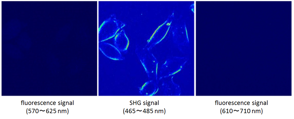
SHG signal and no fluorescence with Ap3 treatment
Upon irradiation with a 950 nm laser, SHG signal is observed with Ap3, but no fluorescence signal is observed.
Features
- SHG-specific dye. No fluorescence signal.
- Enables multimodal imaging by using Ap3 and other fluorescent dyes simultaneously.
- Photostable
- Low phototoxity
- Laser illumination : 950 nm / SHG signal detection : 465 - 485 nm
*Two-photon excitation microscopy system and special filter for SHG imaging is required.
*For SHG imaging, the detection system is required to set on the opposition side of the objective lens. Besides, the installation of photomultiplier tube(PMT) is preferable on the detection side.
Outline Protocol
- Cultivate cells at appropriate cell density on glass bottom dishes.
- Dilute Ap3 stock solution in an appropriate buffer.
- Remove culture medium from glass bottom dishes and add diluted Ap3 (e.g. 200 µL - 1,000 µL), to cells at a final concentration of 20 µM.
- Incubate cells for few minutes.
- Generate SHG signals with 950 nm laser illumination and observe it with 465 - 485 nm band pass filter through two-photon excitation microscopy system.
Performance Data
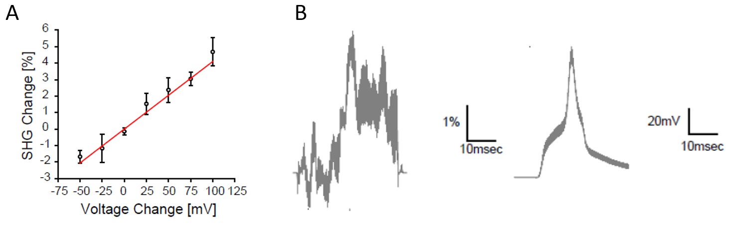
Membrane potential changes by SHG imaging in brain slices.
a: Membrane potential sensitivity of SHG signals from Ap3 assessed by voltage-clamp.
b: SHG signal changes upon action potential in neurons. SHG signals were monitored by a point-scan protocol at the soma of patch-clamped neurons in brain slices.
Nuriya M., et al, Nat. Commun., 7:11557 (2016).
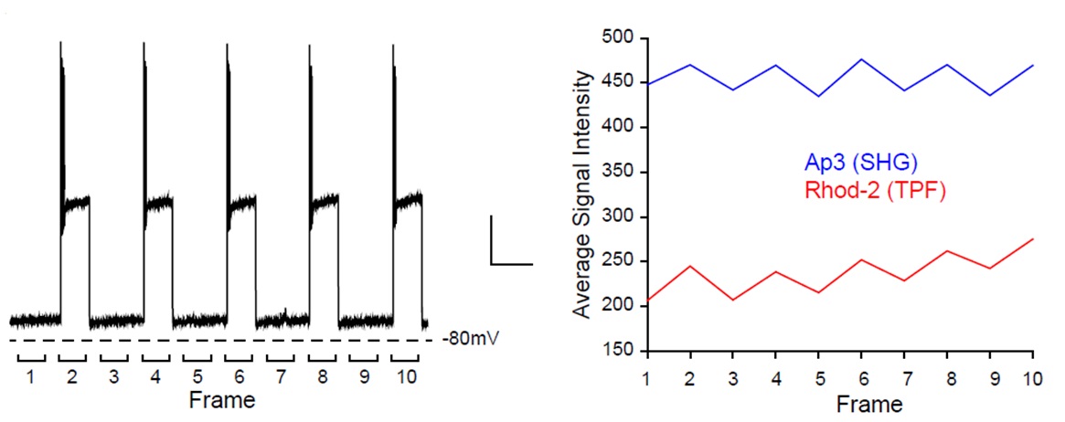
Simultaneous imaging of membrane potential and intracellular calcium dynamics in cortical neurons.
Nuriya M., et al, Nat. Commun., 7:11557 (2016).
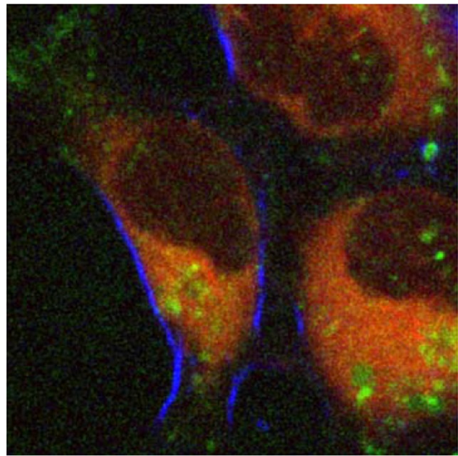
Multimodal two-photon imaging of cell membrane and fluorescence marker.
Blue:Ap3、Green:pHrodo Green、Red:ER-Tracker Red
Reference
- Mizuguchi T., et al, iScience., Nov 30 ; 9 :359~366 (2018).(
 )
)
Product Information
[Date : July 27 2026 00:07]
| Detail | Product Name | Product Code | Supplier | Size | Price | ||||||||||||||||||||||||||||||
|---|---|---|---|---|---|---|---|---|---|---|---|---|---|---|---|---|---|---|---|---|---|---|---|---|---|---|---|---|---|---|---|---|---|---|---|
|
Ap3, SHG Imaging Dye DatasheetThis may not be the latest data sheet. |
FDV-0008 | FNAFunakoshi Co.,Ltd. | 1 mg | $400 | |||||||||||||||||||||||||||||||
|
|
|
||||||||||||||||||||||||||||||||||
[Date : July 27 2026 00:07]
Ap3, SHG Imaging Dye
DatasheetThis may not be the latest data sheet.
- Product Code: FDV-0008
- Supplier: FNA
- Size: 1mg
- Price: $400
| Description |
Ap3 is the world first Non-fluorescent, photostable and membrane potential-sensitive SHG-active organic dye. Second harmonic generation (SHG) imaging is a powerful tool to visualize cell / tissue structure and function, and widely used. However, dyes which have been used so far emit strong fluorescence signals besides SHG signals, and this fluorescence disturbs multimodal imaging with SHG signal. Ap3 overcomes this disadvantage, and enables true SHG signals in multimodal imaging by no interference of signals from other fluorescent molecules. |
||
|---|---|---|---|
| Storage | -20°C | CAS | |
| Link |
|
||
CONTACT
export@funakoshi.co.jp
- ※Prices on our website are for your reference only. Please inquire your distributor for your prices.
- ※Please note that Product Information or Price may change without notice.