
抗53BP1抗体 | Anti-53BP1 Antibody
掲載日情報:2018/07/09 現在Webページ番号:46834

世界最大級の抗体製品数を取り扱うNovus Biologicals社の53BP1に対する抗体(anti-53BP1 | antibody 53BP1)です。Novus Biologicals社の抗体は数多くの学術論文で使用実績があります。
※本製品は研究用です。研究用以外には使用できません。

追加しました。
価格
[在庫・価格 :2026年07月17日 10時15分現在]
| 詳細 | 商品名 |
|
文献数 | ||||||||||||||||||||||||||||||||||||||||||||||||||||||||||||||||||||||||||||||||||
|---|---|---|---|---|---|---|---|---|---|---|---|---|---|---|---|---|---|---|---|---|---|---|---|---|---|---|---|---|---|---|---|---|---|---|---|---|---|---|---|---|---|---|---|---|---|---|---|---|---|---|---|---|---|---|---|---|---|---|---|---|---|---|---|---|---|---|---|---|---|---|---|---|---|---|---|---|---|---|---|---|---|---|---|---|---|
|
Anti-53BP1, Rabbit-Poly <Anti-p53 Binding Protein 1> |
|
502 | |||||||||||||||||||||||||||||||||||||||||||||||||||||||||||||||||||||||||||||||||||
|
|||||||||||||||||||||||||||||||||||||||||||||||||||||||||||||||||||||||||||||||||||||


[在庫・価格 :2026年07月17日 10時15分現在]
Anti-53BP1, Rabbit-Poly <Anti-p53 Binding Protein 1>
文献数: 502
- 商品コード:NB100-304
- メーカー:NOV
- 包装:100μl
- 価格:¥113,000
- 在庫:無(未発注)
- 納期:3~4週間 ※※ 表示されている納期は弊社に在庫がなく、取り寄せた場合の目安納期となります。
- 法規制等:
| 説明文 | レビューあり。抗原:exon11,12,Keywords:53BP1|MGC138366|p202|p53-binding protein 1|p53BP1|53BP1FLJ41424|tumor protein 53-binding protein|1|tumor protein p53 binding protein 1|tumor protein p53-binding protein|tumor suppressor p53-binding protein 1 Genbank No: 7158 Protein Accession No: Q12888 |
||||||
|---|---|---|---|---|---|---|---|
| 別包装品 | 別包装品あり | ||||||
| 法規制等 | |||||||
| 保存条件 | -20℃ | 法規備考 | |||||
| 抗原種 | Human | 免疫動物 | Rabbit | ||||
| 交差性 | Bat/Bovine/Canine/Fish/Goat/Human/Mouse/Primate/Rat | 適用 | ChIP,FCM,IC,IF,IHC,IP,Immunoblotting,Western Blot,in situ hybridization | ||||
| 標識 | Unlabeled | 性状 | Antigen Affinity Purified | ||||
| 吸収処理 | クラス | IgG | |||||
| クロナリティ | Polyclonal | フォーマット | |||||
| 掲載カタログ |
|
||||||
| 製品記事 | 抗53BP1抗体 | Anti- 53BP1 antibody |
||||||
| 関連記事 | |||||||
追加しました。
Image
 | Western Blot: 53BP1 Antibody [NB100-304] - Total protein from HeLa, A431, Neuro2A, and PC12 was separated on a 12% gel by SDS-PAGE, transferred to PVDF membrane and blocked in 5% non-fat milk in TBST. The membrane was probed with 1.0 ug/ml anti-53BP1 in 5% block buffer and detected with an anti-rabbit HRP secondary antibody using chemiluminescence. |
 | Biological Strategies Validation. Immunohistochemistry-Frozen: 53BP1 Antibody [NB100-304] - Irradiated cochlear spiral ganglion cells, mouse, frozen section of fixed material. Data and image from confirmed customer review. |
 | Biological Strategies Validation. Immunocytochemistry/Immunofluorescence: 53BP1 Antibody [NB100-304] - 53BP1 foci in proliferating MEFs; both normal and exposed to 10 Gy of IR. |
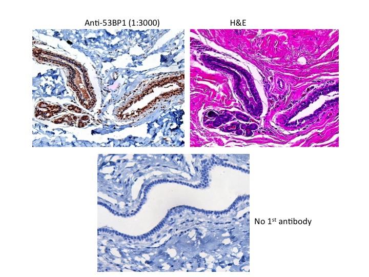
 | Immunohistochemistry-Paraffin: 53BP1 Antibody [NB100-304] - Detection of Human and Mouse 53BP1 by IHC. Sample: FFPE sections of human ovarian carcinoma (left) and mouse teratoma (right). Antibody: NB100-304 used at a dilution of 1:1,000 (1ug/ml). Detection: DAB. |
 | Flow Cytometry: 53BP1 Antibody [NB100-304] - 1 million Jurkat cells were fixed, permeabilized, and stained with 1.5 ug/ml anti-53BP1 NB100-304 in a 150 ul reaction. Isotype control (black), anti-53BP1 (red). |
 | Immunohistochemistry-Paraffin: 53BP1 Antibody [NB100-304] - Human breast tumors stained with 53BP1 antibody. Image from verified customer review. |
 | Western Blot: 53BP1 Antibody [NB100-304] - Detection of 53BP1 by Western Blot Sample: Whole cell lysate from U2OS or 293T cells. |
 | Immunocytochemistry/Immunofluorescence: 53BP1 Antibody [NB100-304] - Upper Panel: Control untreated cells; Lower Panel: Cells exposed to Irradiation (10 Gy) and probed for 53BP1 foci. Cells were grown on coverslips, fixed with 4% paraformaldehyde, methanol permeabilized, blocked for 1 h, RT. Incubated with primary antibody (1:200) overnight, washed 3x with PBS, probed with tubulin (Alexa fluor 594) antibody for 2 h, RT. Washed 3x with PBS, mounted on slides using prolong gold, imaged using Nikon confocal microscope (100x oil). This image was submitted via customer Review. |
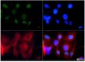
 | Immunocytochemistry/Immunofluorescence: 53BP1 Antibody [NB100-304] - 53BP1 antibody was tested at 1:100 in HeLa cells with FITC (green). alpha-Tubulin antibody NB100-690 was tested at a 1:500 dilution using Mouse IgG secondary Antibody NB710-94914 at 1:1000 [HiLyte Fluor 555] (red). Nuclei were counterstained with Dapi (blue). |
 | Immunocytochemistry/Immunofluorescence: 53BP1 Antibody [NB100-304] - 53BP1 was detected in immersion fixed HeLa human cervical epithelial carcinoma cell line using 1 ug/mL of rabbit anti- 53BP1 polyclonal (NB100-304, Novus Biologicals). Cells were stained donkey anti-goat IgGNL557 and counterstained with DAPI (blue). |
 | Immunocytochemistry/Immunofluorescence: 53BP1 Antibody [NB100-304] - A431 cells were fixed for 10 minutes using 10% formalin and then permeabilized for 5 minutes using 1X PBS + 0.5% Triton-X100. The cells were incubated with anti-53BP1 at 2 ug/ml overnight at 4C and detected with an anti-rabbit Dylight 488 (Green) at a 1:500 dilution. Alpha tubulin (DM1A) NB100-690 was used as a co-stain at a 1:1000 dilution and detected with an anti-mouse Dylight 550 (Red) at a 1:500 dilution. Nuclei were counterstained with DAPI (Blue). Cells were imaged using a 40X objective. |
 | Immunocytochemistry/Immunofluorescence: 53BP1 Antibody [NB100-304] - HeLa cells were fixed for 10 minutes using 10% formalin and then permeabilized for 5 minutes using 1X PBS + 0.5% Triton-X100. The cells were incubated with anti-53BP1 at 2 ug/ml overnight at 4C and detected with an anti-rabbit Dylight 488 (Green) at a 1:500 dilution. Alpha tubulin (DM1A) NB100-690 was used as a co-stain at a 1:1000 dilution and detected with an anti-mouse Dylight 550 (Red) at a 1:500 dilution. Nuclei were counterstained with DAPI (Blue). Cells were imaged using a 40X objective. |
 | Immunocytochemistry/Immunofluorescence: 53BP1 Antibody [NB100-304] - Neuro2a cells were fixed for 10 minutes using 10% formalin and then permeabilized for 5 minutes using 1X PBS + 0.5% Triton-X100. The cells were incubated with anti-53BP1 at 2 ug/ml overnight at 4C and detected with an anti-rabbit Dylight 488 (Green) at a 1:500 dilution. Alpha tubulin (DM1A) NB100-690 was used as a co-stain at a 1:1000 dilution and detected with an anti-mouse Dylight 550 (Red) at a 1:500 dilution. Nuclei were counterstained with DAPI (Blue). Cells were imaged using a 40X objective. |
 | Immunocytochemistry/Immunofluorescence: 53BP1 Antibody [NB100-304] - Human medulloblastoma (DAOY) and mouse astrocyte (C8D1A) cell lines were exposed for 48 hours to DMSO or 1ug/mL of the DNA damaging agent Etoposide.Cells were immunostained for 53BP1 (green). The nuclei were counterstained with Dapi (blue). This image was submitted via customer Review. |
 | Immunocytochemistry/Immunofluorescence: 53BP1 Antibody [NB100-304] - Embryonic Fibroblast cells Pre-extraction - 5mins with CSK buffer Fixed with 4% PFA and 75% Ethanol Primary Antibody - 1:1000 Secondary Antibody - 1:1000. This image was submitted via customer Review. |
 | Immunocytochemistry/Immunofluorescence: 53BP1 Antibody [NB100-304] - HeLa cells were fixed for 10 minutes using 10% formalin and then permeabilized for 5 minutes using 1X PBS + 0.5% Triton-X100. The cells were incubated with anti-53BP1 conjugated to DyLight 550 [NB100-304R] at 10ug/ml for 1 hour at room temperature. Nuclei were counterstained with DAPI (Blue). Cells were imaged using a 40X objective. |
 | Immunohistochemistry-Paraffin: 53BP1 Antibody [NB100-304] - IHC staining of 53BP1 in human colon cancer using DAB with hematoxylin counterstain. |
追加しました。
Background
53BP1 (p53 binding protein 1) plays a key role in response to DNA damage, checkpoint signaling during mitosis and enhancing TP53-mediated transcriptional activation. Originally identified as p53's transcriptional enhancing partner, 53BP1 now has been established as a substrate for ATM (ataxia telangiectasia mutated) signaling and that it relocalizes to discrete foci overlapping with gamma H2AX (phosphorylated histone H2AX); demarcating DNA double strand breaks (DSBs) sites following exposure to radiation. 53BP1 functions downstream of gamma H2AX-dependent hierarchy of proteins that collectively establish IRIF (ionizing radiation induced foci) at DSBs; this hierarchy includes Mre11/Rad50/NBS1 (MRN complex), ATM, MDC1, RNF8, RNF168 and HERC2. With the exception of ATM, whose function to generate gamma H2AX may be partially compensated by the activity of DNA-PK (DNA-dependent kinase), all of these proteins are physically and functionally required to recruit 53BP1 to the DSB site. Briefly, this process involves DSB recognition by MRN, ATM activation, gamma H2AX-formation, MDC1-recruitment, MRN-retention (leading to further ATM-activation and gamma H2AX spreading) and RNF8/RNF168/HERC2-mediated histone H2A and H2AX mono and poly-ubiquitination.追加しました。
製品情報は掲載時点のものですが、価格表内の価格については随時最新のものに更新されます。お問い合わせいただくタイミングにより製品情報・価格などは変更されている場合があります。
表示価格に、消費税等は含まれていません。一部価格が予告なく変更される場合がありますので、あらかじめご了承下さい。