
抗HIF-1 alpha抗体 | Anti-HIF-1 alpha Antibody
掲載日情報:2018/07/09 現在Webページ番号:46320

世界最大級の抗体製品数を取り扱うNovus Biologicals社のHIF-1 alphaに対する抗体(anti-HIF-1 alpha | antibody HIF-1 alpha)です。Novus Biologicals社の抗体は数多くの学術論文で使用実績があります。
※本製品は研究用です。研究用以外には使用できません。

追加しました。
価格
[在庫・価格 :2026年07月17日 00時01分現在]
| 詳細 | 商品名 |
|
文献数 | ||||||||||||||||||||||||||||||||||||||||||||||||||||||||||||||||||||||||||||||||||
|---|---|---|---|---|---|---|---|---|---|---|---|---|---|---|---|---|---|---|---|---|---|---|---|---|---|---|---|---|---|---|---|---|---|---|---|---|---|---|---|---|---|---|---|---|---|---|---|---|---|---|---|---|---|---|---|---|---|---|---|---|---|---|---|---|---|---|---|---|---|---|---|---|---|---|---|---|---|---|---|---|---|---|---|---|---|
|
Anti-HIF-1α, Rabbit-Poly <Anti-Hypoxia-Inducible Factor 1α> |
|
180 | |||||||||||||||||||||||||||||||||||||||||||||||||||||||||||||||||||||||||||||||||||
|
|||||||||||||||||||||||||||||||||||||||||||||||||||||||||||||||||||||||||||||||||||||


[在庫・価格 :2026年07月17日 00時01分現在]
Anti-HIF-1α, Rabbit-Poly <Anti-Hypoxia-Inducible Factor 1α>
文献数: 180
- 商品コード:NB100-134
- メーカー:NOV
- 包装:100μl
- 価格:¥108,000
- 在庫:無(未発注)
- 納期:3~4週間 ※※ 表示されている納期は弊社に在庫がなく、取り寄せた場合の目安納期となります。
- 法規制等:
| 説明文 | レビューあり。Simple Western対応抗体。抗原:aa 432~528,Keywords:ARNT-interacting protein|Basic-helix-loop-helix-PAS protein MOP1|BHLHE78|Class E basic helix-loop-helix protein 78|HIF-1 alpha|HIF1A|HIF-1-alpha|HIF1-alpha|hypoxia inducible factor 1|alpha subunit (basic helix-loop-helix transcriptionfactor) Genbank No: 3091 Protein Accession No: Q16665 |
||||||
|---|---|---|---|---|---|---|---|
| 別包装品 | 別包装品あり | ||||||
| 法規制等 | |||||||
| 保存条件 | -20℃ | 法規備考 | |||||
| 抗原種 | Human | 免疫動物 | Rabbit | ||||
| 交差性 | Bovine/Canine/Guinea Pig/Human/Mouse/Primate/Rat/Xenopus/Zebrafish | 適用 | ChIP,ChIP-seq,ELISA,EMSA,IC,IF,IHC,IP,Immunoblotting,Simple Western,Western Blot | ||||
| 標識 | Unlabeled | 性状 | Antigen Affinity Purified | ||||
| 吸収処理 | クラス | IgG | |||||
| クロナリティ | Polyclonal | フォーマット | |||||
| 掲載カタログ |
|
||||||
| 製品記事 | お勧め低酸素応答因子抗体 抗HIF抗体(Anti-HIF-1/HIF-2 antibody) 抗HIF-1 alpha抗体 | Anti- HIF-1 alpha antibody |
||||||
| 関連記事 | |||||||
追加しました。
Image
 | Independent Antibodies Validation and Genetic Strategies Validation.Western Blot: HIF-1 alpha Antibody [NB100-134] - Analysis of HIF-1 alpha in overexpression and endogenous HIF1 alpha & HIF2 alpha using anti-HIF-1 alpha antibody. The data showed that HIF-1 alpha antibody did not react to HIF-2 alpha overexpression. Image from verified customer review. |
 | Biological Strategies Validation. Western Blot: HIF-1 alpha Antibody [NB100-134] - Analysis of NB100-134, anti-HIF-1 alpha, on normoxic and hypoxic nuclear rat cell lysates. |
 | Independent Antibodies Validation and Biological Strategies Validation.Immunoprecipitation: HIF-1 alpha Antibody [NB100-134] - PC-3 Cell Lysates run with Cobalt Chloride treatments. Immunoprecipitation was performed with protein A/G Agarose beads. This data was provided courtesy of Kelie Reece, Figg lab, NCI. |
 | Biological Strategies Validation. Immunocytochemistry/Immunofluorescence: HIF-1 alpha Antibody [NB100-134] - Analysis of HIF-1 alpha in ARPE-19 cells using anti-HIF-1 alpha antibody. Image from verified customer review. |
 | Immunohistochemistry-Paraffin: HIF-1 alpha Antibody [NB100-134] - Staining in the canine CL on days 10 to 70 after ovulation. PC = positive control (human placenta). NC = negative control. Image from confirmed customer review. |
 | Simple Western: HIF-1 alpha Antibody [NB100-134] - Simple Western lane view shows a specific band for HIF-1 alpha in 0.2 mg/ml of Hypoxic HeLa lysate. This experiment was performed under reducing conditions using the 12-230 kDa separation system. |
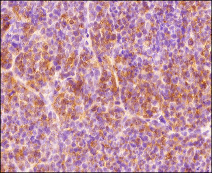 | Immunohistochemistry-Paraffin: HIF-1 alpha Antibody [NB100-134] - IHC analysis of a formalin-fixed paraffin-embedded tissue section of human endometrium carcinoma AN3CA cell line based xenograft using rabbit polyclonal HIF1 alpha antibody at 1:300 dilution. The signal was developed using HRP-labelled secondary antibody and DAB reagent, and the section was further counterstained using hematoxylin. The tested section depicted mainly a diffused cytoplasmic staining but there were some cells which showed nuclear signal also (representing hypoxic cells). |
 | Immunohistochemistry-Paraffin: HIF-1 alpha Antibody [NB100-134] - Analysis of HIF-1 alpha on Zebrafish retina tissue using HIF-1 alpha antibody. Mouse tissue was used as a control. Image from verified customer review. |
 | Western Blot: HIF-1 alpha Antibody [NB100-134] - 20 ug whole cell lysates (human ovarian cancer cell lines). Image from verified customer review. |
 | Biological Strategies Validation. Western Blot: HIF-1 alpha Antibody [NB100-134] - Analysis of HIF-1 alpha in human Retinal and Choroidal primary endothelia cells using anti-HIF-1 alpha antibody. Image from verified customer review. |
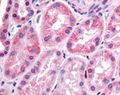
 | Immunohistochemistry: HIF-1 alpha Antibody [NB100-134] - Staining of human kidney, renal tubular epithelium in Cortex. |
追加しました。
Background
HIF-1 alpha (HIF1A) is a nuclear protein involved in mammalian oxygen homeostasis. This occurs as a posttranslational modification by prolyl hydroxylation. HIF-1 is a heterodimer composed of HIF-1 alpha and HIF-1 beta subunits. Both subunits are constantly translated. However, under normoxic conditions, human HIF-1 alpha is hydroxylated at Pro402 or Pro564 by a set of HIF prolyl hydroxylases, is polyubiquinated, and eventually degraded in proteosomes. Under hypoxic conditions, the lack of hydroxylation prevents HIF degradation and increases transcriptional activity. Therefore, the concentration of HIF-1 alpha increases in the cell.追加しました。
製品情報は掲載時点のものですが、価格表内の価格については随時最新のものに更新されます。お問い合わせいただくタイミングにより製品情報・価格などは変更されている場合があります。
表示価格に、消費税等は含まれていません。一部価格が予告なく変更される場合がありますので、あらかじめご了承下さい。