
抗PINK1抗体 | Anti-PINK1 Antibody
掲載日情報:2018/07/09 現在Webページ番号:46206

世界最大級の抗体製品数を取り扱うNovus Biologicals社のPINK1に対する抗体(anti-PINK1 | antibody PINK1)です。Novus Biologicals社の抗体は数多くの学術論文で使用実績があります。
※本製品は研究用です。研究用以外には使用できません。

追加しました。
価格
[在庫・価格 :2026年07月14日 12時55分現在]
| 詳細 | 商品名 |
|
文献数 | ||||||||||||||||||||||||||||||||||||||||||||||||||||||||||||||||||||||||||||||||||
|---|---|---|---|---|---|---|---|---|---|---|---|---|---|---|---|---|---|---|---|---|---|---|---|---|---|---|---|---|---|---|---|---|---|---|---|---|---|---|---|---|---|---|---|---|---|---|---|---|---|---|---|---|---|---|---|---|---|---|---|---|---|---|---|---|---|---|---|---|---|---|---|---|---|---|---|---|---|---|---|---|---|---|---|---|---|
|
Anti-PINK1, Rabbit-Poly <Anti-PTEN Induced Putative Kinase Protein 1> |
|
184 | |||||||||||||||||||||||||||||||||||||||||||||||||||||||||||||||||||||||||||||||||||
|
|||||||||||||||||||||||||||||||||||||||||||||||||||||||||||||||||||||||||||||||||||||


[在庫・価格 :2026年07月14日 12時55分現在]
Anti-PINK1, Rabbit-Poly <Anti-PTEN Induced Putative Kinase Protein 1>
文献数: 184
- 商品コード:BC100-494
- メーカー:NOV
- 包装:0.1ml
- 価格:¥113,000
- 在庫:無(未発注)
- 納期:3~4週間 ※※ 表示されている納期は弊社に在庫がなく、取り寄せた場合の目安納期となります。
- 法規制等:
| 説明文 | レビューあり。Keywords:Anti-PINK1 BRPK EC 2.7.11.1|FLJ27236|Mitochondrial PARK6|Parkinson disease (autosomal recessive) 6|PINK1 PINK1 mouse|PINK1 polyclonal|protein kinase BRPK|PTEN induced putative kinase 1|PTEN-induced putative kinase protein 1 Genbank No: 65018 Protein Accession No: Q9BXM7 |
||||||
|---|---|---|---|---|---|---|---|
| 別包装品 | 別包装品あり | ||||||
| 法規制等 | |||||||
| 保存条件 | -20℃ | 法規備考 | |||||
| 抗原種 | Human | 免疫動物 | Rabbit | ||||
| 交差性 | Human/Mouse/Rabbit/Rat | 適用 | Electron Microscopy,IC,IF,IHC,IP,Immunoblotting,Peptide ELISA,SDS-PAGE,Western Blot | ||||
| 標識 | Unlabeled | 性状 | Antigen Affinity Purified | ||||
| 吸収処理 | クラス | IgG | |||||
| クロナリティ | Polyclonal | フォーマット | |||||
| 掲載カタログ |
|
||||||
| 製品記事 | 神経変性疾患研究用抗体 |
||||||
| 関連記事 | |||||||
追加しました。
Image
 | Biological Strategies Validation. Western Blot: PINK1 Antibody [BC100-494] - Analysis of PINK1 in HeLa whole cell lysate with and without treatment of 10 uM CCCP. Image courtesy of an anonymous customer review. |
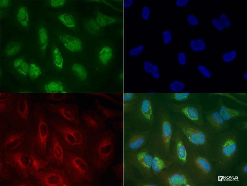 | Biological Strategies Validation. Immunocytochemistry/Immunofluorescence: PINK1 Antibody [BC100-494] - Immunocytochemistry of PINK1 antibody (BC100-494 Lot G). HeLa cells were treated with valinomycin (1 uM for 24h) prior to being fixed in 10% buffered formalin for 10 min and permeabilized in 0.1% Triton X-100 in PBS for 10 min. Cells were incubated with BC100-494 at 20 ug/ml for 1h at room temperature, washed 3x in PBS and incubated with Alexa-Fluor488 anti-rabbit secondary antibody. PINK1 (Green) was detected at the mitochondria. Tubulin (Red) was detected using an anti-tubulin antibody with an anti-mouse DyLight 550 secondary antibody. DNA (Blue) was counterstained with DAPI. Note: mitochondria staining might not be easily observed without treatment with valinomycin or CCCP. |
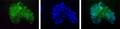
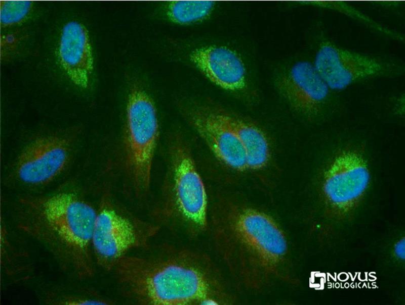 | Biological Strategies Validation. Immunocytochemistry/Immunofluorescence: PINK1 Antibody [BC100-494] - HeLa cells were treated with valinomycin (1 uM for 24h) prior to being fixed in 10% buffered formalin for 10 min and permeabilized in 0.1% triton X-100 in PBS for 10 min. Cells were incubated with BC100-494 at 20 ug/ml for 1h at room temperature, washed 3x in PBS and incubated with Alexa-Fluor488 anti-rabbit secondary antibody. PINK1 (Green) was detected at the mitochondria. Tubulin (Red) was detected using NB100-690 with an anti-mouse DyLight 550 secondary antibody. DNA (Blue) was counterstained with DAPI. Note: mitochondrial staining may only be visible after treatment with valinomycin or CCCP. |
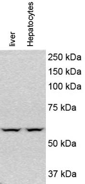 | Western Blot: PINK1 Antibody [BC100-494] - Analysis of PINK1 in mouse liver and hypatocytes using PINK1 antibody. Image from verified customer review. |
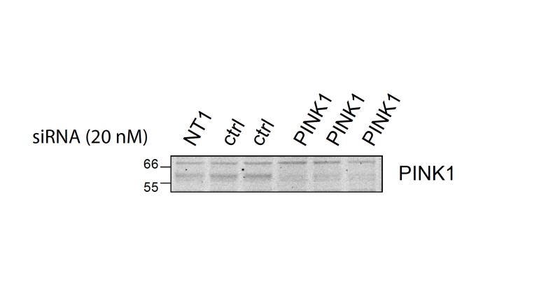 | Genetic Strategies Validation. Western Blot: PINK1 Antibody [BC100-494] - WB analysis of lysates derived from hTERT-RPE1 cells transfected with non-targeting (NT1) or PINK1 targeting siRNA for 72 h. 20 uG were loaded. Image from verified customer review. |
 | Immunohistochemistry-Paraffin: PINK1 Antibody [BC100-494] - Rabbit heart tissue. Image from verified customer review. |
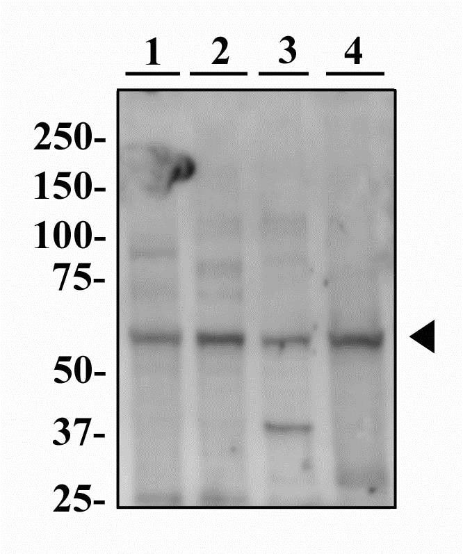 | Western Blot: PINK1 Antibody [BC100-494] - Western blot image of PINK1 antibody (BC100-494) in multiple cells lines. Human HeLa (lane 1), Mouse NIH-3T3 (lane 2), L929 (lane 3) and Rat PC12 (lane 4) whole cell protein were separated by SDS-PAGE on a 7.5% polyacrylamide gel. Protein was transferred to PVDF membrane and probed with 2 ug/ml BC100-494 in 1% BSA and detected with an HRP-conjugated anti-rabbit secondary antibody using chemiluminescence. PINK1 was detected at approximately 60 kDa (arrowhead). |
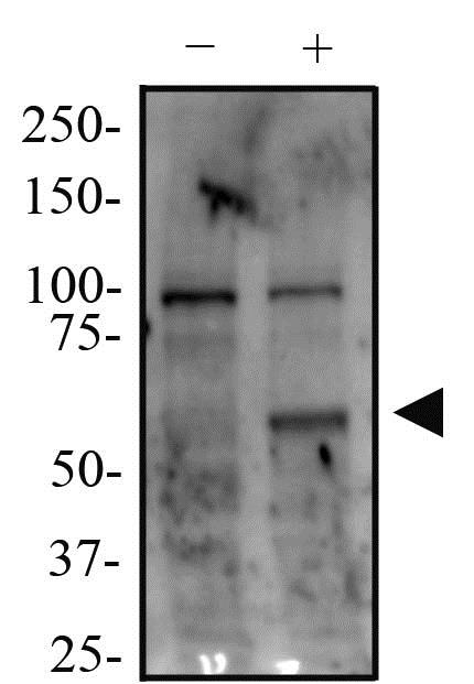 | Biological Strategies Validation. Western Blot: PINK1 Antibody [BC100-494] - Whole cell protein from HeLa cells treated without or with valinomycin (1 uM for 24h) as indicated were separated by SDS-PAGE on a 7.5% polyacrylamide gel. Protein was transferred to PVDF membrane and probed with 1.0 ug/ml BC100-494 in 1% BSA and detected with an HRP-conjugated anti-rabbit secondary antibody using chemiluminescence. PINK1 was detected at approximately 60 kDa in the treated sample(arrowhead). |
追加しました。
Background
PINK1 (PTEN induced putative kinase 1) is a mitochondrial serine/threonine kinase which maintains mitochondrial function/integrity, provides protection against mitochondrial dysfunction during cellular stress by phosphorylating mitochondrial proteins, and is involved in the clearance of damaged mitochondria via selective autophagy (mitophagy). PINK1 is synthesized as a 63 kDa protein which undergoes proteolytic processing to generate at least two cleaved forms (55 kDa and 48 kDa). PINK1 and its cleavage products have been found in the cytosol as well as in different sub-mitochondrial compartments, and according to reports; PINK1 may be targeted to OMM (outer mitochondrial membrane) with its kinase domain facing the cytosol, providing a possible explanation for the observed physical interaction with the cytosolic E3 ubiquitin ligase Parkin. Defective PINK1 may cause alterations in processing, stability, localization and activity as well as binding to substrates/interaction-partners which ultimately leads to differential effects on mitochondrial function and morphology. Mutations in PINK1 are linked to autosomal recessive early onset Parkinson's disease, and are associated with loss of protective function, mitochondrial dysfunction, aggregation of alpha-synuclein, as well as proteasome dysfunction.追加しました。
製品情報は掲載時点のものですが、価格表内の価格については随時最新のものに更新されます。お問い合わせいただくタイミングにより製品情報・価格などは変更されている場合があります。
表示価格に、消費税等は含まれていません。一部価格が予告なく変更される場合がありますので、あらかじめご了承下さい。