
抗E-Cadherin [N3C2], Internal抗体 | Anti-E-Cadherin [N3C2], Internal antibody
掲載日情報:2020/04/02 現在Webページ番号:44968

GeneTex社の抗E-Cadherin [N3C2], Internal抗体 | Anti-E-Cadherin [N3C2], Internal antibodyです。
※ 本製品は研究用です。研究用以外には使用できません。
追加しました。
特長
- 高品質の抗体です。
- 幅広い研究分野に関連する抗体を取り揃えています。
- 使用されたアプリケーションや動物種などの情報が充実しています。
- 多数の論文で使用実績がある信頼性の高い抗体です。
追加しました。
価格
[在庫・価格 :2026年04月23日 10時55分現在]
| 詳細 | 商品名 |
|
文献数 | ||||||||||||||||||||||||||||||||||||||||||||||||||||||||||||||||||||||||||||||||||
|---|---|---|---|---|---|---|---|---|---|---|---|---|---|---|---|---|---|---|---|---|---|---|---|---|---|---|---|---|---|---|---|---|---|---|---|---|---|---|---|---|---|---|---|---|---|---|---|---|---|---|---|---|---|---|---|---|---|---|---|---|---|---|---|---|---|---|---|---|---|---|---|---|---|---|---|---|---|---|---|---|---|---|---|---|---|
|
Anti-E-Cadherin, Rabbit |
|
6 | |||||||||||||||||||||||||||||||||||||||||||||||||||||||||||||||||||||||||||||||||||
|
|||||||||||||||||||||||||||||||||||||||||||||||||||||||||||||||||||||||||||||||||||||
|
Anti-E-Cadherin, Rabbit |
|
6 | |||||||||||||||||||||||||||||||||||||||||||||||||||||||||||||||||||||||||||||||||||
|
|||||||||||||||||||||||||||||||||||||||||||||||||||||||||||||||||||||||||||||||||||||


[在庫・価格 :2026年04月23日 10時55分現在]
Anti-E-Cadherin, Rabbit
文献数: 6
- 商品コード:GTX124178
- メーカー:GNT
- 包装:25μl
- 価格:¥30,000
- 在庫:無(未発注)
- 納期:10日程度 ※※ 表示されている納期は弊社に在庫がなく、取り寄せた場合の目安納期となります。
- 法規制等:
| 説明文 | KO/KDバリデーション済み抗体。 別名:cadherin 1,Arc-1,BCDS1,CD324,CDHE,ECAD,LCAM,UVO Genbank No: 999 |
||||||
|---|---|---|---|---|---|---|---|
| 別包装品 | 別包装品あり | ||||||
| 法規制等 | |||||||
| 保存条件 | -20℃ | 法規備考 | |||||
| 抗原種 | Human | 免疫動物 | Rabbit | ||||
| 交差性 | Dog/Human/Rat | 適用 | IC,IF,IHC,IP,Western Blot | ||||
| 標識 | Unlabeled | 性状 | Purified | ||||
| 吸収処理 | クラス | IgG | |||||
| クロナリティ | Polyclonal | フォーマット | |||||
| 掲載カタログ |
|
||||||
| 製品記事 | 使いっきり抗体 GeneTex社:抗E-カドヘリン抗体 獣医学研究用抗体 VetSignalシリーズ |
||||||
| 関連記事 | GeneTex社における抗体の品質管理 |
||||||
Anti-E-Cadherin, Rabbit
文献数: 6
- 商品コード:GTX124178
- メーカー:GNT
- 包装:100μl
- 価格:¥85,000
- 在庫:1個
- 納期:10日程度 ※※ 表示されている納期は弊社に在庫がなく、取り寄せた場合の目安納期となります。
- 法規制等:
| 説明文 | KO/KDバリデーション済み抗体。 別名:cadherin 1,Arc-1,BCDS1,CD324,CDHE,ECAD,LCAM,UVO Genbank No: 999 |
||||||
|---|---|---|---|---|---|---|---|
| 別包装品 | 別包装品あり | ||||||
| 法規制等 | |||||||
| 保存条件 | -20℃ | 法規備考 | |||||
| 抗原種 | Human | 免疫動物 | Rabbit | ||||
| 交差性 | Dog/Human/Rat | 適用 | IC,IF,IHC,IP,Western Blot | ||||
| 標識 | Unlabeled | 性状 | Purified | ||||
| 吸収処理 | クラス | IgG | |||||
| クロナリティ | Polyclonal | フォーマット | |||||
| 掲載カタログ |
|
||||||
| 製品記事 | GeneTex社:抗E-カドヘリン抗体 獣医学研究用抗体 VetSignalシリーズ |
||||||
| 関連記事 | GeneTex社における抗体の品質管理 |
||||||
追加しました。
DATA IMAGES
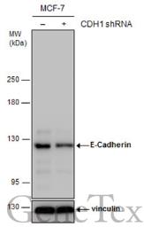 | Non-transfected (–) and transfected (+) MCF-7 whole cell extracts (30 μg) were separated by 5% SDS-PAGE, and the membrane was blotted with E-Cadherin antibody [N3C2], Internal (GTX124178) diluted at 1:10000. The HRP-conjugated anti-rabbit IgG antibody (GTX213110-01) was used to detect the primary antibody. |
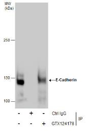 | Immunoprecipitation of E-Cadherin protein from MCF-7 whole cell extracts using 5 μg of E-Cadherin antibody [N3C2], Internal (GTX124178).Western blot analysis was performed using E-Cadherin antibody [N3C2], Internal (GTX124178).EasyBlot anti-Rabbit IgG (GTX221666-01) was used as a secondary reagent. |
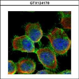 | Confocal immunofluorescence analysis (Olympus FV10i) of methanol-fixed A431, using E-cadherin(GTX124178) antibody (Green) at 1:500 dilution. Alpha-tubulin filaments were labeled with GTX11304 (Red) at 1:2000. |
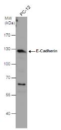 | E-Cadherin antibody [N3C2], Internal detects E-Cadherin protein by western blot analysis. Whole cell extracts (30 μg) was separated by 7.5% SDS-PAGE, and the membrane was blotted with E-Cadherin antibody [N3C2], Internal (GTX124178) diluted at 1:3000. The HRP-conjugated anti-rabbit IgG antibody (GTX213110-01) was used to detect the primary antibody. |
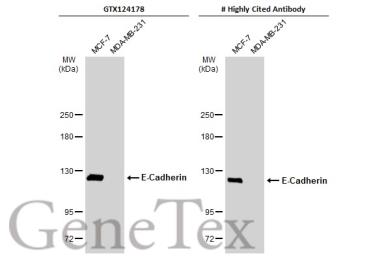 | Various whole cell extracts (30 μg) were separated by 5% SDS-PAGE, and the membranes were blotted with E-Cadherin antibody [N3C2], Internal (GTX124178) diluted at 1:3000 and competitor's antibody diluted at 1:3000. The HRP-conjugated anti-rabbit IgG antibody (GTX213110-01) was used to detect the primary antibody.*The competitor is not affiliated with GeneTex and does not endorse this product. |
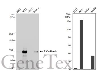 | Various whole cell extracts (30 μg) were separated by 5% SDS-PAGE, and the membrane was blotted with E-Cadherin antibody [N3C2], Internal (GTX124178) diluted at 1:3000. The HRP-conjugated anti-rabbit IgG antibody (GTX213110-01) was used to detect the primary antibody. Corresponding RNA expression data for the same cell lines are based on Human Protein Atlas program. |
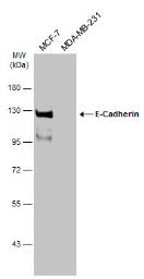 | Various whole cell extracts (30 μg) were separated by 7.5% SDS-PAGE, and the membrane was blotted with E-Cadherin antibody [N3C2], Internal (GTX124178) diluted at 1:3000. The HRP-conjugated anti-rabbit IgG antibody (GTX213110-01) was used to detect the primary antibody.The observed M.W. is based on the publication: PMID: 20356845 |
追加しました。
製品情報
| Host | Rabbit |
|---|---|
| Clonality | Polyclonal |
| Isotype | IgG |
| Application | WB, ICC/IF, IP |
| Reactivity | Human, Rat |
追加しました。
APPLICATION
Application Note
*Optimal dilutions/concentrations should be determined by the researcher.| Application | Dilution |
|---|---|
| WB | 1:500-1:10000 |
| ICC/IF | 1:100-1:1000 |
| IP | 1:100-1:500 |
| Calculated MW | 97 kDa. ( Note ) |
|---|---|
| Positive Control | 293T , PC-12 , MCF-7 |
| Predict Reactivity | Bovine, Pig(>80% identity) |
追加しました。
PROPERTIES
| Form | Liquid |
|---|---|
| Buffer | 1XPBS, 1% BSA, 20% Glycerol (pH7). 0.025% ProClin 300 was added as a preservative. |
| Storage | Store as concentrated solution. Centrifuge briefly prior to opening vial. For short-term storage (1-2 weeks), store at 4ºC. For long-term storage, aliquot and store at -20ºC or below. Avoid multiple freeze-thaw cycles. |
| Concentration | 0.22 mg/ml (Please refer to the vial label for the specific concentration.) |
| Antigen Species | Human |
| Immunogen | Recombinant protein encompassing a sequence within the center region of human E-Cadherin. The exact sequence is proprietary. |
| Purification | Purified by antigen-affinity chromatography. |
| Conjugation | Unconjugated |
| Note | For laboratory use only. Not for any clinical, therapeutic, or diagnostic use in humans or animals. Not for animal or human consumption. |
追加しました。
TARGET
| Synonyms | cadherin 1 , Arc-1 , BCDS1 , CD324 , CDHE , ECAD , LCAM , UVO |
|---|---|
| Cellular Localization | Cell junction , Cell membrane |
| Background | This gene is a classical cadherin from the cadherin superfamily. The encoded protein is a calcium dependent cell-cell adhesion glycoprotein comprised of five extracellular cadherin repeats, a transmembrane region and a highly conserved cytoplasmic tail. Mutations in this gene are correlated with gastric, breast, colorectal, thyroid and ovarian cancer. Loss of function is thought to contribute to progression in cancer by increasing proliferation, invasion, and/or metastasis. The ectodomain of this protein mediates bacterial adhesion to mammalian cells and the cytoplasmic domain is required for internalization. Identified transcript variants arise from mutation at consensus splice sites. [provided by RefSeq] |
| Database | ・ Gene ID: 999 CDH1 ・ UniProt: P12830 CDH1 |
追加しました。
REFERENCEE(抜粋)
| Application Reference | Application/Reactivity |
|---|---|
| Ling HH et al. Exp Cell Res 2016; Elevation of YAP promotes the epithelial-mesenchymal transition and tumor aggressiveness in colorectal cancer. | Application : WB Reactivity : Human |
| Wai-Theng Lee et al. Food and Chemical Toxicology 2015; 78 : 33-41 Antroquinonol from Antrodia Camphorata suppresses breast tumor migration/invasion through inhibiting ERK-AP-1- and AKT-NF-κBdependent MMP-9 and epithelial-mesenchymal transition expressions | Application : WB Reactivity : Human |
追加しました。
製品情報は掲載時点のものですが、価格表内の価格については随時最新のものに更新されます。お問い合わせいただくタイミングにより製品情報・価格などは変更されている場合があります。
表示価格に、消費税等は含まれていません。一部価格が予告なく変更される場合がありますので、あらかじめご了承下さい。