
抗DAG1抗体 | Anti-DAG1 antibody
掲載日情報:2020/04/02 現在Webページ番号:37170

GeneTex社の抗DAG1抗体 | Anti-DAG1 antibodyです。
※ 本製品は研究用です。研究用以外には使用できません。
追加しました。
特長
- 高品質の抗体です。
- 幅広い研究分野に関連する抗体を取り揃えています。
- 使用されたアプリケーションや動物種などの情報が充実しています。
- 多数の論文で使用実績がある信頼性の高い抗体です。
追加しました。
価格
[在庫・価格 :2026年04月01日 00時00分現在]
| 詳細 | 商品名 |
|
文献数 | ||||||||||||||||||||||||||||||||||||||||||||||||||||||||||||||||||||||||||||||||||
|---|---|---|---|---|---|---|---|---|---|---|---|---|---|---|---|---|---|---|---|---|---|---|---|---|---|---|---|---|---|---|---|---|---|---|---|---|---|---|---|---|---|---|---|---|---|---|---|---|---|---|---|---|---|---|---|---|---|---|---|---|---|---|---|---|---|---|---|---|---|---|---|---|---|---|---|---|---|---|---|---|---|---|---|---|---|
|
Anti-DAG1, Rabbit-Poly |
|
5 | |||||||||||||||||||||||||||||||||||||||||||||||||||||||||||||||||||||||||||||||||||

|
|||||||||||||||||||||||||||||||||||||||||||||||||||||||||||||||||||||||||||||||||||||
|
Anti-DAG1, Rabbit-Poly |
|
5 | |||||||||||||||||||||||||||||||||||||||||||||||||||||||||||||||||||||||||||||||||||
|
|||||||||||||||||||||||||||||||||||||||||||||||||||||||||||||||||||||||||||||||||||||


[在庫・価格 :2026年04月01日 00時00分現在]
Anti-DAG1, Rabbit-Poly
文献数: 5
- 商品コード:GTX105038
- メーカー:GNT
- 包装:25μl
- 価格:¥30,000
- 在庫:無(未発注)
- 納期:10日程度 ※※ 表示されている納期は弊社に在庫がなく、取り寄せた場合の目安納期となります。
- 法規制等:
| 説明文 | 別名:dystroglycan 1,156DAG,A3a,AGRNR,DAG,LGMDR16,MDDGA9,MDDGC7,MDDGC9 Genbank No: 1605 |
||||||
|---|---|---|---|---|---|---|---|
| 別包装品 | 別包装品あり | ||||||
| 法規制等 | |||||||
| 保存条件 | -20℃ | 法規備考 | |||||
| 抗原種 | Human | 免疫動物 | Rabbit | ||||
| 交差性 | Horse/Human/Mouse/Rat | 適用 | IC,IF,IHC,Western Blot | ||||
| 標識 | Unlabeled | 性状 | Purified | ||||
| 吸収処理 | クラス | IgG | |||||
| クロナリティ | Polyclonal | フォーマット | |||||
| 掲載カタログ |
|
||||||
| 製品記事 | 使いっきり抗体 |
||||||
| 関連記事 | GeneTex社における抗体の品質管理 |
||||||
Anti-DAG1, Rabbit-Poly
文献数: 5
- 商品コード:GTX105038
- メーカー:GNT
- 包装:100μl
- 価格:¥85,000
- 在庫:1個
- 納期:10日程度 ※※ 表示されている納期は弊社に在庫がなく、取り寄せた場合の目安納期となります。
- 法規制等:
| 説明文 | 別名:dystroglycan 1,156DAG,A3a,AGRNR,DAG,LGMDR16,MDDGA9,MDDGC7,MDDGC9 Genbank No: 1605 |
||||||
|---|---|---|---|---|---|---|---|
| 別包装品 | 別包装品あり | ||||||
| 法規制等 | |||||||
| 保存条件 | -20℃ | 法規備考 | |||||
| 抗原種 | Human | 免疫動物 | Rabbit | ||||
| 交差性 | Horse/Human/Mouse/Rat | 適用 | IC,IF,IHC,Western Blot | ||||
| 標識 | Unlabeled | 性状 | Purified | ||||
| 吸収処理 | クラス | IgG | |||||
| クロナリティ | Polyclonal | フォーマット | |||||
| 掲載カタログ |
|
||||||
| 製品記事 | |||||||
| 関連記事 | GeneTex社における抗体の品質管理 |
||||||
追加しました。
DATA IMAGES
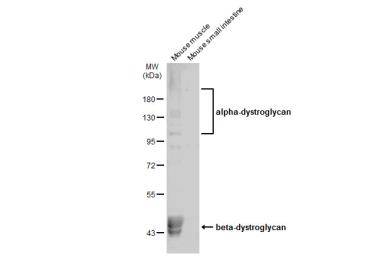 | Various tissue extracts (30 μg) were separated by 7.5% SDS-PAGE, and the membrane was blotted with DAG1 antibody (GTX105038) diluted at 1:500. The HRP-conjugated anti-rabbit IgG antibody (GTX213110-01) was used to detect the primary antibody. |
 | MCF-7 whole cell and membrane extracts (30 μg) were separated by 7.5% SDS-PAGE, and the membrane was blotted with DAG1 antibody (GTX105038) diluted at 1:500. The HRP-conjugated anti-rabbit IgG antibody (GTX213110-01) was used to detect the primary antibody. |
 | 293T whole cell and membrane extracts (30 μg) were separated by 7.5% SDS-PAGE, and the membrane was blotted with DAG1 antibody (GTX105038) diluted at 1:500. The HRP-conjugated anti-rabbit IgG antibody (GTX213110-01) was used to detect the primary antibody. |
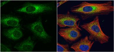 | alpha Dystroglycan antibody detects alpha Dystroglycan protein at cytoplasm by immunofluorescent analysis.Sample: HeLa cells were fixed in 4% paraformaldehyde at RT for 15 min.Green: alpha Dystroglycan protein stained by alpha Dystroglycan antibody (GTX105038) diluted at 1:1000.Red: alpha Tubulin, a cytoskeleton marker, stained by alpha Tubulin antibody [B-5-1-2] (GTX11304) diluted at 1:10000.Blue: Hoechst 33342 staining. |
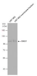 | DAG1 antibody detects DAG1 protein by western blot analysis. U87-MG whole cell extracts and membrane extracts (30 μg) were separated by 7.5% SDS-PAGE, and the membrane was blotted with DAG1 antibody (GTX105038) diluted at 1:500. The HRP-conjugated anti-rabbit IgG antibody (GTX213110-01) was used to detect the primary antibody. |
 | DAG1 antibody detects DAG1 protein by immunofluorescent analysis.Sample: DIV10 rat E18 primary hippocampal neuron cells were fixed in 4% paraformaldehyde at RT for 15 min.Green: DAG1 stained by DAG1 antibody (GTX105038) diluted at 1:500.Red: Tau, stained by Tau antibody [GT287] (GTX634809) diluted at 1:500.Blue: Fluoroshield with DAPI (GTX30920). |
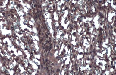 | DAG1 antibody detects DAG1 protein at cytoplasm by immunohistochemical analysis.Sample: Paraffin-embedded mouse placenta.DAG1 stained by DAG1 antibody (GTX105038) diluted at 1:500.Antigen Retrieval: Citrate buffer, pH 6.0, 15 min |
追加しました。
製品情報
| Host | Rabbit |
|---|---|
| Clonality | Polyclonal |
| Isotype | IgG |
| Application | WB, ICC/IF, IHC-P |
| Reactivity | Human, Mouse, Rat, Horse |
追加しました。
APPLICATION
Application Note
*Optimal dilutions/concentrations should be determined by the researcher.| Application | Dilution |
|---|---|
| WB | 1:500-1:3000 |
| ICC/IF | 1:100-1:1000 |
| IHC-P | 1:100-1:1000 |
| Calculated MW | 97 kDa. ( Note ) |
|---|---|
| Positive Control | U87-MG , U87-MG membrane fraction extract |
| Predict Reactivity | Rabbit, Bovine, Cat, Dog, Rhesus Monkey(>80% identity) |
追加しました。
PROPERTIES
| Form | Liquid |
|---|---|
| Buffer | 1XPBS, 20% Glycerol (pH7). 0.025% ProClin 300 was added as a preservative. |
| Storage | Store as concentrated solution. Centrifuge briefly prior to opening vial. For short-term storage (1-2 weeks), store at 4ºC. For long-term storage, aliquot and store at -20ºC or below. Avoid multiple freeze-thaw cycles. |
| Concentration | 1.06 mg/ml (Please refer to the vial label for the specific concentration.) |
| Antigen Species | Human |
| Immunogen | Recombinant protein encompassing a sequence within the center region of human alpha Dystroglycan. The exact sequence is proprietary. |
| Purification | Purified by antigen-affinity chromatography. |
| Conjugation | Unconjugated |
| Note | For laboratory use only. Not for any clinical, therapeutic, or diagnostic use in humans or animals. Not for animal or human consumption. |
追加しました。
TARGET
| Synonyms | dystroglycan 1 , 156DAG , A3a , AGRNR , DAG , LGMDR16 , MDDGA9 , MDDGC7 , MDDGC9 |
|---|---|
| Cellular Localization | Alpha-dystroglycan: Secreted , extracellular space , Beta-dystroglycan: Cell membrane , Cytoplasm , cytoskeleton |
| Background | Dystroglycan is a laminin binding component of the dystrophin-glycoprotein complex which provides a linkage between the subsarcolemmal cytoskeleton and the extracellular matrix. Dystroglycan 1 is a candidate gene for the site of the mutation in autosomal recessive muscular dystrophies. The dramatic reduction of dystroglycan 1 in Duchenne muscular dystrophy leads to a loss of linkage between the sarcolemma and extracellular matrix, rendering muscle fibers more susceptible to necrosis. Dystroglycan also functions as dual receptor for agrin and laminin-2 in the Schwann cell membrane. The muscle and nonmuscle isoforms of dystroglycan differ by carbohydrate moieties but not protein sequence. [provided by RefSeq] |
| Database | ・ Gene ID: 1605 DAG1 ・ UniProt: Q14118 DAG1 |
追加しました。
REFERENCEE(抜粋)
追加しました。
製品情報は掲載時点のものですが、価格表内の価格については随時最新のものに更新されます。お問い合わせいただくタイミングにより製品情報・価格などは変更されている場合があります。
表示価格に、消費税等は含まれていません。一部価格が予告なく変更される場合がありますので、あらかじめご了承下さい。