
Anti-HMGB1, Rabbit-Poly | GeneTex International Corporation
掲載日情報:2026/06/03 現在Webページ番号:35500

GeneTex International CorporationのAnti-HMGB1, Rabbit-Poly商品情報ページです。
※本製品は研究用です。研究用以外には使用できません。
カートに商品を
追加しました。
追加しました。
価格
[在庫・価格 :2026年07月10日 12時35分現在]
※ 表示されている納期は弊社に在庫が無く、取り寄せた場合の納期目安となります。
| 詳細 | 商品名 |
|
文献数 | ||||||||||||||||||||||||||||||||||||||||||||||||||||||||||||||||||||||||||||||||||
|---|---|---|---|---|---|---|---|---|---|---|---|---|---|---|---|---|---|---|---|---|---|---|---|---|---|---|---|---|---|---|---|---|---|---|---|---|---|---|---|---|---|---|---|---|---|---|---|---|---|---|---|---|---|---|---|---|---|---|---|---|---|---|---|---|---|---|---|---|---|---|---|---|---|---|---|---|---|---|---|---|---|---|---|---|---|
|
Anti-HMGB1, Rabbit-Poly |
|
22 | |||||||||||||||||||||||||||||||||||||||||||||||||||||||||||||||||||||||||||||||||||

|
|||||||||||||||||||||||||||||||||||||||||||||||||||||||||||||||||||||||||||||||||||||


[在庫・価格 :2026年07月10日 12時35分現在]
※ 表示されている納期は弊社に在庫が無く、取り寄せた場合の納期目安となります。
Anti-HMGB1, Rabbit-Poly
文献数: 22
- 商品コード:GTX101277
- メーカー:GNT
- 包装:100μl
- 価格:¥85,000
- 在庫:1個
- 納期:10日程度 ※※ 表示されている納期は弊社に在庫がなく、取り寄せた場合の目安納期となります。
- 法規制等:
| 説明文 | レビューあり。KO/KDバリデーション済み抗体。 別名:high mobility group box 1,HMG-1,HMG1,HMG3,SBP-1 Genbank No: 3146 |
||||||
|---|---|---|---|---|---|---|---|
| 別包装品 | 別包装品あり | ||||||
| 法規制等 | |||||||
| 保存条件 | -20℃ | 法規備考 | |||||
| 抗原種 | Human | 免疫動物 | Rabbit | ||||
| 交差性 | Human/Mouse/Pig/Rat/Zebrafish | 適用 | IC,IF,IHC,Western Blot | ||||
| 標識 | Unlabeled | 性状 | Purified | ||||
| 吸収処理 | クラス | IgG | |||||
| クロナリティ | Polyclonal | フォーマット | |||||
| 掲載カタログ |
|
||||||
| 製品記事 | アポトーシス・オートファジー・ネクローシス関連抗体 筋萎縮性側索硬化症研究用抗体 |
||||||
| 関連記事 | GeneTex社における抗体の品質管理 |
||||||
カートに商品を
追加しました。
追加しました。
ラインナップ
カートに商品を
追加しました。
追加しました。
画像
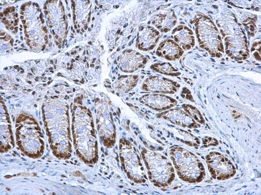
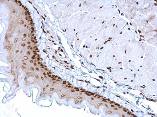
GTX101277 IHC-P Image
HMGB1 antibody detects HMGB1 protein at nucleus on mouse colon by immunohistochemical analysis.
Sample: Paraffin-embedded mouse colon.
HMGB1 antibody (GTX101277) dilution: 1:1000.
HMGB1 antibody detects HMGB1 protein at nucleus on mouse colon by immunohistochemical analysis.
Sample: Paraffin-embedded mouse colon.
HMGB1 antibody (GTX101277) dilution: 1:1000.
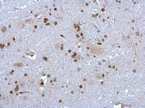
GTX101277 IHC-P Image
HMGB1 antibody detects HMGB1 protein at nucleus on rat brain stem by immunohistochemical analysis.
Sample: Paraffin-embedded rat brain stem.
HMGB1 antibody (GTX101277) dilution: 1:1000.
HMGB1 antibody detects HMGB1 protein at nucleus on rat brain stem by immunohistochemical analysis.
Sample: Paraffin-embedded rat brain stem.
HMGB1 antibody (GTX101277) dilution: 1:1000.
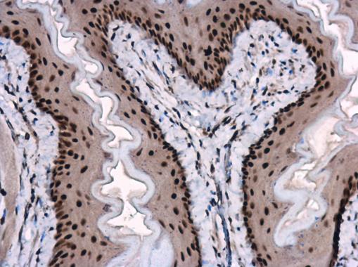
GTX101277 IHC-P Image
HMGB1 antibody detects HMGB1 protein at nucleus in mouse esophagus by immunohistochemical analysis.
Sample: Paraffin-embedded mouse esophagus.
HMGB1 antibody (GTX101277) diluted at 1:1000.
HMGB1 antibody detects HMGB1 protein at nucleus in mouse esophagus by immunohistochemical analysis.
Sample: Paraffin-embedded mouse esophagus.
HMGB1 antibody (GTX101277) diluted at 1:1000.
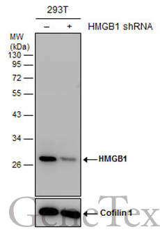
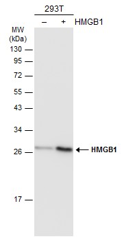
GTX101277 WB Image
Non-transfected (?) and transfected (+) 293T whole cell extracts (30 ug) were separated by 12% SDS-PAGE, and the membrane was blotted with HMGB1 antibody (GTX101277) diluted at 1:5000. The HRP-conjugated anti-rabbit IgG antibody (GTX213110-01) was used to detect the primary antibody.
Non-transfected (?) and transfected (+) 293T whole cell extracts (30 ug) were separated by 12% SDS-PAGE, and the membrane was blotted with HMGB1 antibody (GTX101277) diluted at 1:5000. The HRP-conjugated anti-rabbit IgG antibody (GTX213110-01) was used to detect the primary antibody.
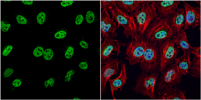
GTX101277 ICC/IF Image
HMGB1 antibody detects HMGB1 protein at nucleus by immunofluorescent analysis.
Sample: HeLa cells were fixed in 4% paraformaldehyde at RT for 15 min.
Green: HMGB1 protein stained by HMGB1 antibody (GTX101277) diluted at 1:1000.
Red: phalloidin, a cytoskeleton marker, stained by phalloidin (invitrogen, A12380) diluted at 1:200.
Blue: Hoechst 33342 staining.
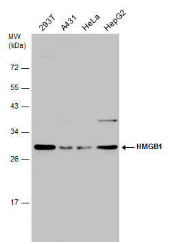
GTX101277 WB Image
Various whole cell extracts (30 ug) were separated by 12% SDS-PAGE, and the membrane was blotted with HMGB1 antibody (GTX101277) diluted at 1:3000.
Various whole cell extracts (30 ug) were separated by 12% SDS-PAGE, and the membrane was blotted with HMGB1 antibody (GTX101277) diluted at 1:3000.
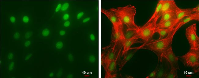
GTX101277 ICC/IF Image
HMGB1 antibody detects HMGB1 protein at nucleus by immunofluorescent analysis.
Sample: NIH/3T3 cells were fixed in 4% paraformaldehyde at RT for 15 min.
Green: HMGB1 protein stained by HMGB1 antibody (GTX101277) diluted at 1:500.
Red: phalloidin, a cytoskeleton marker, diluted at 1:50.
Scale bar = 10 um.
GTX101277 IHC-P Image
HMGB1 antibody detects HMGB1 protein at nucleus on mouse esophagus by immunohistochemical analysis.
Sample: Paraffin-embedded mouse esophagus.
HMGB1 antibody (GTX101277) dilution: 1:1000.
HMGB1 antibody detects HMGB1 protein at nucleus on mouse esophagus by immunohistochemical analysis.
Sample: Paraffin-embedded mouse esophagus.
HMGB1 antibody (GTX101277) dilution: 1:1000.
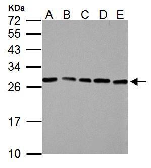
GTX101277 WB Image
HMGB1 antibody detects HMGB1 protein by western blot analysis.
A. 30 ug NIH-3T3 whole cell lysate/extract
B. 30 ug JC whole cell lysate/extract
C. 30 ug BCL-1 whole cell lysate/extract
D. 30 ug C2C12 whole cell lysate/extract
E. 30 ug Raw264.7 whole cell lysate/extract
12% SDS-PAGE
HMGB1 antibody (GTX101277) dilution: 1:3000
The HRP-conjugated anti-rabbit IgG antibody (GTX213110-01) was used to detect the primary antibody.
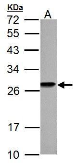
GTX101277 WB Image
HMGB1 antibody detects HMGB1 protein by western blot analysis.
A. 30 ug PC-12 whole cell lysate/extract
12% SDS-PAGE
HMGB1 antibody (GTX101277) dilution: 1:3000
The HRP-conjugated anti-rabbit IgG antibody (GTX213110-01) was used to detect the primary antibody.
HMGB1 antibody detects HMGB1 protein by western blot analysis.
A. 30 ug PC-12 whole cell lysate/extract
12% SDS-PAGE
HMGB1 antibody (GTX101277) dilution: 1:3000
The HRP-conjugated anti-rabbit IgG antibody (GTX213110-01) was used to detect the primary antibody.
![GTX101277 ICC/IF Image<br>HMGB1 antibody detects HMGB1 protein at cytoplasm and nucleus by immunofluorescent analysis.<br>Sample: SK-N-SH cells were fixed in 4% paraformaldehyde at RT for 15 min.<br>Green: HMGB1 protein stained by HMGB1 antibody (GTX101277) diluted at 1:1000.<br>Red: beta Tubulin 3/ Tuj1, a cytoskeleton marker, stained by beta Tubulin 3/ Tuj1 antibody [GT11710] (GTX631836) diluted at 1:500.<br>Scale bar = 10 um.](/domestic/data/graphics/GNT/graphics/GTX101277_42536_20161109_IFA.jpg)
GTX101277 ICC/IF Image
HMGB1 antibody detects HMGB1 protein at cytoplasm and nucleus by immunofluorescent analysis.
Sample: SK-N-SH cells were fixed in 4% paraformaldehyde at RT for 15 min.
Green: HMGB1 protein stained by HMGB1 antibody (GTX101277) diluted at 1:1000.
Red: beta Tubulin 3/ Tuj1, a cytoskeleton marker, stained by beta Tubulin 3/ Tuj1 antibody [GT11710] (GTX631836) diluted at 1:500.
Scale bar = 10 um.
GTX101277 WB Image
Non-transfected (?) and transfected (+) 293T whole cell extracts (30 ug) were separated by 12% SDS-PAGE, and the membrane was blotted with HMGB1 antibody (GTX101277) diluted at 1:5000. The HRP-conjugated anti-rabbit IgG antibody (GTX213110-01) was used to detect the primary antibody.
Non-transfected (?) and transfected (+) 293T whole cell extracts (30 ug) were separated by 12% SDS-PAGE, and the membrane was blotted with HMGB1 antibody (GTX101277) diluted at 1:5000. The HRP-conjugated anti-rabbit IgG antibody (GTX213110-01) was used to detect the primary antibody.
カートに商品を
追加しました。
追加しました。
商品情報
| 商品説明 | レビューあり。KO/KDバリデーション済み抗体 |
|---|---|
| 抗原動物 | Human |
| 交差性 | Human/Mouse/Pig/Rat/Zebrafish |
| 免疫動物 | Rabbit |
| 性状 | Purified |
| 適用 | IC, IF, IHC, Western Blot |
| クラス | IgG |
| 標識 | Unlabeled |
| クロナリティ | Polyclonal |
| 別名 | high mobility group box 1, HMG-1, HMG1, HMG3, SBP-1 |
| Genbank No | 3146 |
| メーカーサイト | メーカーサイト |
| 使用文献 | 使用文献 |
| 保存条件 | -20℃ |
カートに商品を
追加しました。
追加しました。
製品情報は掲載時点のものですが、価格表内の価格については随時最新のものに更新されます。お問い合わせいただくタイミングにより製品情報・価格などは変更されている場合があります。
表示価格に、消費税等は含まれていません。一部価格が予告なく変更される場合がありますので、あらかじめご了承下さい。