
Anti-SQSTM1, Rabbit-Poly | GeneTex International Corporation
掲載日情報:2026/06/03 現在Webページ番号:35236

GeneTex International CorporationのAnti-SQSTM1, Rabbit-Poly商品情報ページです。
※本製品は研究用です。研究用以外には使用できません。
カートに商品を
追加しました。
追加しました。
価格
[在庫・価格 :2026年07月10日 11時15分現在]
※ 表示されている納期は弊社に在庫が無く、取り寄せた場合の納期目安となります。
| 詳細 | 商品名 |
|
文献数 | ||||||||||||||||||||||||||||||||||||||||||||||||||||||||||||||||||||||||||||||||||
|---|---|---|---|---|---|---|---|---|---|---|---|---|---|---|---|---|---|---|---|---|---|---|---|---|---|---|---|---|---|---|---|---|---|---|---|---|---|---|---|---|---|---|---|---|---|---|---|---|---|---|---|---|---|---|---|---|---|---|---|---|---|---|---|---|---|---|---|---|---|---|---|---|---|---|---|---|---|---|---|---|---|---|---|---|---|
|
Anti-SQSTM1, Rabbit-Poly |
|
88 | |||||||||||||||||||||||||||||||||||||||||||||||||||||||||||||||||||||||||||||||||||
|
|||||||||||||||||||||||||||||||||||||||||||||||||||||||||||||||||||||||||||||||||||||


[在庫・価格 :2026年07月10日 11時15分現在]
※ 表示されている納期は弊社に在庫が無く、取り寄せた場合の納期目安となります。
Anti-SQSTM1, Rabbit-Poly
文献数: 88
- 商品コード:GTX100685
- メーカー:GNT
- 包装:100μl
- 価格:¥85,000
- 在庫:1個
- 納期:10日程度 ※※ 表示されている納期は弊社に在庫がなく、取り寄せた場合の目安納期となります。
- 法規制等:
| 説明文 | レビューあり。KO/KDバリデーション済み抗体。 別名:sequestosome 1,A170,DMRV,FTDALS3,NADGP,OSIL,PDB3,ZIP3,p60,p62,p62B Genbank No: 8878 |
||||||
|---|---|---|---|---|---|---|---|
| 別包装品 | 別包装品あり | ||||||
| 法規制等 | |||||||
| 保存条件 | -20℃ | 法規備考 | |||||
| 抗原種 | Human | 免疫動物 | Rabbit | ||||
| 交差性 | Bovine/Honeybee/Human/Mosquito/Mouse/Rat/Zebrafish | 適用 | FCM,IC,IF,IHC,IP,PLA,Western Blot | ||||
| 標識 | Unlabeled | 性状 | Purified | ||||
| 吸収処理 | クラス | IgG | |||||
| クロナリティ | Polyclonal | フォーマット | |||||
| 掲載カタログ |
|
||||||
| 製品記事 | アポトーシス・オートファジー・ネクローシス関連抗体 GeneTex社 オルガネラマーカー抗体 ゼブラフィッシュ(zebrafish)研究用製品特集 |
||||||
| 関連記事 | GeneTex社における抗体の品質管理 |
||||||
カートに商品を
追加しました。
追加しました。
ラインナップ
カートに商品を
追加しました。
追加しました。
画像
![GTX100685 WB Image<br>Untreated (?) and treated (+) HepG2 whole cell extracts (30 ug) were separated by 10% SDS-PAGE, and the membrane was blotted with SQSTM1 antibody [N3C1], Internal (GTX100685) diluted at 1:1000. The HRP-conjugated anti-rabbit IgG antibody (GTX213110-01) was used to detect the primary antibody.](/domestic/data/graphics/GNT/graphics/GTX100685_42824_20170504_WB_Thapsigargin.jpg)
GTX100685 WB Image
Untreated (?) and treated (+) HepG2 whole cell extracts (30 ug) were separated by 10% SDS-PAGE, and the membrane was blotted with SQSTM1 antibody [N3C1], Internal (GTX100685) diluted at 1:1000. The HRP-conjugated anti-rabbit IgG antibody (GTX213110-01) was used to detect the primary antibody.
Untreated (?) and treated (+) HepG2 whole cell extracts (30 ug) were separated by 10% SDS-PAGE, and the membrane was blotted with SQSTM1 antibody [N3C1], Internal (GTX100685) diluted at 1:1000. The HRP-conjugated anti-rabbit IgG antibody (GTX213110-01) was used to detect the primary antibody.
![GTX100685 ICC/IF Image<br>SQSTM1 antibody [N3C1], Internal detects SQSTM1 protein at autophagosome by immunofluorescent analysis.<br>Samples: HepG2 cells treated with 3uM thapsigargin 12 hrs (rigtht) and mock (left) were fixed in ice-cold MeOH for 10 min, permeabilize with cooled acetone for 1 min .<br>Green: SQSTM1 protein stained by SQSTM1 antibody [N3C1], Internal (GTX100685) diluted at 1:500.<br>Blue: Hoechst 33342 staining.<br>Scale bar = 10 um.](/domestic/data/graphics/GNT/graphics/GTX100685_39988_IFA.jpg)
GTX100685 ICC/IF Image
SQSTM1 antibody [N3C1], Internal detects SQSTM1 protein at autophagosome by immunofluorescent analysis.
Samples: HepG2 cells treated with 3uM thapsigargin 12 hrs (rigtht) and mock (left) were fixed in ice-cold MeOH for 10 min, permeabilize with cooled acetone for 1 min .
Green: SQSTM1 protein stained by SQSTM1 antibody [N3C1], Internal (GTX100685) diluted at 1:500.
Blue: Hoechst 33342 staining.
Scale bar = 10 um.
![GTX100685 IP Image<br>Immunoprecipitation of SQSTM1 protein from HeLa whole cell extracts using 5 ug of SQSTM1 antibody [N3C1], Internal (GTX100685).<br>Western blot analysis was performed using SQSTM1 antibody [N3C1], Internal (GTX100685).<br>EasyBlot anti-Rabbit IgG (GTX221666-01) was used as a secondary reagent.</br></br></br>](/domestic/data/graphics/GNT/graphics/GTX100685_41052_20150209_IP.jpg)
GTX100685 IP Image
Immunoprecipitation of SQSTM1 protein from HeLa whole cell extracts using 5 ug of SQSTM1 antibody [N3C1], Internal (GTX100685).
Western blot analysis was performed using SQSTM1 antibody [N3C1], Internal (GTX100685).
EasyBlot anti-Rabbit IgG (GTX221666-01) was used as a secondary reagent.
Immunoprecipitation of SQSTM1 protein from HeLa whole cell extracts using 5 ug of SQSTM1 antibody [N3C1], Internal (GTX100685).
Western blot analysis was performed using SQSTM1 antibody [N3C1], Internal (GTX100685).
EasyBlot anti-Rabbit IgG (GTX221666-01) was used as a secondary reagent.
![GTX100685 ICC/IF Image<br>SQSTM1 antibody [N3C1], Internal detects SQSTM1 protein at autophagosome by immunofluorescent analysis.<br>Samples: HeLa cells mock (left) and treated with 50uM Chloroquine for 24 hr (right) were fixed in 4% paraformaldehyde at RT for 15 min.<br>Green: SQSTM1 protein stained by SQSTM1 antibody [N3C1], Internal (GTX100685) diluted at 1:1000.<br>Red: Phalloidin, a F-actin marker.<br></br>](/domestic/data/graphics/GNT/graphics/GTX100685_41052_20141219_IFA.jpg)
GTX100685 ICC/IF Image
SQSTM1 antibody [N3C1], Internal detects SQSTM1 protein at autophagosome by immunofluorescent analysis.
Samples: HeLa cells mock (left) and treated with 50uM Chloroquine for 24 hr (right) were fixed in 4% paraformaldehyde at RT for 15 min.
Green: SQSTM1 protein stained by SQSTM1 antibody [N3C1], Internal (GTX100685) diluted at 1:1000.
Red: Phalloidin, a F-actin marker.
![GTX100685 IHC-P Image<br>SQSTM1 antibody [N3C1], Internal detects SQSTM1 protein at cytoplasm on human ovarian carcinoma by immunohistochemical analysis.<br>Sample: Paraffin-embedded human ovarian carcinoma.<br>SQSTM1 antibody [N3C1], Internal (GTX100685) diluted at 1:500.<br></br>](/domestic/data/graphics/GNT/graphics/GTX100685_41052_20141226_IHC.jpg)
GTX100685 IHC-P Image
SQSTM1 antibody [N3C1], Internal detects SQSTM1 protein at cytoplasm on human ovarian carcinoma by immunohistochemical analysis.
Sample: Paraffin-embedded human ovarian carcinoma.
SQSTM1 antibody [N3C1], Internal (GTX100685) diluted at 1:500.
SQSTM1 antibody [N3C1], Internal detects SQSTM1 protein at cytoplasm on human ovarian carcinoma by immunohistochemical analysis.
Sample: Paraffin-embedded human ovarian carcinoma.
SQSTM1 antibody [N3C1], Internal (GTX100685) diluted at 1:500.
![GTX100685 FACS Image<br>SQSTM1 antibody [N3C1], Internal (GTX100685) detects SQSTM1 protein by flow cytometry analysis.<br>Sample: HeLa cell fixed in 4% paraformaldehyde at 4oC for 5 min.<br>Brown: Unlabelled sample was also used as a control.<br>Blue: SQSTM1 antibody [N3C1], Internal] (GTX100685) dilution: 1:100.<br>Acquisition of >20,000 events were collected using Argon ion laser (488nm) and 525/30 bandpass filter.</br></br></br></br></br>](/domestic/data/graphics/GNT/graphics/GTX100685_41052_20150212_FACS.jpg)
GTX100685 FACS Image
SQSTM1 antibody [N3C1], Internal (GTX100685) detects SQSTM1 protein by flow cytometry analysis.
Sample: HeLa cell fixed in 4% paraformaldehyde at 4oC for 5 min.
Brown: Unlabelled sample was also used as a control.
Blue: SQSTM1 antibody [N3C1], Internal] (GTX100685) dilution: 1:100.
Acquisition of >20,000 events were collected using Argon ion laser (488nm) and 525/30 bandpass filter.
![GTX100685 WB Image<br>Non-transfected (?) and transfected (+) HepG2 whole cell extracts (30 ug) were separated by 10% SDS-PAGE, and the membrane was blotted with SQSTM1 antibody [N3C1], Internal (GTX100685) diluted at 1:500. The HRP-conjugated anti-rabbit IgG antibody (GTX213110-01) was used to detect the primary antibody.</br>](/domestic/data/graphics/GNT/graphics/GTX100685_41052_20160804_WB_shRNA_watermark.jpg)
GTX100685 WB Image
Non-transfected (?) and transfected (+) HepG2 whole cell extracts (30 ug) were separated by 10% SDS-PAGE, and the membrane was blotted with SQSTM1 antibody [N3C1], Internal (GTX100685) diluted at 1:500. The HRP-conjugated anti-rabbit IgG antibody (GTX213110-01) was used to detect the primary antibody.
Non-transfected (?) and transfected (+) HepG2 whole cell extracts (30 ug) were separated by 10% SDS-PAGE, and the membrane was blotted with SQSTM1 antibody [N3C1], Internal (GTX100685) diluted at 1:500. The HRP-conjugated anti-rabbit IgG antibody (GTX213110-01) was used to detect the primary antibody.
![GTX100685 WB Image<br>Untreated (?) and treated (+) Huh-7 whole cell extracts (30 ug) were separated by 10% SDS-PAGE, and the membrane was blotted with SQSTM1 antibody [N3C1], Internal (GTX100685) diluted at 1:1000. The HRP-conjugated anti-rabbit IgG antibody (GTX213110-01) was used to detect the primary antibody.](/domestic/data/graphics/GNT/graphics/GTX100685_41052_20160331_WB_thapsigargin.jpg)
GTX100685 WB Image
Untreated (?) and treated (+) Huh-7 whole cell extracts (30 ug) were separated by 10% SDS-PAGE, and the membrane was blotted with SQSTM1 antibody [N3C1], Internal (GTX100685) diluted at 1:1000. The HRP-conjugated anti-rabbit IgG antibody (GTX213110-01) was used to detect the primary antibody.
Untreated (?) and treated (+) Huh-7 whole cell extracts (30 ug) were separated by 10% SDS-PAGE, and the membrane was blotted with SQSTM1 antibody [N3C1], Internal (GTX100685) diluted at 1:1000. The HRP-conjugated anti-rabbit IgG antibody (GTX213110-01) was used to detect the primary antibody.
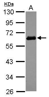
GTX100685 WB Image
Sample (30 ug of whole cell lysate)
A: A549
10% SDS PAGE
GTX100685 diluted at 1:1000
The HRP-conjugated anti-rabbit IgG antibody (GTX213110-01) was used to detect the primary antibody.
Sample (30 ug of whole cell lysate)
A: A549
10% SDS PAGE
GTX100685 diluted at 1:1000
The HRP-conjugated anti-rabbit IgG antibody (GTX213110-01) was used to detect the primary antibody.
![GTX100685 WB Image<br>SQSTM1 antibody [N3C1], Internal detects SQSTM1 protein by western blot analysis.<br>A. 30 ug NIH-3T3 whole cell lysate/extract<br>B. 30 ug JC whole cell lysate/extract<br>C. 30 ug BCL-1 whole cell lysate/extract<br>12% SDS-PAGE<br>SQSTM1 antibody [N3C1], Internal (GTX100685) dilution: 1:1000<br>The HRP-conjugated anti-rabbit IgG antibody (GTX213110-01) was used to detect the primary antibody.](/domestic/data/graphics/GNT/graphics/GTX100685_39988_WB_M.jpg)
GTX100685 WB Image
SQSTM1 antibody [N3C1], Internal detects SQSTM1 protein by western blot analysis.
A. 30 ug NIH-3T3 whole cell lysate/extract
B. 30 ug JC whole cell lysate/extract
C. 30 ug BCL-1 whole cell lysate/extract
12% SDS-PAGE
SQSTM1 antibody [N3C1], Internal (GTX100685) dilution: 1:1000
The HRP-conjugated anti-rabbit IgG antibody (GTX213110-01) was used to detect the primary antibody.
![GTX100685 WB Image<br>SQSTM1 antibody [N3C1], Internal detects SQSTM1 protein by western blot analysis.<br>A. 30 ug PC-12 whole cell lysate/extract<br>B. 30 ug Rat2 whole cell lysate/extract<br>10% SDS-PAGE<br>SQSTM1 antibody [N3C1], Internal (GTX100685) dilution: 1:1000<br>The HRP-conjugated anti-rabbit IgG antibody (GTX213110-01) was used to detect the primary antibody.](/domestic/data/graphics/GNT/graphics/GTX100685_39988_WB_R.jpg)
GTX100685 WB Image
SQSTM1 antibody [N3C1], Internal detects SQSTM1 protein by western blot analysis.
A. 30 ug PC-12 whole cell lysate/extract
B. 30 ug Rat2 whole cell lysate/extract
10% SDS-PAGE
SQSTM1 antibody [N3C1], Internal (GTX100685) dilution: 1:1000
The HRP-conjugated anti-rabbit IgG antibody (GTX213110-01) was used to detect the primary antibody.
カートに商品を
追加しました。
追加しました。
商品情報
| 商品説明 | レビューあり。KO/KDバリデーション済み抗体 |
|---|---|
| 抗原動物 | Human |
| 交差性 | Bovine/Honeybee/Human/Mosquito/Mouse/Rat/Zebrafish |
| 免疫動物 | Rabbit |
| 性状 | Purified |
| 適用 | FCM, IC, IF, IHC, IP, PLA, Western Blot |
| クラス | IgG |
| 標識 | Unlabeled |
| クロナリティ | Polyclonal |
| 別名 | sequestosome 1, A170, DMRV, FTDALS3, NADGP, OSIL, PDB3, ZIP3, p60, p62, p62B |
| Genbank No | 8878 |
| データシート | データシート |
| メーカーサイト | メーカーサイト |
| 使用文献 | 使用文献 |
| 保存条件 | -20℃ |
カートに商品を
追加しました。
追加しました。
製品情報は掲載時点のものですが、価格表内の価格については随時最新のものに更新されます。お問い合わせいただくタイミングにより製品情報・価格などは変更されている場合があります。
表示価格に、消費税等は含まれていません。一部価格が予告なく変更される場合がありますので、あらかじめご了承下さい。