
抗EPCR抗体(Anti-EPCR, Goat-Poly, Biotin antibody)
掲載日情報:2018/11/26 現在Webページ番号:30728

EPCRに対する抗体(Anti-EPCR, Goat-Poly, Biotin )です。
※ 本製品は研究用です。研究用以外には使用できません。
カートに商品を
追加しました。
追加しました。
価格
[在庫・価格 :2024年05月20日 00時00分現在]
※ 表示されている納期は弊社に在庫が無く、取り寄せた場合の納期目安となります。
| 詳細 | 商品名 |
|
文献数 | ||
|---|---|---|---|---|---|
|
Anti-EPCR, Goat-Poly, Biotin |
|
3 | |||
[在庫・価格 :2024年05月20日 00時00分現在]
※ 表示されている納期は弊社に在庫が無く、取り寄せた場合の納期目安となります。
Anti-EPCR, Goat-Poly, Biotin
文献数: 3
- 商品コード:BAF2245
- メーカー:RSD
- 包装:50μg
- 価格:¥111,000
- 在庫:無(未発注)
- 納期:10日程度 ※※ 表示されている納期は弊社に在庫がなく、取り寄せた場合の目安納期となります。
- 法規制等:
| 説明文 |
別名:APC receptor Genbank No: 10544 Protein Accession No: Q9UNN8 |
||
|---|---|---|---|
| 法規制等 | |||
| 保存条件 | 法規備考 | ||
| 抗原種 | Human | 免疫動物 | Goat |
| 交差性 | Human | 適用 | FCM,IHC,Western Blot |
| 標識 | Biotin | 性状 | Antigen Affinity Purified |
| 吸収処理 | クラス | IgG | |
| クロナリティ | Polyclonal | フォーマット | |
| 掲載カタログ |
|
||
| 製品記事 |
免疫染色システム ImmPRESS® Reagent Anti-Goat IgG |
||
| 関連記事 | |||
カートに商品を
追加しました。
追加しました。
Product Details
| Species Reactivity | Human |
|---|---|
| Label | Biotin |
| Immunogen | Mouse myeloma cell line NS0-derived recombinant human EPCRSer18-Ser210Accession # Q9UNN8 |
| Source | Polyclonal Goat IgG |
| Purification | Antigen Affinity-purified |
| Specificity | Detects human EPCR in Western blots. In Western blots, approximately 15% cross-reactivity with recombinant mouse EPCR is observed. |
カートに商品を
追加しました。
追加しました。
Applications and Data
| Recommended Concentration | Sample | |
| Western Blot | 0.1 µg/mL | Recombinant Human EPCR |
| Flow Cytometry | 0.25 µg/106 cells | HUVEC human umbilical vein endothelial cells |
| Immunohistochemistry | 5-15 µg/mL | See below |
| Immunohistochemistry | |
|---|---|
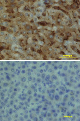 | EPCR in Human Liver. EPCR was detected in immersion fixed paraffin-embedded sections of human liver array using Goat Anti-Human EPCR Biotinylated Antigen Affinity-purified Polyclonal Antibody (Catalog # BAF2245) at 15 µg/mL overnight at 4 °C. Tissue was stained using the Anti-Goat HRP-DAB Cell & Tissue Staining Kit (brown; Catalog # CTS008) and counterstained with hematoxylin (blue). Lower panel shows a lack of labeling if primary antibodies are omitted and tissue is stained only with secondary antibody followed by incubation with detection reagents. View our protocol for Chromogenic IHC Staining of Paraffin-embedded Tissue Sections. |
カートに商品を
追加しました。
追加しました。
Related Product & Information
| Long Name | Endothelial Protein C Receptor |
|---|---|
| Background | EPCR |
| background_content | Background: EPCR Protein C is a vitamin K-dependent serine protease that plays a major role in blood coagulation. Binding of Protein C to EPCR leads to the proteolytic activation of PAR1 (protease-activated receptor 1) on endothelial cells and subsequent up-regulation of Protein C-induced genes. EPCR is a type I transmembrane glycoprotein in the CD1/MHC family. It is expressed most strongly in the endothelial cells of arteries and veins in heart and lung. Membrane bound EPCR is released by metalloproteolytic cleavage to generate the soluble receptor. The extracellular domain of human and mouse EPCR shares approximately 61% amino acid sequence homology. |
カートに商品を
追加しました。
追加しました。
Citations
R&D Systems personnel manually curate a database that contains references using R&D Systems products.
The data collected includes not only links to publications in PubMed,
but also provides information about sample types, species, and experimental conditions.
- Comprehensive analysis of conditioned media from ovarian cancer cell lines identifies novel candidate markers of epithelial ovarian cancer.
Authors: Gunawardana CG, Kuk C, Smith CR
J. Proteome Res., 2009;8(10):4705-13.
Species: Human
Sample Type: Cell Culture Supernates
Application: ELISA Development
カートに商品を
追加しました。
追加しました。
製品情報は掲載時点のものですが、価格表内の価格については随時最新のものに更新されます。お問い合わせいただくタイミングにより製品情報・価格などは変更されている場合があります。
表示価格に、消費税等は含まれていません。一部価格が予告なく変更される場合がありますので、あらかじめご了承下さい。