
抗B7-H3抗体(Anti-B7-H3, Human, Goat-Poly antibody)
掲載日情報:2018/11/26 現在Webページ番号:29586

B7-H3に対する抗体(Anti-B7-H3, Human, Goat-Poly )です。
※ 本製品は研究用です。研究用以外には使用できません。
追加しました。
価格
[在庫・価格 :2026年04月29日 00時00分現在]
| 詳細 | 商品名 |
|
文献数 | ||||||||||||||||||||||||||||||||||||||||||||||||||||||||||||||||||||||||||||||||||
|---|---|---|---|---|---|---|---|---|---|---|---|---|---|---|---|---|---|---|---|---|---|---|---|---|---|---|---|---|---|---|---|---|---|---|---|---|---|---|---|---|---|---|---|---|---|---|---|---|---|---|---|---|---|---|---|---|---|---|---|---|---|---|---|---|---|---|---|---|---|---|---|---|---|---|---|---|---|---|---|---|---|---|---|---|---|
|
Anti-B7-H3, Human, Goat-Poly |
|
22 | |||||||||||||||||||||||||||||||||||||||||||||||||||||||||||||||||||||||||||||||||||
|
|||||||||||||||||||||||||||||||||||||||||||||||||||||||||||||||||||||||||||||||||||||
|
Anti-Human B7-H3 Affinity Purified Polyclonal Ab |
|
16 | |||||||||||||||||||||||||||||||||||||||||||||||||||||||||||||||||||||||||||||||||||
|
|||||||||||||||||||||||||||||||||||||||||||||||||||||||||||||||||||||||||||||||||||||


[在庫・価格 :2026年04月29日 00時00分現在]
Anti-B7-H3, Human, Goat-Poly
文献数: 22
- 商品コード:AF1027
- メーカー:RSD
- 包装:100μg
- 価格:¥81,000
- 在庫:1個
- 納期:10日程度 ※※ 表示されている納期は弊社に在庫がなく、取り寄せた場合の目安納期となります。
- 法規制等:
| 説明文 | 別名:B7H3 Genbank No: 80381 Protein Accession No: NP_079516 |
||||||
|---|---|---|---|---|---|---|---|
| 別包装品 | 別包装品あり | ||||||
| 法規制等 | |||||||
| 保存条件 | -20℃ | 法規備考 | |||||
| 抗原種 | Human | 免疫動物 | Goat | ||||
| 交差性 | Human | 適用 | FCM,IHC,Simple Western,Western Blot | ||||
| 標識 | Unlabeled | 性状 | Antigen Affinity Purified | ||||
| 吸収処理 | クラス | IgG | |||||
| クロナリティ | Polyclonal | フォーマット | |||||
| 掲載カタログ |
|
||||||
| 製品記事 | 免疫染色システム ImmPRESS® Reagent Anti-Goat IgG |
||||||
| 関連記事 | |||||||
Anti-Human B7-H3 Affinity Purified Polyclonal Ab
文献数: 16
- 商品コード:AF1027-SP
- メーカー:RSD
- 包装:25μg
- 価格:¥28,000
- 在庫:1個
- 納期:2~3週間 ※※ 表示されている納期は弊社に在庫がなく、取り寄せた場合の目安納期となります。
- 法規制等:
| 説明文 | ※受注発注品。形状:溶液または凍結乾燥 別名:B7H3 Genbank No: 80381 Protein Accession No: NP_079516 |
||||||
|---|---|---|---|---|---|---|---|
| 別包装品 | 別包装品あり | ||||||
| 法規制等 | |||||||
| 保存条件 | -20℃ | 法規備考 | |||||
| 抗原種 | 免疫動物 | Goat | |||||
| 交差性 | Human | 適用 | FCM,IHC,Simple Western,Western Blot | ||||
| 標識 | Unlabeled | 性状 | Antigen Affinity Purified | ||||
| 吸収処理 | クラス | IgG | |||||
| クロナリティ | Polyclonal | フォーマット | |||||
| 掲載カタログ |
|
||||||
| 製品記事 | 免疫染色システム ImmPRESS® Reagent Anti-Goat IgG 使いっきり抗体 |
||||||
| 関連記事 | |||||||
追加しました。
Product Details
| Species Reactivity | Human |
|---|---|
| Label | Unconjugated |
| Immunogen | Mouse myeloma cell line NS0-derived recombinant human B7‑H3Leu29-Pro245Accession # NP_079516 |
| Source | Polyclonal Goat IgG |
| Purification | Antigen Affinity-purified |
| Specificity | Detects human B7-H3 in direct ELISAs and Western blots. In direct ELISAs, approximately 5% cross-reactivity with recombinant human (rh) B7‑H2 is observed, and less than 1% cross-reactivity with rhB7-1, rhB7-2, rhB7-H1, and rhPD-L2 is observed. |
追加しました。
Applications and Data
| Recommended Concentration | Sample | |
| Western Blot | 1 µg/mL | See below |
| Simple Western | 10 µg/mL | See below |
| Flow Cytometry | 0.25 µg/106 cells | See below |
| Immunohistochemistry | 5-15 µg/mL | See below |
| CyTOF-ready | Ready to be labeled using established conjugation methods. No BSA or other carrier proteins that could interfere with conjugation. | |
| Knockout Validated | B7‑H3 is specifically detected in U2OS human osteosarcoma parental cell line but is not detectable in B7‑H3 knockout U2OS cell line. | |
| Western Blot | |
|---|---|
 | Detection of Human B7‑H3 by Western Blot. Western blot shows lysates of LNCaP human prostate cancer cell line and U2OS human osteosarcoma cell line. PVDF membrane was probed with 1 µg/mL of Goat Anti-Human B7‑H3 Antigen Affinity-purified Polyclonal Antibody (Catalog # AF1027) followed by HRP-conjugated Anti-Goat IgG Secondary Antibody (Catalog # HAF017). A specific band was detected for B7‑H3 at approximately 90-110 kDa (as indicated). This experiment was conducted under reducing conditions and using Immunoblot Buffer Group 1. |
| Flow Cytometry | |
 | Detection of B7‑H3 in PC‑3 Human Cell Line by Flow Cytometry. PC-3 human prostate carcinoma cells were stained with Goat Anti-Human B7‑H3 Antigen Affinity‑purified Polyclonal Antibody (Catalog # AF1027, filled histogram) or control antibody (Catalog #AB‑108‑C, open histogram), followed by Phycoerythrin-conjugated Anti-Goat IgG Secondary Antibody (Catalog # F0107). |
| Immunohistochemistry | |
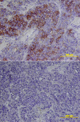 | B7-H3 in Human Melanoma. B7-H3 was detected in immersion fixed paraffin-embedded sections of human melanoma using Goat Anti-Human B7-H3 Antigen Affinity-purified Polyclonal Antibody (Catalog # AF1027) at 15 µg/mL overnight at 4 °C. Tissue was stained using the Anti-Goat HRP-DAB Cell & Tissue Staining Kit (brown; Catalog # CTS008) and counterstained with hematoxylin (blue). Lower panel shows a lack of labeling if primary antibodies are omitted and tissue is stained only with secondary antibody followed by incubation with detection reagents. View our protocol for Chromogenic IHC Staining of Paraffin-embedded Tissue Sections. |
| Simple Western | |
 | Detection of Human B7‑H3 by Simple WesternTM. Simple Western lane view shows lysates of U2OS human osteosarcoma cell line, loaded at 0.2 mg/mL. A specific band was detected for B7‑H3 at approximately120-160 kDa (as indicated) using 10 µg/mL of Goat Anti-Human B7‑H3 Antigen Affinity-purified Polyclonal Antibody (Catalog # AF1027) followed by 1:50 dilution of HRP-conjugated Anti-Goat IgG Secondary Antibody (Catalog # HAF109). This experiment was conducted under reducing conditions and using the 12-230 kDa separation system. |
| Knockout Validated | |
 | Western Blot Shows Human B7‑H3 Specificity by Using Knockout Cell Line. Western blot shows lysates of U2OS human osteosarcoma parental cell line and B7-H3 knockout U2OS cell line (KO). PVDF membrane was probed with 1 µg/mL of Goat Anti-Human B7‑H3 Antigen Affinity-purified Polyclonal Antibody (Catalog # AF1027) followed by HRP-conjugated Anti-Goat IgG Secondary Antibody (Catalog # HAF017). A specific band was detected for B7‑H3 at approximately 95 kDa (as indicated) in the parental U2OS cell line, but is not detectable in knockout U2OS cell line. GAPDH (Catalog # AF5718) is shown as a loading control. This experiment was conducted under reducing conditions and using Immunoblot Buffer Group 1. |
追加しました。
Related Product & Information
| Background | B7-H3 |
|---|---|
| background_content | Background: B7-H3 Human B7 homolog 3 (B7-H3) is a member of the B7 family of immune proteins that provide signals for regulating immune responses (1‑3). Other family members include B7-1, B7-2, B7-H2, PD-L1 (B7-H1), and PD-L2. B7 proteins are immunoglobulin (Ig) superfamily members with extracellular Ig-V-like and Ig-C-like domains and short cytoplasmic domains. Among the family members, they share about 20‑40% amino acid (aa) sequence identity. The cloned human B7-H3 cDNA encodes a 316 aa type I membrane precursor protein with a putative 28 aa signal peptide, a 217 aa extracellular region containing one V-like and one C-like Ig domain, a transmembrane region, and a 45 aa cytoplasmic domain. An isoform of human B7-H3 containing a four-Ig-like domain extracellular region has also been identified. Human B7-H3 is not expressed on resting B cells, T cells, monocytes or dendritic cells, but is induced on dendritic cells and monocytes by inflammatory cytokines. B7-H3 expression is also detected on various normal tissues and in some tumor cell lines. Human B7-H3 does not bind any known members of the CD28 family of immunoreceptors. However, B7-H3 has been shown to bind an unidentified counter-receptor on activated T cells to costimulate the proliferation of CD4+ or CD8+ T cells. B7-H3 has also been found to enhance the induction of primary cytotoxic T lymphocytes and stimulate IFN-gamma production (1‑3). |
追加しました。
Citations
- B7-H3 Promotes the Migration and Invasion of Human Bladder Cancer Cells via the PI3K/Akt/STAT3 Signaling Pathway
Authors: Y Li, G Guo, J Song, Z Cai, J Yang, Z Chen, Y Wang, Y Huang, Q Gao
J Cancer, 2017;8(5):816-824.
Species: Human
Sample Type: Whole Tissue
Application: IHC Paraffin-embedded - Spectroscopic Photoacoustic Molecular Imaging of Breast Cancer using a B7-H3-targeted ICG Contrast Agent
Authors: KE Wilson, SV Bachawal, L Abou-Elkac, K Jensen, S Machtaler, L Tian, JK Willmann
Theranostics, 2017;7(6):1463-1476.
Species: Human
Sample Type: Whole Tissue
Application: IHC - High constitutive B7-H3 expression on human keratinocytes supports T cell immunity
Authors: D Quandt, E Fiedler, A Müller, WC Marsch, B Seliger
J. Dermatol. Sci., 2017;0(0):.
Species: Human
Sample Type: Whole Tissue
Application: IHC - Preferential Induction of the T Cell Auxiliary Signaling Molecule B7-H3 on Synovial Monocytes in Rheumatoid Arthritis
Authors: BR Yoon, YH Chung, SJ Yoo, K Kawara, J Kim, IS Yoo, CG Park, SW Kang, WW Lee
J. Biol. Chem, 2016;291(8):4048-57.
Species: Human
Sample Type: Cell Lysates
Application: WB - Breast Cancer Detection by B7-H3-Targeted Ultrasound Molecular Imaging.
Authors: Bachawal S, Jensen K, Wilson K, Tian L, Lutz A, Willmann J
Cancer Res, 2015;75(12):2501-9.
Species: Human
Sample Type: Whole Tissue
Application: IHC Paraffin-embedded - Origination of new immunological functions in the costimulatory molecule B7-H3: the role of exon duplication in evolution of the immune system.
Authors: Sun J, Fu F, Gu W, Yan R, Zhang G, Shen Z, Zhou Y, Wang H, Shen B, Zhang X
PLoS ONE, 2011;6(9):e24751.
Species: Human
Sample Type: Cell Lysates
Application: WB - Tumor cell and tumor vasculature expression of B7-H3 predict survival in clear cell renal cell carcinoma.
Authors: Crispen PL, Sheinin Y, Roth TJ, Lohse CM, Kuntz SM, Frigola X, Thompson RH, Boorjian SA, Dong H, Leibovich BC, Blute ML, Kwon ED
Clin. Cancer Res., 2008;14(16):5150-7.
Species: Human
Sample Type: Whole Tissue
Application: IHC Paraffin-embedded - Anterior pituitary progenitor cells express costimulatory molecule 4Ig-B7-H3.
Authors: Nagai Y, Aso H, Ogasawara H, Tanaka S, Taketa Y, Watanabe K, Ohwada S, Rose MT, Kitazawa H, Yamaguchi T
J. Immunol., 2008;181(9):6073-81.
Species: Bovine
Sample Type: Cell Lysates
Application: WB - Interactions of T cells with fibroblast-like synoviocytes: role of the B7 family costimulatory ligand B7-H3.
Authors: Tran CN, Thacker SG, Louie DM, Oliver J, White PT, Endres JL, Urquhart AG, Chung KC, Fox DA
J. Immunol., 2008;180(5):2989-98.
Species: Human
Sample Type: Whole Cells
Application: ICC - 12-O-tetradecanoyl phorbol 13-acetate induces the expression of B7-DC, -H1, -H2, and -H3 in K562 cells.
Authors: Jang BC, Park YK, Choi IH, Kim SP, Hwang JB, Baek WK, Suh MH, Mun KC, Suh SI
Int. J. Oncol., 2007;31(6):1439-47.
Species: Human
Sample Type: Whole Cells
Application: Flow
追加しました。
製品情報は掲載時点のものですが、価格表内の価格については随時最新のものに更新されます。お問い合わせいただくタイミングにより製品情報・価格などは変更されている場合があります。
表示価格に、消費税等は含まれていません。一部価格が予告なく変更される場合がありますので、あらかじめご了承下さい。