抗B7-H1抗体(Anti-B7-H1, Mouse, Goat-Poly antibody)
掲載日情報:2018/11/26 現在Webページ番号:29580
B7-H1に対する抗体(Anti-B7-H1, Mouse, Goat-Poly )です。
※ 本製品は研究用です。研究用以外には使用できません。
追加しました。
-
2025/12/01
試薬 特別価格
[RSD]厳選 抗体&リコンビナントタンパク質 30%OFFキャンペーン[~2026/02/27]
期間:2025/12/01 ~2026/02/27
R&D Systems, Inc.(RSD)
価格
[在庫・価格 :2026年02月23日 00時00分現在]
| 詳細 | 商品名 |
|
文献数 | ||||||||||||||||||||||||||||||||||||||||||||||||||||||||||||||||||||||||||||||||||||||||||
|---|---|---|---|---|---|---|---|---|---|---|---|---|---|---|---|---|---|---|---|---|---|---|---|---|---|---|---|---|---|---|---|---|---|---|---|---|---|---|---|---|---|---|---|---|---|---|---|---|---|---|---|---|---|---|---|---|---|---|---|---|---|---|---|---|---|---|---|---|---|---|---|---|---|---|---|---|---|---|---|---|---|---|---|---|---|---|---|---|---|---|---|---|---|
|
Anti-B7-H1, Mouse, Goat-Poly |
|
20 | |||||||||||||||||||||||||||||||||||||||||||||||||||||||||||||||||||||||||||||||||||||||||||
|
|||||||||||||||||||||||||||||||||||||||||||||||||||||||||||||||||||||||||||||||||||||||||||||
|
Anti-Mouse B7-H1 Affinity Purified Polyclonal Ab |
|
18 | |||||||||||||||||||||||||||||||||||||||||||||||||||||||||||||||||||||||||||||||||||||||||||
|
|||||||||||||||||||||||||||||||||||||||||||||||||||||||||||||||||||||||||||||||||||||||||||||
[在庫・価格 :2026年02月23日 00時00分現在]
Anti-B7-H1, Mouse, Goat-Poly
文献数: 20
- 商品コード:AF1019
- メーカー:RSD
- 包装:100μg
-
価格:
¥81,000特別価格 ¥56,700 - 在庫:1個
- 納期:10日程度 ※※ 表示されている納期は弊社に在庫がなく、取り寄せた場合の目安納期となります。
- 法規制等:
| キャンペーン期間 |
2025/12/01 ~ 2026/02/27
[RSD]厳選 抗体&リコンビナントタンパク質 30%OFFキャンペーン[~2026/02/27] |
||||||
|---|---|---|---|---|---|---|---|
| 説明文 | 別名:B7-H,PD-L1,CD274 Genbank No: 29126 Protein Accession No: Q9EP73 |
||||||
| 別包装品 | 別包装品あり | ||||||
| 法規制等 | |||||||
| 保存条件 | -20℃ | 法規備考 | |||||
| 抗原種 | Mouse | 免疫動物 | Goat | ||||
| 交差性 | Mouse | 適用 | FCM,IHC,Western Blot | ||||
| 標識 | Unlabeled | 性状 | Antigen Affinity Purified | ||||
| 吸収処理 | クラス | IgG | |||||
| クロナリティ | Polyclonal | フォーマット | |||||
| 掲載カタログ |
|
||||||
| 製品記事 | 免疫染色システム ImmPRESS® Reagent Anti-Goat IgG R&D SystemsのB7-H1/PD-L1関連製品 |
||||||
| 関連記事 | |||||||
Anti-Mouse B7-H1 Affinity Purified Polyclonal Ab
文献数: 18
- 商品コード:AF1019-SP
- メーカー:RSD
- 包装:25μg
- 価格:¥28,000
- 在庫:無(未発注)
- 納期:2~3週間 ※※ 表示されている納期は弊社に在庫がなく、取り寄せた場合の目安納期となります。
- 法規制等:
| 説明文 | ※受注発注品。形状:溶液または凍結乾燥 別名:B7-H,PD-L1,CD274 Genbank No: 29126 Protein Accession No: Q9EP73 |
||||||
|---|---|---|---|---|---|---|---|
| 別包装品 | 別包装品あり | ||||||
| 法規制等 | |||||||
| 保存条件 | -20℃ | 法規備考 | |||||
| 抗原種 | 免疫動物 | Goat | |||||
| 交差性 | Mouse | 適用 | FCM,IHC,Western Blot | ||||
| 標識 | Unlabeled | 性状 | Antigen Affinity Purified | ||||
| 吸収処理 | クラス | IgG | |||||
| クロナリティ | Polyclonal | フォーマット | |||||
| 掲載カタログ |
|
||||||
| 製品記事 | 免疫染色システム ImmPRESS® Reagent Anti-Goat IgG 使いっきり抗体 |
||||||
| 関連記事 | |||||||
追加しました。
Product Details
| Species Reactivity | Mouse |
|---|---|
| Label | Unconjugated |
| Immunogen | Mouse myeloma cell line NS0-derived recombinant mouse PD-L1/B7-H1Phe19-Thr238Accession # Q9EP73 |
| Source | Polyclonal Goat IgG |
| Purification | Antigen Affinity-purified |
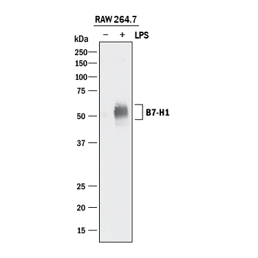
| Specificity | Detects mouse PD-L1/B7-H1 in direct ELISAs and Western blots. In direct ELISAs, less than 20% cross-reactivity with recombinant human PD-L1/B7-H1 is observed. |
追加しました。
Applications and Data
| Recommended Concentration | Sample | |
| Western Blot | 0.5 µg/mL | See below |
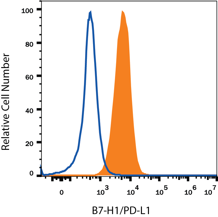
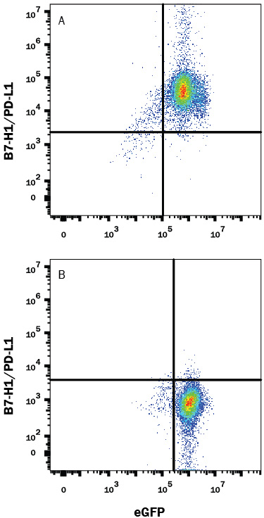
| Flow Cytometry | 0.25 µg/106 cells | See below |
| Immunohistochemistry | 5-15 µg/mL | Perfusion fixed frozen sections of mouse small intestine (Peyer's patch) and thymus |
| CyTOF-ready | Ready to be labeled using established conjugation methods. No BSA or other carrier proteins that could interfere with conjugation. | |
追加しました。
Related Product & Information
| Background | PD-L1/B7-H1 |
|---|---|
| background_content | Background: PD-L1/B7-H1 Mouse B7 homolog 1(B7-H1), also called programmed death ligand 1 (PD-L1) and programmed cell death 1 ligand 1 (PDCD1L1), is a member of the B7 family of proteins that provide signals for regulating T-cell activation and tolerance (1‑4). Other family members include B7-1, B7-2, B7-H2, B7-H3 and PD-L2. B7 proteins are immunoglobulin (Ig) superfamily members with extracellular Ig-V-like and Ig-C-like domains and a short cytoplasmic region. Among the family members, they share from 20‑40% amino acid (aa) sequence identity. The cloned mouse B7-H1/PD-L1 cDNA encodes a 290 aa type I membrane precursor protein with a putative 18 aa signal peptide, a 220 aa extracellular region containing one V-like and one C-like Ig domain, a 22 aa transmembrane region, and a 30 aa cytoplasmic domain. Mouse and human B7-H1/PD-L1 share approximately 70% aa sequence identity. B7-H1/PD-L1 is one of two ligands for programmed death-1 (PD-1), a member of the CD28 family of immunoreceptors. The other identified ligand is PD-L2. Mouse B7-H1/PD-L1 and PD-L2 share approximately 34% aa sequence identity and have similar functions. B7-H1/PD-L1 is constitutively expressed in various lymphoid and non-lymphoid organs including placenta, heart, pancreas, lung, liver, and endothelium (1‑4). The expression of B7-H1/PD-L1 is detected on B cells, T cells, monocytes, dendritic cells and thymic epithelial cells. IFN-gamma treatment induces B7‑H1/PD‑L1 expression in monocytes, dendritic cells, and endothelial cells. B7-H1/PD-L1 expression is also upregulated in a variety of tumor cell lines. On previously activated T cells, B7-H1/PD-L1 interaction with PD-1 inhibits TCR-mediated proliferation and cytokine production, suggesting an inhibitory role in regulating immune responses. In contrast, a costimulatory function for the PD-1 ligands on resting T cells has also been reported (1‑4). |
追加しました。
Citations
- Interferon-gamma drives programmed death-ligand 1 expression on islet ? cells to limit T cell function during autoimmune diabetes
Authors: KC Osum, AL Burrack, T Martinov, NL Sahli, JS Mitchell, CG Tucker, KE Pauken, K Papas, B Appakalai, JA Spanier, BT Fife
Sci Rep, 2018;8(1):8295.
Species: Mouse
Sample Type: Whole Tissue
Application: IHC - PD-L1 checkpoint inhibition and anti-CTLA-4 whole tumor cell vaccination counter adaptive immune resistance: A mouse neuroblastoma model that mimics human disease
Authors: P Srinivasan, X Wu, M Basu, C Rossi, AD Sandler
PLoS Med., 2018;15(1):e1002497.
Species: Mouse
Sample Type: Whole Tissue
Application: IHC-P - Dependence of Glomerulonephritis Induction on Novel Intraglomerular Alternatively Activated Bone Marrow-Derived Macrophages and Mac-1 and PD-L1 in Lupus-Prone NZM2328 Mice
Authors: SJ Sung, Y Ge, C Dai, H Wang, SM Fu, R Sharma, YS Hahn, J Yu, TH Le, MD Okusa, WK Bolton, JR Lawler
J. Immunol, 2017;0(0):.
Species: Mouse
Sample Type: Whole Cells
Application: Flow - Tumor localized secretion of soluble PD1 enhances oncolytic virotherapy
Authors: MY Bartee, KM Dunlap, E Bartee
Cancer Res, 2017;0(0):.
Species: Mouse
Sample Type: Cell Lysates
Application: WB - Hormonal vitamin D upregulates tissue-specific PD-L1 and PD-L2 surface glycoprotein expression in human but not mouse
Authors: V Dimitrov, M Bouttier, G Boukhaled, R Salehi-Tab, R Avramescu, B Memari, B Hasaj, GL Lukacs, CM Krawczyk, JH White
J. Biol. Chem., 2017;0(0):.
Species: Human
Sample Type: Cell Lysates
Application: WB - PD-1/PD-L1 blockade enhances T cell activity and antitumor efficacy of imatinib in gastrointestinal stromal tumors
Clin Cancer Res, 2016;0(0):.
Species: Mouse
Sample Type: Whole Tissue
Application: IHC - Paraffin embedded - Cell autonomous or systemic EGFR blockade alters the immune-environment in squamous cell carcinomas
Int J Cancer, 2016;0(0):.
Species: Mouse
Sample Type: Whole Tissue
Application: IHC - Paraffin embedded - beta-Cell-targeted blockage of PD1 and CTLA4 pathways prevents development of autoimmune diabetes and acute allogeneic islets rejection.
Authors: El Khatib M, Sakuma T, Tonne J, Mohamed M, Holditch S, Lu B, Kudva Y, Ikeda Y
Gene Ther, 2015;22(5):430-8.
Species: Human
Sample Type: Cell Lysates
Application: WB - IFN-gamma from lymphocytes induces PD-L1 expression and promotes progression of ovarian cancer.
Authors: Abiko K, Matsumura N, Hamanishi J, Horikawa N, Murakami R, Yamaguchi K, Yoshioka Y, Baba T, Konishi I, Mandai M
Br J Cancer, 2015;112(9):1501-9. - Endogenous retinoids in the pathogenesis of alopecia areata.
Authors: Duncan F, Silva K, Johnson C, King B, Szatkiewicz J, Kamdar S, Ong D, Napoli J, Wang J, King L, Whiting D, McElwee K, Sundberg J, Everts H
J Invest Dermatol, 2013;133(2):334-43.
Species: Mouse
Sample Type: Whole Tissue
Application: IHC - Not specified
追加しました。
製品情報は掲載時点のものですが、価格表内の価格については随時最新のものに更新されます。お問い合わせいただくタイミングにより製品情報・価格などは変更されている場合があります。
表示価格に、消費税等は含まれていません。一部価格が予告なく変更される場合がありますので、あらかじめご了承下さい。


