
抗Lipocalin-2抗体(Anti-Lipocalin-2, Human, Goat-Poly antibody)
掲載日情報:2018/11/26 現在Webページ番号:28999

Lipocalin-2に対する抗体(Anti-Lipocalin-2, Human, Goat-Poly )です。
※ 本製品は研究用です。研究用以外には使用できません。
追加しました。
価格
[在庫・価格 :2024年05月18日 08時15分現在]
| 詳細 | 商品名 |
|
文献数 | ||||||||||||||||||||||||||||||||||||||||||||||||||||||||||||||||||||||||||||||||||
|---|---|---|---|---|---|---|---|---|---|---|---|---|---|---|---|---|---|---|---|---|---|---|---|---|---|---|---|---|---|---|---|---|---|---|---|---|---|---|---|---|---|---|---|---|---|---|---|---|---|---|---|---|---|---|---|---|---|---|---|---|---|---|---|---|---|---|---|---|---|---|---|---|---|---|---|---|---|---|---|---|---|---|---|---|---|
|
Anti-Lipocalin-2, Human, Goat-Poly <Anti-NGAL> |
|
19 | |||||||||||||||||||||||||||||||||||||||||||||||||||||||||||||||||||||||||||||||||||
|
|||||||||||||||||||||||||||||||||||||||||||||||||||||||||||||||||||||||||||||||||||||
|
Anti-Human Lipocalin-2/NGAL Affinity Purified Polyclonal Ab |
|
4 | |||||||||||||||||||||||||||||||||||||||||||||||||||||||||||||||||||||||||||||||||||
|
|||||||||||||||||||||||||||||||||||||||||||||||||||||||||||||||||||||||||||||||||||||
[在庫・価格 :2024年05月18日 08時15分現在]
Anti-Lipocalin-2, Human, Goat-Poly <Anti-NGAL>
文献数: 19
- 商品コード:AF1757
- メーカー:RSD
- 包装:100μg
- 価格:¥106,000
- 在庫:無(未発注)
- 納期:10日程度 ※※ 表示されている納期は弊社に在庫がなく、取り寄せた場合の目安納期となります。
- 法規制等:
| 説明文 |
別名:24p3 Genbank No: 3934 Protein Accession No: P80188 |
||||||
|---|---|---|---|---|---|---|---|
| 別包装品 | 別包装品あり | ||||||
| 法規制等 | |||||||
| 保存条件 | -20℃ | 法規備考 | |||||
| 抗原種 | Human | 免疫動物 | Goat | ||||
| 交差性 | Mouse | 適用 | IHC,Simple Western,Western Blot | ||||
| 標識 | Unlabeled | 性状 | Antigen Affinity Purified | ||||
| 吸収処理 | クラス | IgG | |||||
| クロナリティ | Polyclonal | フォーマット | |||||
| 掲載カタログ |
|
||||||
| 製品記事 |
免疫染色システム ImmPRESS® Reagent Anti-Goat IgG |
||||||
| 関連記事 | |||||||
Anti-Human Lipocalin-2/NGAL Affinity Purified Polyclonal Ab
文献数: 4
- 商品コード:AF1757-SP
- メーカー:RSD
- 包装:25μg
- 価格:¥30,000
- 在庫:無(未発注)
- 納期:2~3週間 ※※ 表示されている納期は弊社に在庫がなく、取り寄せた場合の目安納期となります。
- 法規制等:
| 説明文 |
※受注発注品。形状:溶液または凍結乾燥 別名:24p3 Genbank No: 3934 Protein Accession No: P80188 |
||||||
|---|---|---|---|---|---|---|---|
| 別包装品 | 別包装品あり | ||||||
| 法規制等 | |||||||
| 保存条件 | -20℃ | 法規備考 | |||||
| 抗原種 | 免疫動物 | Goat | |||||
| 交差性 | Mouse | 適用 | IHC,Simple Western,Western Blot | ||||
| 標識 | Unlabeled | 性状 | Antigen Affinity Purified | ||||
| 吸収処理 | クラス | IgG | |||||
| クロナリティ | Polyclonal | フォーマット | |||||
| 掲載カタログ |
|
||||||
| 製品記事 |
免疫染色システム ImmPRESS® Reagent Anti-Goat IgG |
||||||
| 関連記事 | |||||||
追加しました。
Product Details
| Species Reactivity | Human, Mouse, Rat |
|---|---|
| Label | Unconjugated |
| Immunogen | Mouse myeloma cell line NS0-derived Recombinant Human Lipocalin‑2/NGALGln21-Gly198Accession # Q6FGL5 |
| Source | Polyclonal Goat IgG |
| Purification | Antigen Affinity-purified |
| Specificity | Detects human Lipocalin-2/NGAL in direct ELISAs and Western blots. |
追加しました。
Applications and Data
| Recommended Concentration | Sample | |
| Western Blot | 0.2 µg/mL | See below |
| Simple Western | 2 µg/mL | See below |
| Immunohistochemistry | 5-15 µg/mL | See below |
| Western Blot | |
|---|---|
 | Detection of Human Lipocalin‑2/NGAL by Western Blot. Western blot shows lysates of Capan‑1 human pancreatic adenocarcinoma cell line and human pancreas tissue. PVDF membrane was probed with 0.2 µg/mL of Goat Anti-Human/Mouse/Rat Lipocalin‑2/NGAL Antigen Affinity-purified Polyclonal Antibody (Catalog # AF1757) followed by HRP-conjugated Anti-Goat IgG Secondary Antibody (Catalog # HAF109). A specific band was detected for Lipocalin‑2/NGAL at approximately 22 kDa (as indicated). This experiment was conducted under reducing conditions and using Immunoblot Buffer Group 1. |
| Western Blot | |
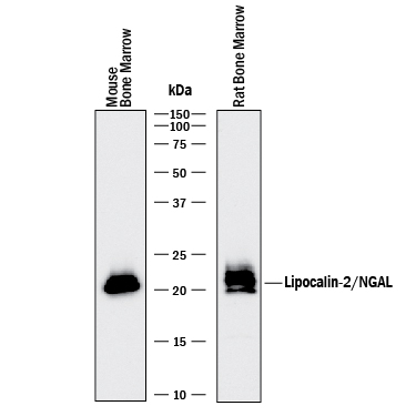 | Detection of Mouse and Rat Lipocalin‑2/NGAL by Western Blot. Western blot shows lysates of mouse and rat bone marrow. PVDF membrane was probed with 0.2 µg/mL of Goat Anti-Human/Mouse/Rat Lipocalin‑2/NGAL Antigen Affinity-purified Polyclonal Antibody (Catalog # AF1757) followed by HRP-conjugated Anti-Goat IgG Secondary Antibody (Catalog # HAF017). A specific band was detected for Lipocalin‑2/NGAL at approximately 22 kDa (as indicated). This experiment was conducted under reducing conditions and using Immunoblot Buffer Group 1. |
| Immunohistochemistry | |
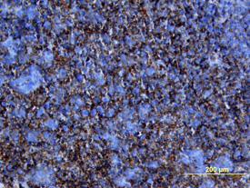 | Lipocalin‑2/NGAL in Human Pancreatic Cancer Tissue. Lipocalin‑2/NGAL was detected in immersion fixed paraffin-embedded sections of human pancreatic cancer tissue using Goat Anti-Human/Mouse/Rat Lipocalin‑2/NGAL Antigen Affinity-purified Polyclonal Antibody (Catalog # AF1757) at 15 µg/mL overnight at 4 °C. Tissue was stained using the Anti-Goat HRP-DAB Cell & Tissue Staining Kit (brown; Catalog # CTS008) and counterstained with hematoxylin (blue). View our protocol for Chromogenic IHC Staining of Paraffin-embedded Tissue Sections. |
| Immunohistochemistry | |
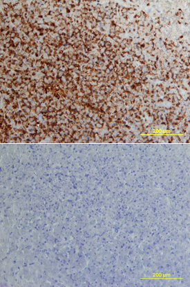 | Lipocalin‑2/NGAL in Human Pancreatic Cancer Tissue. Lipocalin‑2/NGAL was detected in immersion fixed paraffin-embedded sections of human pancreatic cancer tissue using Goat Anti-Human/Mouse/Rat Lipocalin‑2/NGAL Antigen Affinity-purified Polyclonal Antibody (Catalog # AF1757) at 15 µg/mL overnight at 4 °C. Tissue was stained using the Anti-Goat HRP-DAB Cell & Tissue Staining Kit (brown; Catalog # CTS008) and counterstained with hematoxylin (blue). Lower panel shows a lack of labeling if primary antibodies are omitted and tissue is stained only with secondary antibody followed by incubation with detection reagents. View our protocol for Chromogenic IHC Staining of Paraffin-embedded Tissue Sections. |
| Simple Western | |
 | Detection of Human Lipocalin‑2/NGAL by Simple WesternTM. Simple Western lane view shows lysates of Capan‑1 human pancreatic adenocarcinoma cell line, loaded at 0.2 mg/mL. A specific band was detected for Lipocalin‑2/NGAL at approximately 34 kDa (as indicated) using 2 µg/mL of Goat Anti-Human/Mouse/Rat Lipocalin‑2/NGAL Antigen Affinity-purified Polyclonal Antibody (Catalog # AF1757) followed by 1:50 dilution of HRP-conjugated Anti-Goat IgG Secondary Antibody (Catalog # HAF109). This experiment was conducted under reducing conditions and using the 12-230 kDa separation system. |
追加しました。
Related Product & Information
| Long Name | Neutrophil Gelatinase-associated Lipocalin |
|---|---|
| Background | Lipocalin-2/NGAL |
| background_content | Background: Lipocalin-2/NGAL Members of Lipocalin family share a highly conserved fold with an eight-stranded antiparallel beta barrel, and act as a transporters, carrying small molecules to specific cells. Lipocalin-2, also known as Neutrophil Gelatinase-Associated Lipocalin (NGAL), was originally identified as a component of neutrophil granules. It is a 25 kDa protein existing in monomeric and homo- and heterodimeric forms, the latter as a dimer with human neutrophil gelatinases (MMP-9). Its expression has been observed in most tissues normally exposed to microorganism, and its synthesis is induced in epithelial cells during inflammation. Lipocalin-2 has been implicated in a variety of processes including cell differentiation, tumorigenesis, and apoptosis. Studies indicate that Lipocalin-2 binds a bacterial catecholate sidropore bound to ferric ion such as enterobactin with a subnanomolar dissociation constant (Kd = 0.41 nM). The bound ferric enterobactin complex breaks down slowly in a month into dihydroxybenzoyl serine and dihydroxybenzoic acid (DHBA). It also binds to a ferric DHBA complex with much less Kd values (7.9 nM). Secretion of Lipocalin‑2 in immune cells increases by stimulation of Toll-like receptor as an acute phase response to infection. As a result, it acts as a potent bacteriostatic reagent by sequestering iron. Moreover, Lipocalin-2 can alter the invasive and metastatic behavior of Ras-transformed breast cancer cells in vitro and in vivo by reversing epithelial to mesenchymal transition inducing activity of Ras, through restoration of E-cadherin expression, via effects on the Ras-MAPK signaling pathway. |
追加しました。
Citations
- Cilostazol attenuates kainic acid-induced hippocampal cell death
Authors: YS Park, Z Jin, EA Jeong, CO Yi, JY Lee, IS Park, GS Roh
Korean J. Physiol. Pharmacol., 2018;22(1):63-70.
Species: Mouse
Sample Type: Tissue Lysates
Application: WB - Unexpected kidney-restricted role for IL-17 receptor signaling in defense against systemic Candida albicans infection
Authors: K Ramani, CV Jawale, AH Verma, BM Coleman, JK Kolls, PS Biswas
JCI Insight, 2018;3(9):.
Species: Mouse
Sample Type: Whole Tissue
Application: IHC-P - Metformin alleviates nickel-induced autophagy and apoptosis via inhibition of hexokinase-2, activating lipocalin-2, in human bronchial epithelial cells
Authors: YT Kang, WC Hsu, CH Wu, IL Hsin, PR Wu, KT Yeh, JL Ko
Oncotarget, 2017;8(62):105536-105552.
Species: Human
Sample Type: Cell Lysates
Application: WB - The adipokine lipocalin-2 in the context of the osteoarthritic osteochondral junction
Sci Rep, 2016;6(0):29243.
Species: Human
Sample Type: Cell Culture Supernates
Application: WB - Expression of uterine lipocalin 2 and its receptor during early- to mid-pregnancy period in mares
Authors: Shingo Haneda
J. Reprod. Dev, 2016;0(0):.
Species: Equine
Sample Type: Luminal Fluid
Application: WBy - Lipocalin2 promotes invasion, tumorigenicity and gemcitabine resistance in pancreatic ductal adenocarcinoma.
Authors: Leung, Lisa, Radulovich, Nikolina, Zhu, Chang-Qi, Organ, Shawna, Bandarchi, Bizhan, Pintilie, Melania, To, Christin, Panchal, Devang, Tsao, Ming Sou
PLoS ONE, 2012;7(10):e46677.
Species: Human
Sample Type: Cell Lysates
Application: WB - Cellular settings mediating Src Substrate switching between focal adhesion kinase tyrosine 861 and CUB-domain-containing protein 1 (CDCP1) tyrosine 734.
Authors: Wortmann A, He Y, Christensen ME, Linn M, Lumley JW, Pollock PM, Waterhouse NJ, Hooper JD
J. Biol. Chem., 2011;286(49):42303-15.
Species: Human
Sample Type: Cell Lysates
Application: WB - Urinary neutrophil gelatinase-associated lipocalin levels reflect damage to glomeruli, proximal tubules, and distal nephrons.
Authors: Kuwabara T, Mori K, Mukoyama M, Kasahara M, Yokoi H, Saito Y, Yoshioka T, Ogawa Y, Imamaki H, Kusakabe T, Ebihara K, Omata M, Satoh N, Sugawara A, Barasch J, Nakao K
Kidney Int., 2009;75(3):285-94.
Species: Human
Sample Type: Whole Tissue
Application: IHC Frozen
追加しました。
製品情報は掲載時点のものですが、価格表内の価格については随時最新のものに更新されます。お問い合わせいただくタイミングにより製品情報・価格などは変更されている場合があります。
表示価格に、消費税等は含まれていません。一部価格が予告なく変更される場合がありますので、あらかじめご了承下さい。