
抗E-Cadherin抗体(Anti-E-Cadherin, Human, Goat-Poly antibody)
掲載日情報:2018/11/26 現在Webページ番号:27187

E-Cadherinに対する抗体(Anti-E-Cadherin, Human, Goat-Poly )です。
※ 本製品は研究用です。研究用以外には使用できません。
追加しました。
価格
[在庫・価格 :2026年05月02日 20時55分現在]
| 詳細 | 商品名 |
|
文献数 | ||||||||||||||||||||||||||||||||||||||||||||||||||||||||||||||||||||||||||||||||||
|---|---|---|---|---|---|---|---|---|---|---|---|---|---|---|---|---|---|---|---|---|---|---|---|---|---|---|---|---|---|---|---|---|---|---|---|---|---|---|---|---|---|---|---|---|---|---|---|---|---|---|---|---|---|---|---|---|---|---|---|---|---|---|---|---|---|---|---|---|---|---|---|---|---|---|---|---|---|---|---|---|---|---|---|---|---|
|
Anti-E-Cadherin, Human, Goat-Poly |
|
29 | |||||||||||||||||||||||||||||||||||||||||||||||||||||||||||||||||||||||||||||||||||
|
|||||||||||||||||||||||||||||||||||||||||||||||||||||||||||||||||||||||||||||||||||||
|
Anti-Human E-Cadherin Affinity Purified Polyclonal Ab |
|
24 | |||||||||||||||||||||||||||||||||||||||||||||||||||||||||||||||||||||||||||||||||||
|
|||||||||||||||||||||||||||||||||||||||||||||||||||||||||||||||||||||||||||||||||||||


[在庫・価格 :2026年05月02日 20時55分現在]
Anti-E-Cadherin, Human, Goat-Poly
文献数: 29
- 商品コード:AF648
- メーカー:RSD
- 包装:100μg
- 価格:¥89,000
- 在庫:無(未発注)
- 納期:10日程度 ※※ 表示されている納期は弊社に在庫がなく、取り寄せた場合の目安納期となります。
- 法規制等:
| 説明文 | 別名:Arc-1 Genbank No: 999 Protein Accession No: P12830 |
||||||
|---|---|---|---|---|---|---|---|
| 別包装品 | 別包装品あり | ||||||
| 法規制等 | |||||||
| 保存条件 | -20℃ | 法規備考 | |||||
| 抗原種 | Human | 免疫動物 | Goat | ||||
| 交差性 | Human | 適用 | FCM,IC,IHC,Simple Western,Western Blot | ||||
| 標識 | Unlabeled | 性状 | Antigen Affinity Purified | ||||
| 吸収処理 | クラス | IgG | |||||
| クロナリティ | Polyclonal | フォーマット | |||||
| 掲載カタログ |
|
||||||
| 製品記事 | 抗幹細胞マーカー抗体/抗胚性幹細胞マーカー抗体 免疫染色システム ImmPRESS® Reagent Anti-Goat IgG |
||||||
| 関連記事 | |||||||
Anti-Human E-Cadherin Affinity Purified Polyclonal Ab
文献数: 24
- 商品コード:AF648-SP
- メーカー:RSD
- 包装:25μg
- 価格:¥30,000
- 在庫:無(未発注)
- 納期:2~3週間 ※※ 表示されている納期は弊社に在庫がなく、取り寄せた場合の目安納期となります。
- 法規制等:
| 説明文 | ※受注発注品。形状:溶液または凍結乾燥 別名:Arc-1 Genbank No: 999 Protein Accession No: P12830 |
||||||
|---|---|---|---|---|---|---|---|
| 別包装品 | 別包装品あり | ||||||
| 法規制等 | |||||||
| 保存条件 | -20℃ | 法規備考 | |||||
| 抗原種 | 免疫動物 | Goat | |||||
| 交差性 | Human | 適用 | FCM,IC,IHC,Simple Western,Western Blot | ||||
| 標識 | Unlabeled | 性状 | Antigen Affinity Purified | ||||
| 吸収処理 | クラス | IgG | |||||
| クロナリティ | Polyclonal | フォーマット | |||||
| 掲載カタログ |
|
||||||
| 製品記事 | 免疫染色システム ImmPRESS® Reagent Anti-Goat IgG 使いっきり抗体 |
||||||
| 関連記事 | |||||||
追加しました。
Product Details
| Species Reactivity | Human, Mouse |
|---|---|
| Label | Unconjugated |
| Immunogen | Mouse myeloma cell line NS0-derived recombinant human E-CadherinAsp155-Ile707Accession # P12830 |
| Source | Polyclonal Goat IgG |
| Purification | Antigen Affinity-purified |
| Specificity | Detects human E-Cadherin in direct ELISAs and Western blots. |
追加しました。
Applications and Data
| Recommended Concentration | Sample | |
| Western Blot | 0.5 µg/mL | See below |
| Simple Western | 5 µg/mL | See below |
| Flow Cytometry | 0.25 µg/106 cells | See below |
| Immunohistochemistry | 0.3-15 µg/mL | See below |
| CyTOF-ready | Ready to be labeled using established conjugation methods. No BSA or other carrier proteins that could interfere with conjugation. | |
| Immunocytochemistry | 5-15 µg/mL | See below |
| Western Blot | |
|---|---|
 | Detection of Human and MouseE‑Cadherin by Western Blot. Western blot shows lysates of A431 human epithelial carcinoma cell line, A549 human lung carcinoma cell line, HepG2 human hepatocellular carcinoma cell line, P19 mouse embryonal carcinoma cell line, and 4T1 mouse breast cancer cell line. PVDF membrane was probed with 0.5 µg/mL of Goat Anti-Human/Mouse E‑Cadherin Antigen Affinity-purified Polyclonal Antibody (Catalog # AF648) followed by HRP-conjugated Anti-Goat IgG Secondary Antibody (Catalog # HAF017). A specific band was detected for E‑Cadherin at approximately 110 kDa (as indicated). This experiment was conducted under reducing conditions and using Immunoblot Buffer Group 1. |
| Flow Cytometry | |
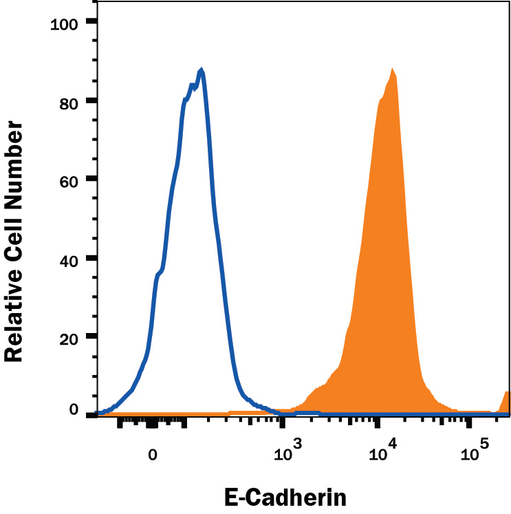 | Detection of E‑Cadherin in MCF‑7 Human Cell Line by Flow Cytometry. MCF‑7 human breast cancer cell line was stained with Goat Anti-Human/Mouse E‑Cadherin Antigen Affinity-purified Polyclonal Antibody (Catalog # AF648, filled histogram) or isotype control antibody (Catalog # AB-108-C, open histogram), followed by Allophycocyanin-conjugated Anti-Goat IgG Secondary Antibody (Catalog # F0108). View our protocol for Staining Membrane-associated Proteins. |
| Immunocytochemistry | |
 | E‑Cadherin in Human Epidermoid Carcinoma Cells. E‑Cadherin was detected in immersion fixed human epidermoid carcinoma cells using 10 µg/mL Goat Anti-Human/Mouse E‑Cadherin Antigen Affinity-purified Polyclonal Antibody (Catalog # AF648) for 3 hours at room temperature. Cells were stained with the NorthernLights™ 557-conjugated Anti-Goat IgG Secondary Antibody (red; Catalog # NL001) and counterstained with DAPI (blue). View our protocol for Fluorescent ICC Staining of Cells on Coverslips. |
| Immunocytochemistry | |
 | E‑Cadherin and SOX2 in BG01V Human Stem Cells. E‑Cadherin and SOX2 were detected in BG01V human embryonic stem cells using 10 µg/mL Goat Anti-Human/Mouse E‑Cadherin Antigen Affinity-purified Polyclonal Antibody (Catalog # AF648) and 10 µg/mL Human/Mouse SOX2 Monoclonal Antibody (Catalog # MAB2018). Cells were incubated with primary antibodies for 3 hours at room temperature. Cells were stained for E‑Cadherin using the NorthernLights™ 557-conjugated Anti-Goat IgG Secondary Antibody (green; Catalog # NL001) and for SOX2 using the NorthernLights 493-conjugated Anti-Mouse Secondary Antibody (red; Catalog # NL009). Cells were counterstained with DAPI (blue). View our protocol for Fluorescent ICC Staining of Cells on Coverslips. |
| Immunohistochemistry | |
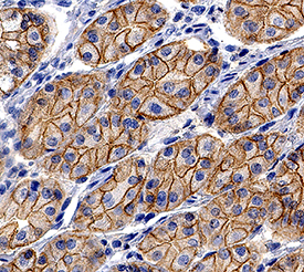 | E‑Cadherin in Human Stomach. E‑Cadherin was detected in immersion fixed paraffin-embedded sections of human stomach using Goat Anti-Human/Mouse E‑Cadherin Antigen Affinity-purified Polyclonal Antibody (Catalog # AF648) at 0.3 µg/mL for 1 hour at room temperature followed by incubation with the Anti-Goat IgG VisUCyte™ HRP Polymer Antibody (Catalog # VC004). Tissue was stained using DAB (brown) and counterstained with hematoxylin (blue). Specific staining was localized to cell membrane and cytoplasm in gastric glands. View our protocol for IHC Staining with VisUCyte HRP Polymer Detection Reagents. |
| Simple Western | |
 | Detection of Human and Mouse E‑Cadherin by Simple WesternTM. Simple Western lane view shows lysates of 4T1 mouse breast cancer cell line, P19 mouse embryonal carcinoma cell line, A431 human epithelial carcinoma cell line, and MCF‑7 human breast cancer cell line, loaded at 0.2 mg/mL. A specific band was detected for E‑Cadherin at approximately 128 kDa (as indicated) using 5 µg/mL of Goat Anti-Human/Mouse E‑Cadherin Antigen Affinity-purified Polyclonal Antibody (Catalog # AF648) followed by 1:50 dilution of HRP-conjugated Anti-Goat IgG Secondary Antibody (Catalog # HAF017). This experiment was conducted under reducing conditions and using the 12-230 kDa separation system. |
追加しました。
Related Product & Information
| Background | E-Cadherin |
|---|---|
| background_content | Background: E-Cadherin Epithelial (E)‑Cadherin (ECAD), also known as cell-CAM120/80 in the human, uvomorulin in the mouse, Arc-1 in the dog, and L-CAM in the chicken, is a member of the Cadherin family of cell adhesion molecules. Cadherins are calcium-dependent transmembrane proteins which bind to one another in a homophilic manner. On their cytoplasmic side, they associate with the three catenins, alpha, beta, and gamma (plakoglobin). This association links the cadherin protein to the cytoskeleton. Without association with the catenins, the cadherins are non-adhesive. Cadherins play a role in development, specifically in tissue formation. They may also help to maintain tissue architecture in the adult. E‑Cadherin may also play a role in tumor development, as loss of E‑Cadherin has been associated with tumor invasiveness. E‑Cadherin is a classical cadherin molecule. Classical cadherins consist of a large extracellular domain which contains DXD and DXNDN repeats responsible for mediating calcium‑dependent adhesion, a single-pass transmembrane domain, and a short carboxy-terminal cytoplasmic domain responsible for interacting with the catenins. E‑Cadherin contains five extracellular calcium‑binding domains of approximately 110 amino acids each. |
追加しました。
Citations
- Placenta-specific protein 1 promotes cell proliferation and invasion in non-small cell lung cancer
Authors: L Yang, TQ Zha, X He, L Chen, Q Zhu, WB Wu, FQ Nie, Q Wang, CS Zang, ML Zhang, J He, W Li, W Jiang, KH Lu
Oncol. Rep., 2018;39(1):53-60.
Species: Human
Sample Type: Cell Lysates
Application: WB - Wnt/?-catenin promotes gastric fundus specification in mice and humans
Authors: KW McCracken, E Aihara, B Martin, CM Crawford, T Broda, J Treguier, X Zhang, JM Shannon, MH Montrose, JM Wells
Nature, 2017;0(0):.
Species: Mouse
Sample Type: Whole Tissue
Application: IHC Paraffin-embedded - Novel role for IL-22 in protection during chronic Mycobacterium tuberculosis HN878 infection
Authors: P Treerat, O Prince, A Cruz-Lagun, M Muñoz-Torr, MA Salazar-Le, M Selman, B Fallert-Ju, TA Reinhardt, JF Alcorn, D Kaushal, J Zuñiga, J Rangel-Mor, JK Kolls, SA Khader
Mucosal Immunol, 2017;0(0):.
Species: Mouse
Sample Type: Whole Tissue
Application: IHC - miR-151a induces partial EMT by regulating E-cadherin in NSCLC cells
Authors: I Daugaard, KJ Sanders, A Idica, K Vittayaruk, M Hamdorf, JD Krog, R Chow, D Jury, LL Hansen, H Hager, P Lamy, CL Choi, D Agalliu, DG Zisoulis, IM Pedersen
Oncogenesis, 2017;6(7):e366.
Species: Human
Sample Type: Whole Cells
Application: ICC - Surface Topography Guides Morphology and Spatial Patterning of Induced Pluripotent Stem Cell Colonies
Authors: G Abagnale, A Sechi, M Steger, Q Zhou, CC Kuo, G Aydin, C Schalla, G Müller-New, M Zenke, IG Costa, P van Rijn, A Gillner, W Wagner
Stem Cell Reports, 2017;9(2):654-666.
Species: Human
Sample Type: Whole Cells
Application: ICC - Directed Differentiation of Human Induced Pluripotent Stem Cells into Fallopian Tube Epithelium
Authors: N Yucer, M Holzapfel, T Jenkins Vo, L Lenaeus, L Ornelas, A Laury, D Sareen, R Barrett, BY Karlan, CN Svendsen
Sci Rep, 2017;7(1):10741.
Species: Human
Sample Type: Whole Cells
Application: ICC - Canine spontaneous head and neck squamous cell carcinomas represent their human counterparts at the molecular level.
Authors: Liu D, Xiong H, Ellis A, Northrup N, Dobbin K, Shin D, Zhao S
PLoS Genet, 2015;11(6):e1005277.
Species: Canine
Sample Type: Whole Cells
Application: IHC Paraffin-embedded - Molecular homology and difference between spontaneous canine mammary cancer and human breast cancer.
Authors: Liu D, Xiong H, Ellis A, Northrup N, Rodriguez C, O'Regan R, Dalton S, Zhao S
Cancer Res, 2014;74(18):5045-56.
Species: Canine
Sample Type: Whole Tissue
Application: IHC Paraffin-embedded - Penton-dodecahedral particles trigger opening of intercellular junctions and facilitate viral spread during adenovirus serotype 3 infection of epithelial cells.
Authors: Lu Z, Wang H, Zhang Y, Cao H, Li Z, Fender P, Lieber A
PLoS Pathog, 2013;9(10):e1003718.
Species: Human
Sample Type: Whole Cells
Application: ICC - Interleukin-15 plays a central role in human kidney physiology and cancer through the gamma c signaling pathway.
Authors: Giron-Michel J, Azzi S, Khawam K, Mortier E, Caignard A, Devocelle A, Ferrini S, Croce M, Francois H, Lecru L, Charpentier B, Chouaib S, Azzarone B, Eid P
PLoS ONE, 2012;7(2):e31624.
Species: Human
Sample Type: Whole Cells
Application: ICC
追加しました。
製品情報は掲載時点のものですが、価格表内の価格については随時最新のものに更新されます。お問い合わせいただくタイミングにより製品情報・価格などは変更されている場合があります。
表示価格に、消費税等は含まれていません。一部価格が予告なく変更される場合がありますので、あらかじめご了承下さい。