
抗PD-1 (PDCD1), Mouse-Mono(UMAB199) UltraMAB抗体 | Anti-PD-1 (PDCD1), Mouse-Mono(UMAB199) UltraMAB Antibody
掲載日情報:2015/11/06 現在Webページ番号:178056

OriGene社の抗PD-1 (PDCD1), Mouse-Mono(UMAB199) UltraMAB抗体です。
本製品は、UltraMAB抗体シリーズ(三次元構造を有する組換え体ヒトタンパク質の全長を抗原として作製したモノクローナル抗体)です。
※本製品は研究用です。研究用以外には使用できません。
追加しました。
価格
[在庫・価格 :2026年07月19日 00時00分現在]
| 詳細 | 商品名 |
|
文献数 | ||||||||||||||||||||||||||||||||||||||||||||||||||||||||||||||||||||||||||
|---|---|---|---|---|---|---|---|---|---|---|---|---|---|---|---|---|---|---|---|---|---|---|---|---|---|---|---|---|---|---|---|---|---|---|---|---|---|---|---|---|---|---|---|---|---|---|---|---|---|---|---|---|---|---|---|---|---|---|---|---|---|---|---|---|---|---|---|---|---|---|---|---|---|---|---|---|---|
|
Anti-PD-1 (PDCD1), Mouse-Mono(UMAB199), UltraMAB |
|
2 | |||||||||||||||||||||||||||||||||||||||||||||||||||||||||||||||||||||||||||
|
|||||||||||||||||||||||||||||||||||||||||||||||||||||||||||||||||||||||||||||


[在庫・価格 :2026年07月19日 00時00分現在]
Anti-PD-1 (PDCD1), Mouse-Mono(UMAB199), UltraMAB
文献数: 2
- 商品コード:UM800091
- メーカー:ORI
- 包装:100μl
- 価格:¥204,000
- 在庫:無(未発注)
- 納期:3~4週間 ※※ 表示されている納期は弊社に在庫がなく、取り寄せた場合の目安納期となります。
- 法規制等:
| 説明文 | クローン:UMAB199 Genbank No: 5133 Gene Accession No: NM_005018 Protein Accession No: Q15116 |
||
|---|---|---|---|
| 法規制等 | |||
| 保存条件 | -20℃ | 法規備考 | |
| 抗原種 | 免疫動物 | Mouse | |
| 交差性 | Dog/Human/Mouse/Rat | 適用 | ChIP,IF,IHC,Western Blot |
| 標識 | Unlabeled | 性状 | Affinity Purified |
| 吸収処理 | クラス | IgG | |
| クロナリティ | Monoclonal | フォーマット | |
| 掲載カタログ |
ニュース2017年11月1日号 p.3
|
||
| 製品記事 | 抗PD-1 (PDCD1) UltraMAB抗体 UltraMAB抗体 |
||
| 関連記事 | |||
追加しました。
UltraMAB抗体について

OriGene社製品の中でも、特にUltraMAB抗体シリーズは、ヒト組織の免疫染色(IHC)に適した非常に特異性の高いモノクローナル抗体です。抗原にはTrueMAB抗体と同様、組換え体ヒト全長タンパク質を使用しています。1万種類を超えるタンパク質をスポットしたマイクロアレイを用い、標的タンパク質以外の分子との交差反応がないことを確認済みの、癌研究・病理学・解剖学向けの製品です。
追加しました。
Product Data
| Immunogen | Full length human recombinant protein of human PDCD1 (NP_005009) produced in HEK293T cell. | ||
|---|---|---|---|
| Clone Name | Clone UMAB199 | Isotype | IgG2a |
| Species Reactivity | Human | Concentration | 0.5~1.0 mg/ml (Lot Dependent) |
| Guaranteed Application | IHC, 10K-CHIP | Suggested Dilutions | IHC 1:1000 |
| Buffer | PBS (PH 7.3) containing 1% BSA, 50% glycerol and 0.02% sodium azide. | ||
| Purification | Purified from cell culture supernatant by affinity chromatography | ||
追加しました。
Reference Data
| Target Name | Homo sapiens programmed cell death 1 (PDCD1) | ||
|---|---|---|---|
| Alternative Name | CD279; hPD-1; hPD-l; hSLE1; PD-1; PD1; SLEB2 | ||
| Database Link | NP_005009 | ||
| Function | This gene encodes a cell surface membrane protein of the immunoglobulin superfamily. This protein is expressed in pro-B-cells and is thought to play a role in their differentiation. In mice, expression of this gene is induced in the thymus when anti-CD3 antibodies are injected and large numbers of thymocytes undergo apoptosis. Mice deficient for this gene bred on a BALB/c background developed dilated cardiomyopathy and died from congestive heart failure. These studies suggest that this gene product may also be important in T cell function and contribute to the prevention of autoimmune diseases. [provided by RefSeq, Jul | ||
| Related Pathway | |||
追加しました。
Image
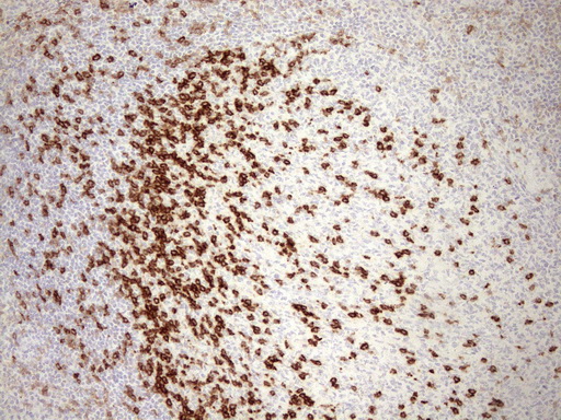
| Immunohistochemical staining of paraffin-embedded Human tonsil using anti-PDCD1 mouse monoclonal antibody.(Heat-induced epitope retrieval by 1mM EDTA in 10mM Tris buffer (pH8.5) at 110C for 10 min, UM800091)(1:1200) | |
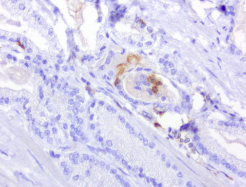
| Immunohistochemical staining of 3 paraffin-embedded human lung carcinomas using anti-PD1 clone UMAB199 mouse monoclonal antibody at 1:800 requires HIER Accel [OriGene/GBI Labs B22-125] in Pressure Chamber for 3 minute on high. Detection of primary antibody was achieved with Polink2 Broad HRP DAB [OriGene/GBI Labs D22]. | |
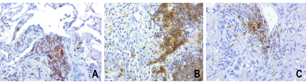
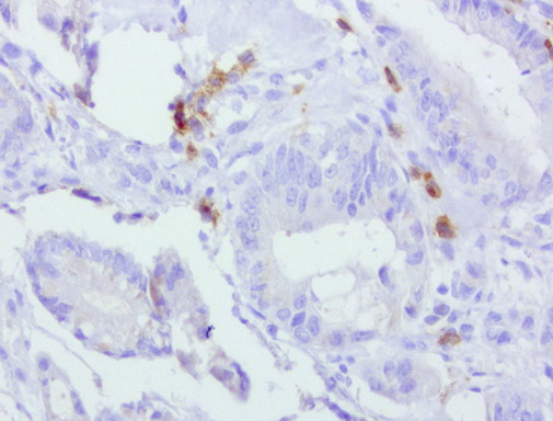
| Immunohistochemical staining of paraffin-embedded human melanoma using anti-PD-1 clone UMAB199 mouse monoclonal antibody at 1:800 dilution of 1mg/mL and detection with Polink2 Broad HRP DAB. UM800091 requires heat-induced epitope retrieval with Accel for 3minutes at110C in pressure chamber . The composite image of 3 melanoma shows the tumor cells are negative for PD-1 however the activated TCells show strong membranous and cytoplasmic staining. | |
| Immunohistochemical staining of paraffin-embedded human ovarian carcinoma using anti-PD-1 clone UMAB199 mouse monoclonal antibody at 1:800 dilution of 1mg/mL and detection with Polink2 Broad HRP DAB. UM800091 requires heat-induced epitope retrieval with Accel for 3minutes at110C in pressure chamber . The image shows the tumor cells are negative for PD-1 however the activated TCells show strong membranous and cytoplasmic staining. | |
| Immunohistochemical staining of paraffin-embedded human prostate carcinoma using anti-PD-1 clone UMAB199 mouse monoclonal antibody at 1:800 dilution of 1mg/mL and detection with Polink2 Broad HRP DAB. UM800091 requires heat-induced epitope retrieval with Accel for 3minutes at110C in pressure chamber . The image shows the tumor cells are negative for PD-1 however the activated TCells show strong membranous and cytoplasmic staining. | |
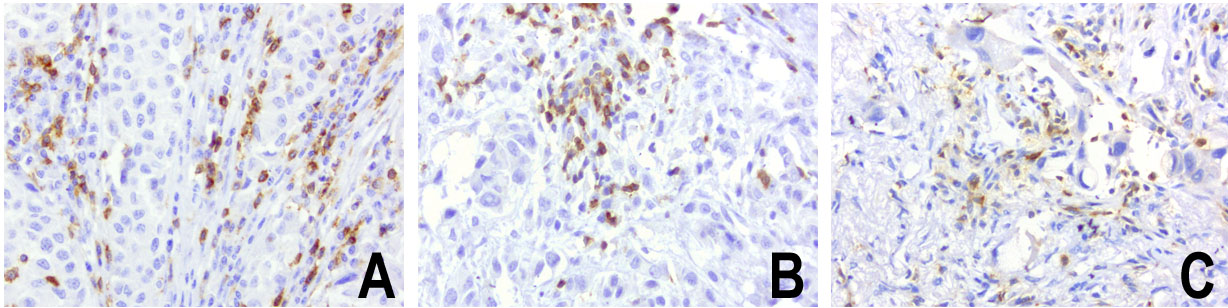
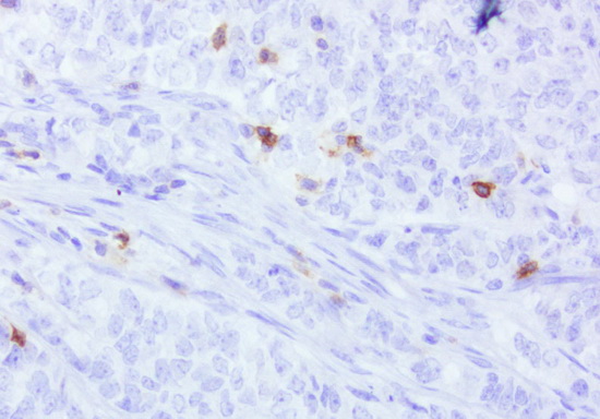
| Immunohistochemical staining of paraffin-embedded human colon cancer using anti-PD-1 clone UMAB199 mouse monoclonal antibody at 1:800 dilution of 1mg/mL and detection with Polink2 Broad HRP DAB. UM800091 requires heat-induced epitope retrieval with Accel for 3minutes at110C in pressure chamber . The image shows the tumor cells are negative for PD-1 however the activated TCells show strong membranous and cytoplasmic staining. | |
| Immunohistochemical staining of paraffin-embedded human endometrial cancer using anti-PD-1 clone UMAB199 mouse monoclonal antibody at 1:800 dilution of 1mg/mL and detection with Polink2 Broad HRP DAB. UM800091 requires heat-induced epitope retrieval with Accel for 3minutes at110C in pressure chamber . The image shows the tumor cells are negative for PD-1 however the activated TCells show strong membranous and cytoplasmic staining. | |
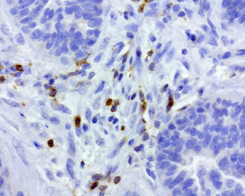
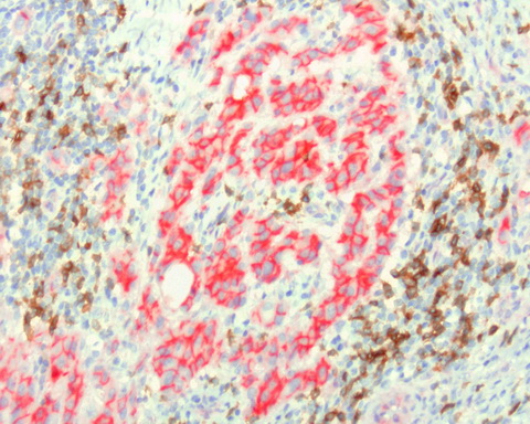
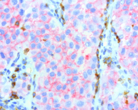
| Sequential double staining of paraffin human lung using anti-b-Catenin UM500015 (red) and anti-PD1 UM800091 (brown). Both antibodies at 1:800 dilution of 1mg/mL. Anti-PD1: heat-induced epitope retrieval with Accel; anti-b-Catenin: citrate pH6.0. Image shows tumor cells are strongly positve for b-catenin (red) and negative for PD1. The arrows point to the activated T cells (brown) showing strong membranous and cytoplasmic staining of PD1 and no staining with b-catenin. | |
| Sequential double staining of paraffin human melanoma using b-catenin UM500015 (red) and PD1 UM800091 (brown). Both abs at 1:800 dilution of 1mg/mL; detection with Polink2 HRP DAB followed by Polink2 Broad AP. Anti-PD1: heat-induced epitope retrieval with Accel; anti-b-catenin: citrate pH6.0 . Image shows tumor cells are strongly positve for b-catenin (red) and negative for PD1. The activated T cells (brown) show strong membranous and cytoplasmic staining for PD1 and no staining with b-catenin. | |
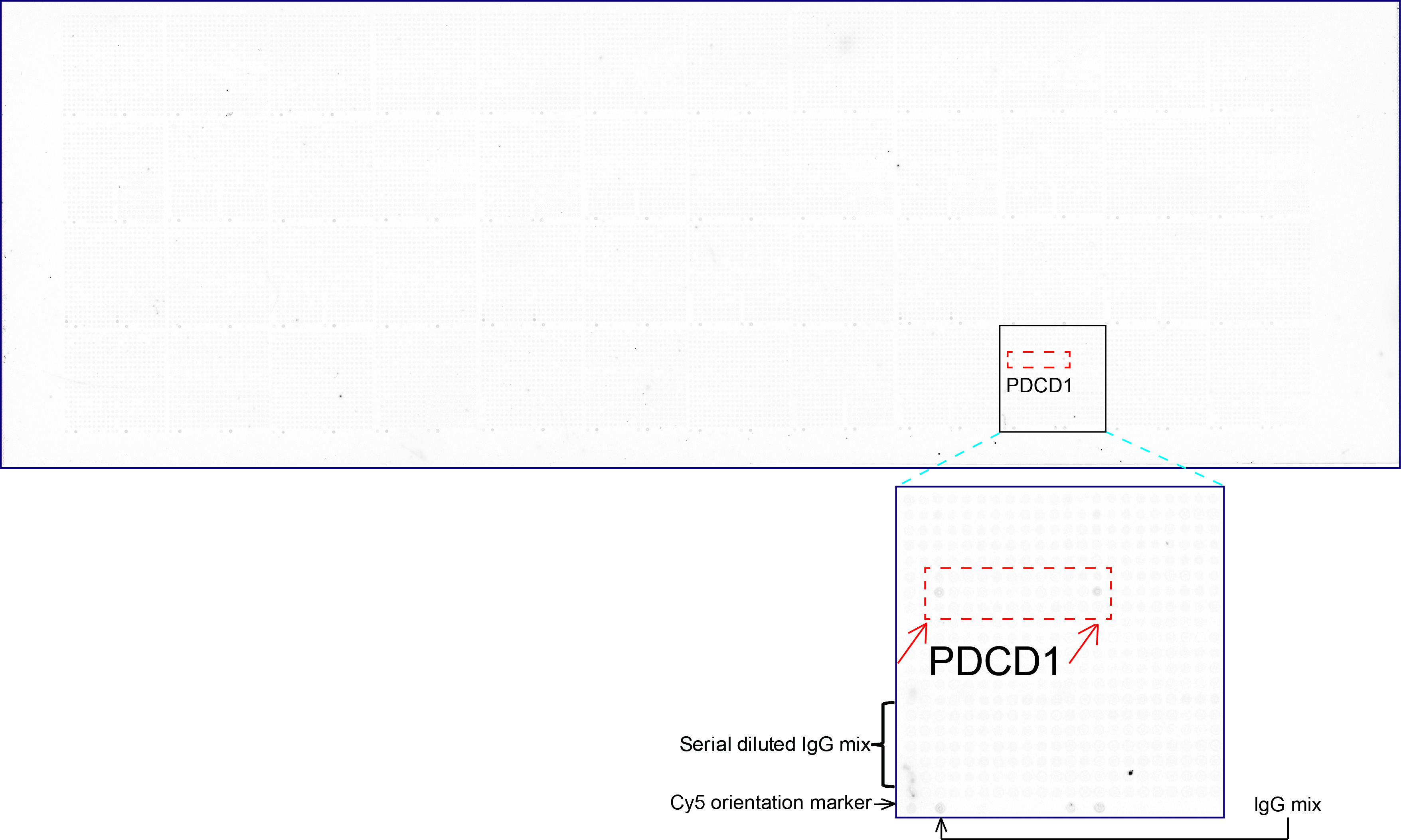
| OriGene overexpression protein microarray chip was immunostained with UltraMAB anti-PDCD1 mouse monoclonal antibody (UM800091). The positive reactive proteins are highlighted with two red arrows in the enlarged subarray. All the positive controls spotted in this subarray are also labeled for clarification.(1:100) |
追加しました。
製品情報は掲載時点のものですが、価格表内の価格については随時最新のものに更新されます。お問い合わせいただくタイミングにより製品情報・価格などは変更されている場合があります。
表示価格に、消費税等は含まれていません。一部価格が予告なく変更される場合がありますので、あらかじめご了承下さい。