
抗Human DC-SIGN/CD209抗体(Anti-Human DC-SIGN/CD209 antibody)
掲載日情報:2019/10/21 現在Webページ番号:177585

Human DC-SIGN/CD209に対する抗体(Anti-Human DC-SIGN/CD209 )です。
※ 本製品は研究用です。研究用以外には使用できません。
追加しました。
価格
[在庫・価格 :2026年06月25日 10時15分現在]
| 詳細 | 商品名 |
|
文献数 | ||||||||||||||||||||||||||||||||||||||||||||||||||||||||||||||||||||||||||||||||||
|---|---|---|---|---|---|---|---|---|---|---|---|---|---|---|---|---|---|---|---|---|---|---|---|---|---|---|---|---|---|---|---|---|---|---|---|---|---|---|---|---|---|---|---|---|---|---|---|---|---|---|---|---|---|---|---|---|---|---|---|---|---|---|---|---|---|---|---|---|---|---|---|---|---|---|---|---|---|---|---|---|---|---|---|---|---|
|
Anti-Human DC-SIGN/CD209 MAb (Clone 120507) |
|
38 | |||||||||||||||||||||||||||||||||||||||||||||||||||||||||||||||||||||||||||||||||||
|
|||||||||||||||||||||||||||||||||||||||||||||||||||||||||||||||||||||||||||||||||||||
|
Anti-Human DC-SIGN/CD209 MAb (Clone 120507) |
|
38 | |||||||||||||||||||||||||||||||||||||||||||||||||||||||||||||||||||||||||||||||||||
|
|||||||||||||||||||||||||||||||||||||||||||||||||||||||||||||||||||||||||||||||||||||
|
Anti-Human DC-SIGN/CD209 MAb (Clone 120507) |
|
38 | |||||||||||||||||||||||||||||||||||||||||||||||||||||||||||||||||||||||||||||||||||
|
|||||||||||||||||||||||||||||||||||||||||||||||||||||||||||||||||||||||||||||||||||||


[在庫・価格 :2026年06月25日 10時15分現在]
Anti-Human DC-SIGN/CD209 MAb (Clone 120507)
文献数: 38
- 商品コード:MAB161-100
- メーカー:RSD
- 包装:100μg
- 価格:¥79,000
- 在庫:1個
- 納期:10日程度 ※※ 表示されている納期は弊社に在庫がなく、取り寄せた場合の目安納期となります。
- 法規制等:
| 説明文 | 別名:CD209 クローン:120507 Genbank No: 30835 |
||||||
|---|---|---|---|---|---|---|---|
| 別包装品 | 別包装品あり | ||||||
| 法規制等 | |||||||
| 保存条件 | -20℃ | 法規備考 | |||||
| 抗原種 | 免疫動物 | Mouse | |||||
| 交差性 | Human | 適用 | Blocking,FCM,Western Blot | ||||
| 標識 | Unlabeled | 性状 | Protein A/G Affinity Purified | ||||
| 吸収処理 | クラス | IgG | |||||
| クロナリティ | Monoclonal | フォーマット | |||||
| 掲載カタログ |
|
||||||
| 製品記事 | M1/M2 Macrophage Activation Marker 抗幹細胞マーカー抗体/抗造血幹細胞マーカー抗体(R&D Systems社) |
||||||
| 関連記事 | |||||||
Anti-Human DC-SIGN/CD209 MAb (Clone 120507)
文献数: 38
- 商品コード:MAB161-500
- メーカー:RSD
- 包装:500μg
- 価格:¥266,000
- 在庫:無(未発注)
- 納期:10日程度 ※※ 表示されている納期は弊社に在庫がなく、取り寄せた場合の目安納期となります。
- 法規制等:
| 説明文 | 別名:CD209 クローン:120507 Genbank No: 30835 |
||||||
|---|---|---|---|---|---|---|---|
| 別包装品 | 別包装品あり | ||||||
| 法規制等 | |||||||
| 保存条件 | -20℃ | 法規備考 | |||||
| 抗原種 | 免疫動物 | Mouse | |||||
| 交差性 | Human | 適用 | Blocking,FCM,Western Blot | ||||
| 標識 | Unlabeled | 性状 | Protein A/G Affinity Purified | ||||
| 吸収処理 | クラス | IgG | |||||
| クロナリティ | Monoclonal | フォーマット | |||||
| 掲載カタログ |
|
||||||
| 製品記事 | M1/M2 Macrophage Activation Marker 抗幹細胞マーカー抗体/抗造血幹細胞マーカー抗体(R&D Systems社) |
||||||
| 関連記事 | |||||||
Anti-Human DC-SIGN/CD209 MAb (Clone 120507)
文献数: 38
- 商品コード:MAB161-SP
- メーカー:RSD
- 包装:25μg
- 価格:¥27,000
- 在庫:無(未発注)
- 納期:2~3週間 ※※ 表示されている納期は弊社に在庫がなく、取り寄せた場合の目安納期となります。
- 法規制等:
| 説明文 | ※受注発注品。形状:溶液または凍結乾燥 別名:CD209 クローン:120507 Genbank No: 30835 |
||||||
|---|---|---|---|---|---|---|---|
| 別包装品 | 別包装品あり | ||||||
| 法規制等 | |||||||
| 保存条件 | -20℃ | 法規備考 | |||||
| 抗原種 | 免疫動物 | Mouse | |||||
| 交差性 | Human | 適用 | Blocking,FCM,Western Blot | ||||
| 標識 | Unlabeled | 性状 | Protein A/G Affinity Purified | ||||
| 吸収処理 | クラス | IgG | |||||
| クロナリティ | Monoclonal | フォーマット | |||||
| 掲載カタログ |
|
||||||
| 製品記事 | M1/M2 Macrophage Activation Marker 抗幹細胞マーカー抗体/抗造血幹細胞マーカー抗体(R&D Systems社) 使いっきり抗体 |
||||||
| 関連記事 | |||||||
追加しました。
Product Details
| Species Reactivity | Human |
|---|---|
| Label | Unconjugated |
| Immunogen | NIH-3T3 mouse embryonic fibroblast cell line transfected with human DC‑SIGN/CD209 |
| Source | Monoclonal Mouse IgG2B Clone # 120507 |
| Purification | Protein A or G purified from hybridoma culture supernatant |
| Specificity | Detects human DC‑SIGN/CD209 on transfected NIH/3T3 cells and on monocyte derived dendritic cells. Does not react with parental mouse cells or irrelevant transfectants, such as human DC-SIGN2. |
追加しました。
Applications and Data
| Recommended Concentration | Sample | |
| Western Blot | 1 µg/mL | Recombinant Human DC-SIGN Fc Chimera (Catalog # 161-DC) |
| Flow Cytometry | 2.5 µg/106 cells | See below |
| Immunohistochemistry | 8-25 µg/mL | Immersion fixed paraffin-embedded sections of human lymph node |
| Adhesion Blockade | The adhesion of NIH-3T3 mouse embryonic fibroblast cells (5 x 10⁴ cells/well) to immobilized Recombinant Human ICAM-3/CD50 Fc Chimera (Catalog # 715-IC, 5 µg/mL, 100 µL/well) was maximally inhibited (80-100%) by 5 µg/mL of the antibody. | |
| CyTOF-ready | Ready to be labeled using established conjugation methods. No BSA or other carrier proteins that could interfere with conjugation. | |
| Immunocytochemistry | 8-25 µg/mL | See below |
| Flow Cytometry | |
|---|---|
 | Detection of DC‑SIGN in Human DC‑SIGN Transfected 3T3 Mouse Cell Line by Flow Cytometry. Human DC‑SIGN and DC‑SIGN2 transfected 3T3 mouse embryonic fibroblast cell line were stained with Mouse Anti-Human DC‑SIGN Monoclonal Antibody (Catalog # MAB161, filled histograms) or isotype control antibody (Catalog # MAB0041, open histogram), followed by Phycoerythrin-conjugated Anti-Mouse IgG F(ab')2 Secondary Antibody (Catalog # F0102B). |
| Flow Cytometry | |
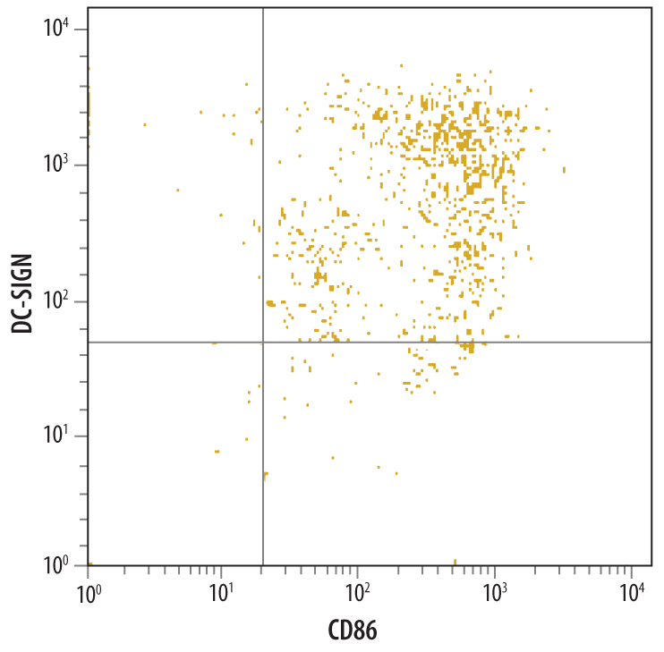 | Detection of DC‑SIGN in Human Monocyte Derived Dendritic Cells by Flow Cytometry. Human monocyte derived dendritic cells were stained with Mouse Anti-Human DC‑SIGN Monoclonal Antibody (Catalog # MAB161) followed by PE-conjugated anti-mouse IgG (Catalog # F0102B) and Anti-Human B7-2/CD86 Fluorescein-conjugated Monoclonal Antibody (Catalog # FAB141F). Quadrant markers were set based on control antibody staining (Catalog # MAB0041). |
| Immunocytochemistry | |
 | DC-SIGN in Human Dendritic Cells. DC-SIGN was detected in immersion fixed mature human dendritic cells using Mouse Anti-Human DC-SIGN Monoclonal Antibody (Catalog # MAB161) at 10 µg/mL for 3 hours at room temperature. Cells were stained using the NorthernLights™ 557-conjugated Anti-Mouse IgG Secondary Antibody (yellow; Catalog # NL007) and counterstained with DAPI (blue). View our protocol for Fluorescent ICC Staining of Non-adherent Cells. |
追加しました。
Related Product & Information
| Long Name | Dendritic Cell-specific ICAM-3-grabbing Non-integrin 1 |
|---|---|
| Entrez Gene IDs | 30835 (Human); 170786 (Mouse) |
| Background | DC-SIGN/CD209 |
| background_content | Background: DC-SIGN/CD209 Human DC-Sign (dendritic cell-specific ICAM-3 grabbing nonintegrin; also CD209) is a member of the chromosome 19 C-type lectin family that includes DC-SIGN, DC-SIGN-related protein, CD23 and LSECtin (1). DC-SIGN was initially reported to be a 46 kDa, 404 amino acid (aa) type II transmembrane protein that contained a 40 aa cytoplasmic N-terminus, a 21 aa transmembrane segment, and a 343 aa extracellular C-terminus (2). The extracellular region contains a distal, 115 aa Ca++-dependent carbohydrate-binding lectin domain and a membrane-proximal linker segment that is composed of seven 23 aa repeats (2, 3). The lectin domain is believed to preferably bind mannose, either within the context of ICAM-3 (on T cells) or ICAM-2 (on endothelial cells) (2, 4, 5). DC-SIGN expression appears to be limited to dendritic cells (DC) and macrophages (6), and DC interaction with the ICAMs both aids DC cell trafficking and immunological synapse formation (7). Since the original report on DC-SIGN, multiple splice forms have been discovered, generating both membrane-bound and soluble forms (3). There are eight type A isoforms, all of which begin with the same 15 aa of exon 1a. Four contain the transmembrane region of exon II, and four do not (i.e., are soluble). Among these eight type A isoforms, only three retain the entire 343 aa found in the full length form described in reference #2 (the full length form is referred to as type I mDC-SIGN1A) (3). Five additional isoforms utilize an alternate start site, and these are referred to as type B isoforms. These all show a 35 aa cytoplasmic domain. One also has a transmembrane segment; four do not. Two of the five contain full, unspliced extracellular regions (3). All of this suggests enormous complexity in DC-SIGN biology. DC-SIGN is not well conserved across species. Human and mouse show little overall aa identity. In the lectin domain, however, human DC-SIGN shares 68% aa identity with mouse DC-SIGN (8). Human and rhesus monkey DC-SIGN share 91% aa identity over the entire extracellular region (8). A detailed description of the additional properties of this monoclonal antibody (MAB161) have been published (9, 10). |
追加しました。
Citations
- Properdin and factor H production by human dendritic cells modulates their T-cell stimulatory capacity and is regulated by IFN-?
Authors: KO Dixon, J O'Flynn, N Klar-Moham, MR Daha, C van Kooten
Eur. J. Immunol, 2017;0(0):.
Species: Human
Sample Type: Whole Cells
Application: Flow - Metformin Uniquely Prevents Thrombosis by Inhibiting Platelet Activation and mtDNA Release
Sci Rep, 2016;6(0):36222.
Species: Rat
Sample Type: Whole Cells
Application: Neut - Kluyveromyces marxianus and Saccharomyces boulardii Induce Distinct Levels of Dendritic Cell Cytokine Secretion and Significantly Different T Cell Responses In Vitro
PLoS ONE, 2016;11(11):e0167410.
Species: Human
Sample Type: Whole Cells
Application: Neut - Clearance of autophagy-associated dying retinal pigment epithelial cells - a possible source for inflammation in age-related macular degeneration
Cell Death Dis, 2016;7(9):e2367.
Species: Human
Sample Type: Whole Cells
Application: Flow - HCV RNA Activates APCs via TLR7/TLR8 While Virus Selectively Stimulates Macrophages Without Inducing Antiviral Responses
Sci Rep, 2016;6(0):29447.
Species: Human
Sample Type: Whole Cells
Application: Functional Assay - Porphyromonas gingivalis evasion of autophagy and intracellular killing by human myeloid dendritic cells involves DC-SIGN-TLR2 crosstalk.
Authors: El-Awady, Ahmed R, Miles, Brodie, Scisci, Elizabet, Kurago, Zoya B, Palani, Chithra, Arce, Roger M, Waller, Jennifer, Genco, Caroline, Slocum, Connie, Manning, Matthew, Schoenlein, Patricia, Cutler, Christop
PLoS Pathog, 2015;10(2):e1004647.
Species: Human
Sample Type: Whole Cells
Application: Flow - Live-attenuated measles virus vaccine targets dendritic cells and macrophages in muscle of nonhuman primates.
Authors: Rennick L, de Vries R, Carsillo T, Lemon K, van Amerongen G, Ludlow M, Nguyen D, Yuksel S, Verburgh R, Haddock P, McQuaid S, Duprex W, de Swart R
J Virol, 2015;89(4):2192-200. - Paracoccidioides brasiliensis interferes on dendritic cells maturation by inhibiting PGE2 production.
Authors: Fernandes R, Bachiega T, Rodrigues D, Golim M, Dias-Melicio L, Balderramas H, Kaneno R, Soares A
PLoS ONE, 2015;10(3):e0120948.
Species: Human
Sample Type: Whole Cells
Application: Blocking - Selective susceptibility of human skin antigen presenting cells to productive dengue virus infection.
Authors: Cerny D, Haniffa M, Shin A, Bigliardi P, Tan B, Lee B, Poidinger M, Tan E, Ginhoux F, Fink K
PLoS Pathog, 2014;10(12):e1004548.
Species: Human
Sample Type: Whole Cells
Application: Blocking - Early biodistribution and persistence of a protective live attenuated SIV vaccine elicits localised innate responses in multiple lymphoid tissues.
Authors: Ferguson D, Mattiuzzo G, Ham C, Stebbings R, Li B, Rose N, Mee E, Smith D, Page M, Cranage M, Almond N, Towers G, Berry N
PLoS ONE, 2014;9(8):e104390.
Species: Primate - Macaca fascicularis (Crab-eating Monkey or Cynomolgus Macaque)
Sample Type: Whole Tissue
Application: IHC Paraffin-embedded
追加しました。
製品情報は掲載時点のものですが、価格表内の価格については随時最新のものに更新されます。お問い合わせいただくタイミングにより製品情報・価格などは変更されている場合があります。
表示価格に、消費税等は含まれていません。一部価格が予告なく変更される場合がありますので、あらかじめご了承下さい。