
抗Human IGF-I R抗体(Anti-Human IGF-I R antibody)
掲載日情報:2021/01/28 現在Webページ番号:177335

Human IGF-I Rに対する抗体(Anti-Human IGF-I R )です。
※ 本製品は研究用です。研究用以外には使用できません。
追加しました。
- 価格
- Product Details
- Applications and Data
- Data Examples
- Related Research Areas
- Related Product & Information
- Citations
価格
[在庫・価格 :2026年03月31日 00時00分現在]
| 詳細 | 商品名 |
|
文献数 | ||||||||||||||||||||||||||||||||||||||||||||||||||||||||||||||||||||||||||||||||||
|---|---|---|---|---|---|---|---|---|---|---|---|---|---|---|---|---|---|---|---|---|---|---|---|---|---|---|---|---|---|---|---|---|---|---|---|---|---|---|---|---|---|---|---|---|---|---|---|---|---|---|---|---|---|---|---|---|---|---|---|---|---|---|---|---|---|---|---|---|---|---|---|---|---|---|---|---|---|---|---|---|---|---|---|---|---|
|
Anti-Human IGF-I R MAb (Clone 33255) |
|
17 | |||||||||||||||||||||||||||||||||||||||||||||||||||||||||||||||||||||||||||||||||||
|
|||||||||||||||||||||||||||||||||||||||||||||||||||||||||||||||||||||||||||||||||||||
|
Anti-Human IGF-I R MAb (Clone 33255) |
|
17 | |||||||||||||||||||||||||||||||||||||||||||||||||||||||||||||||||||||||||||||||||||
|
|||||||||||||||||||||||||||||||||||||||||||||||||||||||||||||||||||||||||||||||||||||
|
Anti-Human IGF-I R MAb (Clone 33255) |
|
17 | |||||||||||||||||||||||||||||||||||||||||||||||||||||||||||||||||||||||||||||||||||
|
|||||||||||||||||||||||||||||||||||||||||||||||||||||||||||||||||||||||||||||||||||||


[在庫・価格 :2026年03月31日 00時00分現在]
Anti-Human IGF-I R MAb (Clone 33255)
文献数: 17
- 商品コード:MAB391-100
- メーカー:RSD
- 包装:100μg
- 価格:¥79,000
- 在庫:無(未発注)
- 納期:10日程度 ※※ 表示されている納期は弊社に在庫がなく、取り寄せた場合の目安納期となります。
- 法規制等:
| 説明文 | 別名:CD221 クローン:33255 Genbank No: 3480 Protein Accession No: P08069 |
||||||
|---|---|---|---|---|---|---|---|
| 別包装品 | 別包装品あり | ||||||
| 法規制等 | |||||||
| 保存条件 | -20℃ | 法規備考 | |||||
| 抗原種 | 免疫動物 | Mouse | |||||
| 交差性 | Human | 適用 | ELISA,FCM,IC,IHC,Neutralising,Western Blot | ||||
| 標識 | Unlabeled | 性状 | Protein A/G Affinity Purified | ||||
| 吸収処理 | クラス | IgG | |||||
| クロナリティ | Monoclonal | フォーマット | |||||
| 掲載カタログ |
|
||||||
| 製品記事 | |||||||
| 関連記事 | R&D Systems(R&Dシステムズ)社 ELISA用ペア抗体を使用したELISA 構築ガイド |
||||||
Anti-Human IGF-I R MAb (Clone 33255)
文献数: 17
- 商品コード:MAB391-500
- メーカー:RSD
- 包装:500μg
- 価格:¥266,000
- 在庫:無(未発注)
- 納期:10日程度 ※※ 表示されている納期は弊社に在庫がなく、取り寄せた場合の目安納期となります。
- 法規制等:
| 説明文 | マッチドペア:Human IGF-I R サンドイッチELISAの補足用抗体として利用可能,検出用抗体として#BAF391,スタンダードとして#391-GR-050を用いる。 別名:CD221 クローン:33255 Genbank No: 3480 Protein Accession No: P08069 |
||||||
|---|---|---|---|---|---|---|---|
| 別包装品 | 別包装品あり | ||||||
| 法規制等 | |||||||
| 保存条件 | -20℃ | 法規備考 | |||||
| 抗原種 | 免疫動物 | Mouse | |||||
| 交差性 | Human | 適用 | ELISA,FCM,IC,IHC,Neutralising,Western Blot | ||||
| 標識 | Unlabeled | 性状 | Protein A/G Affinity Purified | ||||
| 吸収処理 | クラス | IgG | |||||
| クロナリティ | Monoclonal | フォーマット | |||||
| 掲載カタログ |
|
||||||
| 製品記事 | |||||||
| 関連記事 | R&D Systems(R&Dシステムズ)社 ELISA用ペア抗体を使用したELISA 構築ガイド |
||||||
Anti-Human IGF-I R MAb (Clone 33255)
文献数: 17
- 商品コード:MAB391-SP
- メーカー:RSD
- 包装:25μg
- 価格:¥28,000
- 在庫:無(未発注)
- 納期:2~3週間 ※※ 表示されている納期は弊社に在庫がなく、取り寄せた場合の目安納期となります。
- 法規制等:
| 説明文 | ※受注発注品。形状:溶液または凍結乾燥 別名:CD221 クローン:33255 Genbank No: 3480 Protein Accession No: P08069 |
||||||
|---|---|---|---|---|---|---|---|
| 別包装品 | 別包装品あり | ||||||
| 法規制等 | |||||||
| 保存条件 | -20℃ | 法規備考 | |||||
| 抗原種 | 免疫動物 | Mouse | |||||
| 交差性 | Human | 適用 | ELISA,FCM,IC,IHC,Neutralising,Western Blot | ||||
| 標識 | Unlabeled | 性状 | Protein A/G Affinity Purified | ||||
| 吸収処理 | クラス | IgG | |||||
| クロナリティ | Monoclonal | フォーマット | |||||
| 掲載カタログ |
|
||||||
| 製品記事 | 使いっきり抗体 |
||||||
| 関連記事 | R&D Systems(R&Dシステムズ)社 ELISA用ペア抗体を使用したELISA 構築ガイド |
||||||
追加しました。
Product Details
| Species Reactivity | Human, Mouse |
|---|---|
| Label | Unconjugated |
| Immunogen | S. frugiperda insect ovarian cell line Sf 21-derived recombinant human IGF-I RGlu31-Asn932Accession # P08069 |
| Source | Monoclonal Mouse IgG1 Clone # 33255 |
| Purification | Protein A or G purified from hybridoma culture supernatant |
| Specificity | Detects human IGF-I R in sandwich ELISAs and Western blots. Detects mouse IGF-I R in Immunohistochemistry. In sandwich immunoassays, less than 0.15% cross-reactivity or interference was observed with recombinant human (rh) IGF-I, rhIGF-II, rhIL-3 R alpha, rhIL‑9 R, and rhTGF-beta RII. |
追加しました。
Applications and Data
| Recommended Concentration | Sample | |
| Western Blot | 1 µg/mL | See below |
| Flow Cytometry | 0.25 µg/106 cells | See below |
| Immunohistochemistry | 5-25 µg/mL | See below |
| Human IGF-I R Sandwich Immunoassay | Reagent | |
| ELISA Capture (Matched Antibody Pair) | 2-8 µg/mL | Human/Mouse IGF‑I R Antibody (Catalog #MAB391 ) |
| ELISA Detection (Matched Antibody Pair) | 0.1-0.4 µg/mL | Human IGF‑I R Biotinylated Antibody (Catalog #BAF391 ) |
| ELISA Standard | Recombinant Human IGF-I R Protein, CF (Catalog #391-GR ) | |
| Neutralization | Measured by its ability to neutralize IGF‑I-induced proliferation in the MCF‑7 human breast cancer cell line. Karey, K.P. et al. (1988) Cancer Research 48:4083. At 11 µg/mL, this antibody will neutralize approximately 50-75% of the bioactivity due to 6 ng/mL Recombinant Human IGF‑I. | |
| Please Note: Optimal dilutions should be determined by each laboratory for each application.General Protocolsare available in the Technical Information section on our website. | ||
追加しました。
Data Examples
| Western Blot |
![Western Blot IGF-I R Antibody (33255) [Unconjugated]](http://resources.rndsystems.com/images/datasheets/antibody/IGF-I_R_MAB391_Western_Blot_6136.jpg) click image to view larger | Detection of Human IGF‑I R by Western Blot. Western blot shows lysates of NTera‑2 human testicular embryonic carcinoma cell line, SK‑Mel‑28 human malignant melanoma cell line, and G361 human melanoma cell line. PVDF membrane was probed with 1 µg/mL of Mouse Anti-Human/Mouse IGF-I R Monoclonal Antibody (Catalog # MAB391) followed by HRP-conjugated Anti-Mouse IgG Secondary Antibody (Catalog # HAF007). A specific band was detected for IGF-I R at approximately 275 kDa (as indicated). This experiment was conducted under non-reducing conditions and using Immunoblot Buffer Group 2. |
| Flow Cytometry |
![Flow Cytometry IGF-I R Antibody (33255) [Unconjugated]](http://resources.rndsystems.com/images/datasheets/antibody/IGFI_R_MAB391_Flow_Cytometry_22536.jpg) click image to view larger | Detection of IGF‑I R in MCF‑7 Human Cell Line by Flow Cytometry.MCF‑7 human breast cancer cell line was stained with Mouse Anti-Human IGF‑I R Monoclonal Antibody (Catalog # MAB391, filled histogram) or isotype control antibody (Catalog # MAB002, open histogram) followed by anti-mouse IgG PE-conjugated secondary antibody (Catalog # F0102B). View our protocol for Staining Membrane-associated Proteins. |
| Immunohistochemistry |
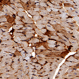
![Immunohistochemistry IGF-I R Antibody (33255) [Unconjugated]](http://resources.rndsystems.com/images/datasheets/antibody/IGFI_R_MAB391_Immunohistochemistry_21743.jpg) click image to view larger | IGF‑I R in Mouse Heart. IGF‑I R was detected in perfusion fixed paraffin-embedded sections of mouse heart using Mouse Anti-Human/Mouse IGF‑I R Monoclonal Antibody (Catalog # MAB391) at 15 µg/mL for 1 hour at room temperature followed by incubation with the Anti-Mouse IgG VisUCyte™ HRP Polymer Antibody (Catalog # VC001). Tissue was stained using DAB (brown) and counterstained with hematoxylin (blue). Specific staining was localized to plasma membrane and cytoplasm. View our protocol for IHC Staining with VisUCyte HRP Polymer Detection Reagents. |
| Neutralization |
![Neutralization IGF-I R Antibody (33255) [Unconjugated]](http://resources.rndsystems.com/images/datasheets/antibody/IGF-I_R_MAB391_Block_Neutralize_9173.jpg) click image to view larger | Cell Proliferation Induced by IGF‑I and Neutralization by Human IGF‑I R Antibody. Recombinant Human IGF‑I (Catalog # 291-G1) stimulates proliferation in the MCF‑7 human breast cancer cell line in a dose-dependent manner (orange line). Proliferation elicited by Recombinant Human IGF‑I (6 ng/mL) is neutralized (green line) by increasing concentrations of Mouse Anti-Human/Mouse IGF-I R Monoclonal Antibody (Catalog # MAB391). At 11 µg/mL, this antibody will neutralize 50-75% rhIGF-1 induced activity. |
| Reconstitution | Reconstitute at 0.5 mg/mL in sterile PBS. | Reconstitution Buffer Available |
| Shipping | The product is shipped at ambient temperature. Upon receipt, store it immediately at the temperature recommended below. *Small pack size (SP) is shipped with polar packs. Upon receipt, store it immediately at -20 to -70 °C | |
| Stability & Storage | Use a manual defrost freezer and avoid repeated freeze-thaw cycles. 12 months from date of receipt, -20 to -70 °C as supplied. | |
| 1 month, 2 to 8 °C under sterile conditions after reconstitution. | ||
| 6 months, -20 to -70 °C under sterile conditions after reconstitution. | ||
| Background: IGF-I R | Insulin-like growth factor I receptor (IGF-I R) is a disulfide-linked heterotetrameric transmembrane protein consisting of two alpha and two beta subunits. Both the alpha and beta subunits are encoded within a single receptor precursor cDNA. The proreceptor polypeptide is proteolytically cleaved and disulfide-linked to yield the mature heterotetrameric receptor. The alpha subunit of IGF-I R is extracellular while the beta subunit has an extracellular domain, a transmembrane domain and a cytoplasmic tyrosine kinase domain. IGF-I R is highly expressed in all cell types and tissues. Essentially all of the biological activities of IGF-I and -II have been shown to be mediated via IGF-I R. |
|
| Long Name: | Insulin-like Growth Factor I Receptor | |
| Entrez Gene IDs: | 3480 (Human) | |
| Alternate Names: | CD221 antigen; CD221; EC 2.7.10; EC 2.7.10.1; IGF1R; IGF-I R; IGF-I receptor; IGFIR; IGF-IR; IGFR; insulin-like growth factor 1 receptor; Insulin-like growth factor I receptor; JTK13; MGC142170; MGC142172; MGC18216; soluble IGF1R variant 1; soluble IGF1R variant 2 | |
追加しました。
Related Research Areas
| Adipocytokines | ||
| Cancer Biomarkers | ||
| Endocrine Regulation of Lipid Metabolism | ||
| Glucose Homeostasis | ||
| IGF Family | ||
| Metabolic Peptide Hormones and Regulators | ||
| Peptide Receptors on VSMC | ||
| Platelet Cytokine and Growth Factor Receptors | ||
| Receptor Tyrosine Kinases (RTKs) | ||
| Receptor Tyrosine Kinases (RTKs) in the Akt Pathway | ||
| Receptors in the Jak/STAT Pathway | ||
| ||
| Western Blot | |
|---|---|
 | Detection of Human IGF‑I R by Western Blot. Western blot shows lysates of NTera‑2 human testicular embryonic carcinoma cell line, SK‑Mel‑28 human malignant melanoma cell line, and G361 human melanoma cell line. PVDF membrane was probed with 1 µg/mL of Mouse Anti-Human/Mouse IGF-I R Monoclonal Antibody (Catalog # MAB391) followed by HRP-conjugated Anti-Mouse IgG Secondary Antibody (Catalog # HAF007). A specific band was detected for IGF-I R at approximately 275 kDa (as indicated). This experiment was conducted under non-reducing conditions and using Immunoblot Buffer Group 2. |
| Flow Cytometry | |
 | Detection of IGF‑I R in MCF‑7 Human Cell Line by Flow Cytometry. MCF‑7 human breast cancer cell line was stained with Mouse Anti-Human IGF‑I R Monoclonal Antibody (Catalog # MAB391, filled histogram) or isotype control antibody (Catalog # MAB002, open histogram) followed by anti-mouse IgG PE-conjugated secondary antibody (Catalog # F0102B). View our protocol for Staining Membrane-associated Proteins. |
| Immunohistochemistry | |
 | IGF‑I R in Mouse Heart. IGF‑I R was detected in perfusion fixed paraffin-embedded sections of mouse heart using Mouse Anti-Human/Mouse IGF‑I R Monoclonal Antibody (Catalog # MAB391) at 15 µg/mL for 1 hour at room temperature followed by incubation with the Anti-Mouse IgG VisUCyte™ HRP Polymer Antibody (Catalog # VC001). Tissue was stained using DAB (brown) and counterstained with hematoxylin (blue). Specific staining was localized to plasma membrane and cytoplasm. View our protocol for IHC Staining with VisUCyte HRP Polymer Detection Reagents. |
| Neutralization | |
 | Cell Proliferation Induced by IGF‑I and Neutralization by Human IGF‑I R Antibody. Recombinant Human IGF‑I (Catalog # 291-G1) stimulates proliferation in the MCF‑7 human breast cancer cell line in a dose-dependent manner (orange line). Proliferation elicited by Recombinant Human IGF‑I (6 ng/mL) is neutralized (green line) by increasing concentrations of Mouse Anti-Human/Mouse IGF-I R Monoclonal Antibody (Catalog # MAB391). At 11 µg/mL, this antibody will neutralize 50-75% rhIGF-1 induced activity. |
追加しました。
Related Product & Information
| Long Name | Insulin-like Growth Factor I Receptor |
|---|---|
| Entrez Gene IDs | 3480 (Human) |
| Background | IGF-I R |
| background_content | Background: IGF-I R Insulin-like growth factor I receptor (IGF-I R) is a disulfide-linked heterotetrameric transmembrane protein consisting of two alpha and two beta subunits. Both the alpha and beta subunits are encoded within a single receptor precursor cDNA. The proreceptor polypeptide is proteolytically cleaved and disulfide-linked to yield the mature heterotetrameric receptor. The alpha subunit of IGF-I R is extracellular while the beta subunit has an extracellular domain, a transmembrane domain and a cytoplasmic tyrosine kinase domain. IGF-I R is highly expressed in all cell types and tissues. Essentially all of the biological activities of IGF-I and -II have been shown to be mediated via IGF-I R. |
追加しました。
Citations
- IGF1R activation and the in vitro antiproliferative efficacy of IGF1R inhibitor are inversely correlated with IGFBP5 expression in bladder cancer
Authors: Y Neuzillet, E Chapeaubla, C Krucker, L De Koning, T Lebret, F Radvanyi, I Bernard-Pi
BMC Cancer, 2017;17(1):636.
Species: Human
Sample Type: Whole Cells
Application: Neut - A SPOPL/Cullin-3 ubiquitin ligase complex regulates endocytic trafficking by targeting EPS15 at endosomes
Authors: M Gschweitl, A Ulbricht, CA Barnes, RI Enchev, I Stoffel-St, N Meyer-Scha, J Huotari, Y Yamauchi, UF Greber, A Helenius, M Peter
Elife, 2016;5(0):.
Species: Human
Sample Type: Cell Lysates
Application: WB - Impact of the putative cancer stem cell markers and growth factor receptor expression on the sensitivity of ovarian cancer cells to treatment with various forms of small molecule tyrosine kinase inhibitors and cytotoxic drugs
Authors: Helmout Modjtahedi
Int. J. Oncol., 2016;49(5):1825-1838.
Species: Human
Sample Type: Cell Lysates
Application: WB - Insulin and IGF1 signalling pathways in human astrocytes in vitro and in vivo; characterisation, subcellular localisation and modulation of the receptors.
Authors: Garwood C, Ratcliffe L, Morgan S, Simpson J, Owens H, Vazquez-Villasenor I, Heath P, Romero I, Ince P, Wharton S
Mol Brain, 2015;8(0):51.
Species: Human
Sample Type: Whole Cells
Application: Neut - Insulin-like growth factor 1 and 2 (IGF1, IGF2) expression in human microglia: differential regulation by inflammatory mediators.
Authors: Suh H, Zhao M, Derico L, Choi N, Lee S
J Neuroinflammation, 2013;10(0):37.
Species: Human
Sample Type: Whole Cells
Application: ICC Frozen - Targeting both IGF-1R and mTOR synergistically inhibits growth of renal cell carcinoma in vitro.
Authors: Cardillo T, Trisal P, Arrojo R, Goldenberg D, Chang C
BMC Cancer, 2013;13(0):170.
Species: Human
Sample Type: Whole Cells
Application: Flow - Proinflammatory actions of visfatin/nicotinamide phosphoribosyltransferase (Nampt) involve regulation of insulin signaling pathway and Nampt enzymatic activity.
Authors: Jacques C, Holzenberger M, Mladenovic Z
J. Biol. Chem., 2012;287(18):15100-8.
Species: Human
Sample Type: Whole Cells
Application: Neut - Expression of the IGF axis is decreased in local prostate cancer but enhanced after benign prostate epithelial differentiation and TGF-beta treatment.
Authors: Massoner P, Ladurner Rennau M, Heidegger I, Kloss-Brandstatter A, Summerer M, Reichhart E, Schafer G, Klocker H
Am. J. Pathol., 2011;179(6):2905-19.
Species: Human
Sample Type: Whole Tissue
Application: IHC Paraffin-embedded - Changes in insulin and IGF-I receptor expression during differentiation of human preadipocytes.
Authors: Back K, Arnqvist HJ
Growth Horm. IGF Res., 2009;19(2):101-11.
Species: Human
Sample Type: Cell Lysates
Application: ELISA Development - Autologous bone marrow stromal cells genetically engineered to secrete an igf-I receptor decoy prevent the growth of liver metastases.
Authors: Wang N, Fallavollita L, Nguyen L, Burnier J, Rafei M, Galipeau J, Yakar S, Brodt P
Mol. Ther., 2009;17(7):1241-9.
Species: Human
Sample Type: Plasma
Application: ELISA Development
追加しました。
製品情報は掲載時点のものですが、価格表内の価格については随時最新のものに更新されます。お問い合わせいただくタイミングにより製品情報・価格などは変更されている場合があります。
表示価格に、消費税等は含まれていません。一部価格が予告なく変更される場合がありますので、あらかじめご了承下さい。