
抗TLR9 (26C593.2)抗体 | Anti- TLR9 (26C593.2) antibody
掲載日情報:2018/08/30 現在Webページ番号:61001

世界最大級の抗体製品数を取り扱うNovus Biologicals社のTLR9 (26C593.2)に対する抗体(anti-TLR9 (26C593.2) | antibody TLR9 (26C593.2))です。Novus Biologicals社の抗体は数多くの学術論文で使用実績があります。
※本製品は研究用です。研究用以外には使用できません。
PE, AlexaFluor, FITC, DyLight などの蛍光を含む、さまざまな標識体も取り扱っています。
追加しました。
Image
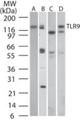
-Western-Blot-NBP2-24729-img0014.jpg) | Western Blot: TLR9 Antibody (26C593.2) [NBP2-24729] - Analysis of TLR9 in A) human PBMC, B) human intestine, C) mouse intestine, and D) rat intestine tissue lysates using Azide free TLR9 antibody at a dilution of 3 ug/ml. |
-Immunohistochemistry-Paraffin-NBP2-24729-img0023.jpg) | Immunohistochemistry-Paraffin: TLR9 Antibody (26C593.2) [NBP2-24729] - Monkey retina tissue. This image was submitted via customer review. |
-Flow-Cytometry-NBP2-24729-img0022.jpg) | Flow Cytometry: TLR9 Antibody (26C593.2) [NBP2-24729] - Flow analysis of TLR9 in human PBMC using 0.5 ug of TLR9 antibody (red) and isotype control antibody (green). |
-Western-Blot-NBP2-24729-img0007.jpg) | Western Blot: TLR9 Antibody (26C593.2) [NBP2-24729] - analysis of TLR9 in human B cells using anti-TLR9 antibody. Image from verified customer review. |
-Immunohistochemistry-Paraffin-NBP2-24729-img0021.jpg) | Immunohistochemistry-Paraffin: TLR9 Antibody (26C593.2) [NBP2-24729] - Analysis using the Azide Free version of NBP2-24729. Staining of human spleen probed with TLR9 antibody at 5 ug/ml. Human tissue was used for this test. Staining of formalin-fixed tissues is enhanced by boiling tissue sections in 10 mM sodium citrate buffer, pH 6.0 for 10-20 min followed by cooling at RT for 20 min. |
-Flow-Cytometry-NBP2-24729-img0010.jpg) | Flow Cytometry: TLR9 Antibody (26C593.2) [NBP2-24729] - Intracellular flow cytometric analysis of TLR9 FITC in human B cells using 1ug/10^6 cells of NBP2-24908. Cells were primarily stained with PE-conjugated CD19 antibody. Cells were then fixed and permeabilized and stained with 1ug of either isotype control |
-Flow-Cytometry-NBP2-24729-img0016.jpg) | Flow Cytometry: TLR9 Antibody (26C593.2) [NBP2-24729] - Analysis using the Azide Free version of NBP2-24729. Staining of TLR9 in Ramos cells using 0.5 ug of this antibody. Shaded histogram represents Ramos cells without antibody; green represents isotype control; purple represents anti-TLR9 antibody. |
-Flow-Cytometry-NBP2-24729-img0018.jpg) | Flow Cytometry: TLR9 Antibody (26C593.2) [NBP2-24729] - Analysis using the FITC conjugate of NBP2-24729. Staining of TLR9 in Ramos cells using 0.5 ug of this antibody. Green represents isotype control ; red represents anti-TLR9 antibody. TLR intracellular flow kit was used for this test. |
-Flow-(Intracellular)-NBP2-24729-img0019.jpg) | Flow (Intracellular): TLR9 Antibody (26C593.2) [NBP2-24729] - Analysis using the PE conjugate of NBP2-24729. Staining of TLR9 in human PBMCs using 0.2 ug of this antibody. Shaded histogram represents cells without antibody; green represents a mouse IgG1-PE isotype control ; red represents anti-TLR9 antibody. |
-Flow-Cytometry-NBP2-24729-img0020.jpg) | Flow Cytometry: TLR9 Antibody (26C593.2) [NBP2-24729] - Analysis using the PE conjugate of NBP2-24729. Staining of TLR9 in Ramos cells using 0.1 ug of antibody. Shaded histogram represents Ramos cells without antibody; green represents a mouse IgG1-PE isotype control red represents anti-TLR9 antibody. |
-Simple-Western-NBP2-24729-img0008.jpg) | Simple Western: TLR9 Antibody (26C593.2) [NBP2-24729] - Simple Western lane view shows a specific band for TLR9 in 0.5 mg/ml of Ramos lysate. This experiment was performed under reducing conditions using the 66-440 kDa separation system. |
追加しました。
価格
[在庫・価格 :2026年07月14日 12時35分現在]
| 詳細 | 商品名 |
|
文献数 | ||||||||||||||||||||||||||||||||||||||||||||||||||||||||||||||||||||||||||||||||||
|---|---|---|---|---|---|---|---|---|---|---|---|---|---|---|---|---|---|---|---|---|---|---|---|---|---|---|---|---|---|---|---|---|---|---|---|---|---|---|---|---|---|---|---|---|---|---|---|---|---|---|---|---|---|---|---|---|---|---|---|---|---|---|---|---|---|---|---|---|---|---|---|---|---|---|---|---|---|---|---|---|---|---|---|---|---|
|
Anti-TLR9, Mouse-Mono(26C593.2) |
|
139 | |||||||||||||||||||||||||||||||||||||||||||||||||||||||||||||||||||||||||||||||||||
|
|||||||||||||||||||||||||||||||||||||||||||||||||||||||||||||||||||||||||||||||||||||
|
Anti-TLR9, Mouse-Mono(26C593.2) |
|
80 | |||||||||||||||||||||||||||||||||||||||||||||||||||||||||||||||||||||||||||||||||||
|
|||||||||||||||||||||||||||||||||||||||||||||||||||||||||||||||||||||||||||||||||||||


[在庫・価格 :2026年07月14日 12時35分現在]
Anti-TLR9, Mouse-Mono(26C593.2)
文献数: 139
- 商品コード:NBP2-24729
- メーカー:NOV
- 包装:0.1mg
- 価格:¥108,000
- 在庫:無(未発注)
- 納期:3~4週間 ※※ 表示されている納期は弊社に在庫がなく、取り寄せた場合の目安納期となります。
- 法規制等:
| 説明文 | レビューあり。Simple Western対応抗体。旧IMGENEX社 商品コード:IMG-305A,Keywords:CD289|CD289 antigen|toll-like receptor 9 クローン:26C593.2 Genbank No: 54106 Protein Accession No: Q9NR96 |
||||||
|---|---|---|---|---|---|---|---|
| 別包装品 | 別包装品あり | ||||||
| 法規制等 | |||||||
| 保存条件 | -20℃ | 法規備考 | |||||
| 抗原種 | 免疫動物 | Mouse | |||||
| 交差性 | Canine/Equine/Human/Monkey/Mouse/Primate/Rat | 適用 | Dot,ELISA,FCM,Functional Assay,IC,IF,IHC,IP,Neutralising,Simple Western,Western Blot,in vitro assay | ||||
| 標識 | Unlabeled | 性状 | Protein A/G Affinity Purified | ||||
| 吸収処理 | クラス | IgG | |||||
| クロナリティ | Monoclonal | フォーマット | |||||
| 掲載カタログ |
|
||||||
| 製品記事 | IHCPro Toll - Like Receptor ( TLR ) 関連抗体 Toll-Like Receptor(TLR) Screening Kit /抗 TLR 抗体 フローサイトメトリー用抗 TLR 抗体 抗TLR9 (26C593.2)抗体 | Anti-TLR9 (26C593.2) Antibody |
||||||
| 関連記事 | |||||||
Anti-TLR9, Mouse-Mono(26C593.2)
文献数: 80
- 商品コード:NBP2-24729SS
- メーカー:NOV
- 包装:0.025mg
- 価格:¥53,000
- 在庫:無(未発注)
- 納期:3~4週間 ※※ 表示されている納期は弊社に在庫がなく、取り寄せた場合の目安納期となります。
- 法規制等:
| 説明文 | Simple Western対応抗体。Keywords:CD289|CD289 antigen|toll-like receptor 9 クローン:26C593.2 Genbank No: 54106 Protein Accession No: Q9NR96 |
||||||
|---|---|---|---|---|---|---|---|
| 別包装品 | 別包装品あり | ||||||
| 法規制等 | |||||||
| 保存条件 | -20℃ | 法規備考 | |||||
| 抗原種 | 免疫動物 | Mouse | |||||
| 交差性 | Canine/Equine/Human/Monkey/Mouse/Primate/Rat | 適用 | Dot,ELISA,FCM,Functional Assay,IC,IF,IHC,IP,Neutralising,Simple Western,Western Blot,in vitro assay | ||||
| 標識 | Unlabeled | 性状 | |||||
| 吸収処理 | クラス | IgG | |||||
| クロナリティ | Monoclonal | フォーマット | |||||
| 掲載カタログ |
|
||||||
| 製品記事 | |||||||
| 関連記事 | |||||||
追加しました。
標識済み抗体価格
[在庫・価格 :2026年07月14日 12時35分現在]
| 詳細 | 商品名 |
|
文献数 | ||||||||||||||||||||||||||||||||||||||||||||||||||||||||||||||||||||||||||
|---|---|---|---|---|---|---|---|---|---|---|---|---|---|---|---|---|---|---|---|---|---|---|---|---|---|---|---|---|---|---|---|---|---|---|---|---|---|---|---|---|---|---|---|---|---|---|---|---|---|---|---|---|---|---|---|---|---|---|---|---|---|---|---|---|---|---|---|---|---|---|---|---|---|---|---|---|---|
|
Anti-TLR9, Mouse-Mono(26C593.2), FITC |
|
1 | |||||||||||||||||||||||||||||||||||||||||||||||||||||||||||||||||||||||||||
|
|||||||||||||||||||||||||||||||||||||||||||||||||||||||||||||||||||||||||||||
[在庫・価格 :2026年07月14日 12時35分現在]
Anti-TLR9, Mouse-Mono(26C593.2), FITC
文献数: 1
- 商品コード:NBP2-24729F
- メーカー:NOV
- 包装:0.1ml
- 価格:¥132,000
- 在庫:無(未発注)
- 納期:3~4週間 ※※ 表示されている納期は弊社に在庫がなく、取り寄せた場合の目安納期となります。
- 法規制等:
| 説明文 | クローン:26C593.2 Genbank No: 54106 |
||
|---|---|---|---|
| 法規制等 | |||
| 保存条件 | 法規備考 | ||
| 抗原種 | 免疫動物 | Mouse | |
| 交差性 | Canine/Equine/Human/Monkey/Mouse/Primate/Rat | 適用 | Dot,ELISA,FCM,Functional Assay,IC,IF,IHC,IP,Neutralising,Simple Western,Western Blot,in vitro assay |
| 標識 | FITC | 性状 | Protein A/G Affinity Purified |
| 吸収処理 | クラス | IgG | |
| クロナリティ | Monoclonal | フォーマット | |
| 掲載カタログ |
|
||
| 製品記事 | |||
| 関連記事 | |||
追加しました。
製品情報は掲載時点のものですが、価格表内の価格については随時最新のものに更新されます。お問い合わせいただくタイミングにより製品情報・価格などは変更されている場合があります。
表示価格に、消費税等は含まれていません。一部価格が予告なく変更される場合がありますので、あらかじめご了承下さい。