
抗CD44 (8E2F3)抗体 | Anti-CD44 (8E2F3) Antibody
掲載日情報:2018/07/09 現在Webページ番号:50292

世界最大級の抗体製品数を取り扱うNovus Biologicals社のCD44 (8E2F3)に対する抗体(anti-CD44 (8E2F3) | antibody CD44 (8E2F3))です。Novus Biologicals社の抗体は数多くの学術論文で使用実績があります。
※本製品は研究用です。研究用以外には使用できません。

追加しました。
価格
[在庫・価格 :2026年07月16日 00時00分現在]
| 詳細 | 商品名 |
|
文献数 | ||||||||||||||||||||||||||||||||||||||||||||||||||||||||||||||||||||||||||||||||||
|---|---|---|---|---|---|---|---|---|---|---|---|---|---|---|---|---|---|---|---|---|---|---|---|---|---|---|---|---|---|---|---|---|---|---|---|---|---|---|---|---|---|---|---|---|---|---|---|---|---|---|---|---|---|---|---|---|---|---|---|---|---|---|---|---|---|---|---|---|---|---|---|---|---|---|---|---|---|---|---|---|---|---|---|---|---|
|
Anti-CD44, Mouse-Mono(8E2F3) |
|
14 | |||||||||||||||||||||||||||||||||||||||||||||||||||||||||||||||||||||||||||||||||||
|
|||||||||||||||||||||||||||||||||||||||||||||||||||||||||||||||||||||||||||||||||||||


[在庫・価格 :2026年07月16日 00時00分現在]
Anti-CD44, Mouse-Mono(8E2F3)
文献数: 14
- 商品コード:NBP1-47386
- メーカー:NOV
- 包装:0.1ml
- 価格:¥110,000
- 在庫:無(未発注)
- 納期:3~4週間 ※※ 表示されている納期は弊社に在庫がなく、取り寄せた場合の目安納期となります。
- 法規制等:
| 説明文 | レビューあり。Keywords:CD44 antigen|CD44 molecule (Indian blood group)|CD44R|CDw44|cell surface glycoprotein CD44|chondroitin sulfate proteoglycan 8|CSPG8|ECMR-III|Epican|Extracellular matrix receptor III|GP90 lymphocyte homing/adhesion receptor|HCELL クローン:8E2F3 Genbank No: 960 Protein Accession No: P16070 |
||||||
|---|---|---|---|---|---|---|---|
| 別包装品 | 別包装品あり | ||||||
| 法規制等 | |||||||
| 保存条件 | -20℃ | 法規備考 | |||||
| 抗原種 | 免疫動物 | Mouse | |||||
| 交差性 | Human/Mouse | 適用 | ELISA,FCM,IC,IF,IHC,IP,Western Blot | ||||
| 標識 | Unlabeled | 性状 | |||||
| 吸収処理 | クラス | IgG | |||||
| クロナリティ | Monoclonal | フォーマット | |||||
| 掲載カタログ |
|
||||||
| 製品記事 | 血液がん関連抗体 |
||||||
| 関連記事 | |||||||
追加しました。
Image
-Immunocytochemistry-Immunofluorescence-NBP1-47386-img0011.jpg) | Immunocytochemistry/Immunofluorescence: CD44 Antibody (8E2F3) [NBP1-47386] - Confocal analysis of paraffin-embedded human lung cancer tissues using anti-CD44 mAb (green), showing membrane localization. Blue: DRAQ5 fluorescent DNA dye. |
-Immunohistochemistry-Paraffin-NBP1-47386-img0014.jpg) | Immunohistochemistry-Paraffin: CD44 Antibody (8E2F3) [NBP1-47386] - IHC analysis of formalin fixed paraffin-embedded (FFPE) human breast carcinoma tissues using CD44 antibody (clone 8E2F3). The signal was developed using DAB based detection and the sections were processed for counterstaining with hematoxylin. The antibody generated mainly a membrane staining representative of CD44 protein. |
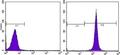
-Flow-Cytometry-NBP1-47386-img0008.jpg) | Flow Cytometry: CD44 Antibody (8E2F3) [NBP1-47386] - Flow cytometric analysis of Hela cells using anti-CD44 mAb (right) and negative control (left). |
-Western-Blot-NBP1-47386-img0013.jpg) | Western Blot: CD44 Antibody (8E2F3) [NBP1-47386] - Western blot analysis using anti-CD44 mAb against Hela (1) and HUVE-12 (2) cell lysates. |
-Western-Blot-NBP1-47386-img0015.jpg) | Genetic Strategies Validation. Western Blot: CD44 Antibody (8E2F3) [NBP1-47386] - Western blot analysis using anti-CD44 mAb against truncated Trx-CD44 recombinant protein (1) and GST-CD44 (aa628-699) recombinant protein (2). |
-Flow-Cytometry-NBP1-47386-img0017.jpg) | Flow Cytometry: CD44 Antibody (8E2F3) [NBP1-47386] - An intracellular stain was performed on HeLa cells with CD44 Antibody (8E2F3) NBP1-47386AF488 (blue) and a matched isotype control (orange). Cells were fixed with 4% PFA and then permeablized with 0.1% saponin. Cells were incubated in an antibody dilution of 5 ug/mL for 30 minutes at room temperature. Both antibodies were conjugated to Alexa Fluor 488. |
-Immunocytochemistry-Immunofluorescence-NBP1-47386-img0009.jpg) | Immunocytochemistry/Immunofluorescence: CD44 Antibody (8E2F3) [NBP1-47386] - Confocal immunofluorescence analysis of methanol-fixed A431 (A), Hela (B), PANC-1 (C) and EC (D) cells using anti-CD44 mAb (green), showing membrane localization. Blue: DRAQ5 fluorescent DNA dye. |
-Immunocytochemistry-Immunofluorescence-NBP1-47386-img0010.jpg) | Immunocytochemistry/Immunofluorescence: CD44 Antibody (8E2F3) [NBP1-47386] - Confocal immunofluorescence analysis of PANC-1 cells using anti-CD44 mAb (green). Red: Actin filaments have been labeled with DY-554 phalloidin. Blue: DRAQ5 fluorescent DNA dye. |
-Flow-(Intracellular)-NBP1-47386-img0016.jpg) | Flow (Intracellular): CD44 Antibody (8E2F3) [NBP1-47386] - An intracellular stain was performed on HeLa cells with CD44 Antibody (8E2F3) NBP1-47386AF647 (blue) and a matched isotype control (orange). Cells were fixed with 4% PFA and then permeablized with 0.1% saponin. Cells were incubated in an antibody dilution of 2.5 ug/mL for 30 minutes at room temperature. Both antibodies were conjugated to Alexa Fluor 647. |
追加しました。
Background
CD44, also known as IN, LHRMIC4, CDW44, HCELL, is a cell-surface glycoprotein involved in cell-cell interactions, cell adhesion and migration. It is a receptor for hyaluronic acid (HA) and can also interact with other ligands, such as osteopontin, collagens, and matrix metalloproteinases (MMPs). This protein participates in a wide variety of cellular functions including lymphocyte activation, recirculation and homing, hematopoiesis, and tumor metastasis. Transcripts for this gene undergo complex alternative splicing that results in many functionally distinct isoforms, however, the full length nature of some of these variants has not been determined. Alternative splicing is the basis for the structural and functional diversity of this protein, and may be related to tumor metastasis.追加しました。
製品情報は掲載時点のものですが、価格表内の価格については随時最新のものに更新されます。お問い合わせいただくタイミングにより製品情報・価格などは変更されている場合があります。
表示価格に、消費税等は含まれていません。一部価格が予告なく変更される場合がありますので、あらかじめご了承下さい。