抗CDK4抗体 | Anti-CDK4 Antibody

掲載日情報:2018/07/09 現在Webページ番号:50016

世界最大級の抗体製品数を取り扱うNovus Biologicals社のCDK4に対する抗体(anti-CDK4 | antibody CDK4)です。Novus Biologicals社の抗体は数多くの学術論文で使用実績があります。

本製品は研究用です。研究用以外には使用できません。

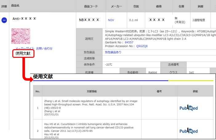
価格表左の「文献」アイコンから、使用文献情報一覧が表示できます。
価格

[在庫・価格 :2025年04月27日 00時00分現在]

※ 表示されている納期は弊社に在庫が無く、取り寄せた場合の納期目安となります。
詳細 商品名
  • 商品コード
  • メーカー
  • 包装
  • 価格
  • 在庫
  • 法規制等
納期 文献数
Anti-CDK4, Rabbit-Poly
3~4週間 ※ 表示されている納期は弊社に在庫がなく、取り寄せた場合の目安納期となります。 5
  • 使用文献
  • メーカーサイト
  • お問い合わせ
説明文
Genbank No: 1019
Protein Accession No: P11802
法規制等
保存条件 法規備考
抗原種 免疫動物 Rabbit クラス IgG 標識 Unlabeled
交差性 Bovine/Feline/Human/Mouse/Rat 適用 IC,IF,IHC,Simple Western,Western Blot
クロナリティ Polyclonal フォーマット 性状 Antigen Affinity Purified 吸収処理
掲載カタログ

製品記事
関連記事

[在庫・価格 :2025年04月27日 00時00分現在]

※ 表示されている納期は弊社に在庫が無く、取り寄せた場合の納期目安となります。

Anti-CDK4, Rabbit-Poly

文献数: 5

説明文 Genbank No: 1019
Protein Accession No: P11802
法規制等
保存条件 法規備考
抗原種 免疫動物 Rabbit
交差性 Bovine/Feline/Human/Mouse/Rat 適用 IC,IF,IHC,Simple Western,Western Blot
標識 Unlabeled 性状 Antigen Affinity Purified
吸収処理 クラス IgG
クロナリティ Polyclonal フォーマット
掲載カタログ

製品記事
関連記事


目次に戻る

Image

Genetic Strategies Validation. Western Blot: CDK4 Antibody [NBP1-31308] - Non-transfected (-) and transfected (+) 293T whole cell extracts (30 ug) were separated by 12% SDS-PAGE, and the membrane was blotted with CDK4 antibody. HRP-conjugated anti-rabbit IgG antibody was used to detect the primary antibody.
Immunocytochemistry/Immunofluorescence: CDK4 Antibody [NBP1-31308] - HeLa cells were fixed in 4% paraformaldehyde at RT for 15 min.Green: CDK4 protein stained by CDK4 antibody diluted at 1:500. Red: phalloidin, a cytoskeleton marker, diluted at 1:200. Scale bar = 10 um.
Immunohistochemistry-Paraffin: Cdk4 Antibody [NBP1-31308] - Paraffin-embedded Gastric ca, using antibody at 1:500 dilution.
Western Blot: Cdk4 Antibody [NBP1-31308] - Sample (30 ug of whole cell lysate) A:NIH-3T3 12% SDS PAGE; antibody diluted at 1:1000.
Western Blot: CDK4 Antibody [NBP1-31308] - Whole cell extract (30 ug) was separated by 12% SDS-PAGE, and the membrane was blotted with CDK4 antibody diluted at 1:500. The HRP-conjugated anti-rabbit IgG antibody (NBP2-19301) was used to detect the primary antibody.
Immunocytochemistry/Immunofluorescence: CDK4 Antibody [NBP1-31308] - MCF7 cells were fixed in 4% paraformaldehyde at RT for 15 min. Green: CDK4 protein stained by CDK4 antibody diluted at 1:1000.
CDK4 Antibody [NBP1-31308] - Various whole cell extracts (30 ug) were separated by 12% SDS-PAGE, and the membrane was blotted with CDK4 antibody diluted at 1:1000. The HRP-conjugated anti-rabbit IgG antibody (NBP2-19301) was used to detect the primary antibody.


目次に戻る

Background

The protein encoded by this gene is a member of the Ser/Thr protein kinase family. This protein is highly similar to the gene products of S. cerevisiae cdc28 and S. pombe cdc2. It is a catalytic subunit of the protein kinase complex that is important for cell cycle G1 phase progression. The activity of this kinase is restricted to the G1-S phase, which is controlled by the regulatory subunits D-type cyclins and CDK inhibitor p16(INK4a). This kinase was shown to be responsible for the phosphorylation of retinoblastoma gene product (Rb). Mutations in this gene as well as in its related proteins including D-type cyclins, p16(INK4a) and Rb were all found to be associated with tumorigenesis of a variety of cancers. Multiple polyadenylation sites of this gene have been reported.

目次に戻る

お問い合わせ先

(テクニカルサポート 試薬担当)

reagent@funakoshi.co.jp

製品情報は掲載時点のものですが、価格表内の価格については随時最新のものに更新されます。お問い合わせいただくタイミングにより製品情報・価格などは変更されている場合があります。
表示価格に、消費税等は含まれていません。一部価格が予告なく変更される場合がありますので、あらかじめご了承下さい。