
抗LC3A抗体 | Anti-LC3A Antibody
掲載日情報:2018/07/09 現在Webページ番号:49815

世界最大級の抗体製品数を取り扱うNovus Biologicals社のLC3Aに対する抗体(anti-LC3A | antibody LC3A)です。Novus Biologicals社の抗体は数多くの学術論文で使用実績があります。
※本製品は研究用です。研究用以外には使用できません。
価格表左の「文献」アイコンから、使用文献情報一覧が表示できます。

カートに商品を
追加しました。
追加しました。
価格
[在庫・価格 :2026年07月15日 20時55分現在]
※ 表示されている納期は弊社に在庫が無く、取り寄せた場合の納期目安となります。
| 詳細 | 商品名 |
|
文献数 | ||||||||||||||||||||||||||||||||||||||||||||||||||||||||||||||||||||||||||||||||||
|---|---|---|---|---|---|---|---|---|---|---|---|---|---|---|---|---|---|---|---|---|---|---|---|---|---|---|---|---|---|---|---|---|---|---|---|---|---|---|---|---|---|---|---|---|---|---|---|---|---|---|---|---|---|---|---|---|---|---|---|---|---|---|---|---|---|---|---|---|---|---|---|---|---|---|---|---|---|---|---|---|---|---|---|---|---|
|
Anti-LC3, Rabbit-Poly |
|
18 | |||||||||||||||||||||||||||||||||||||||||||||||||||||||||||||||||||||||||||||||||||
|
|||||||||||||||||||||||||||||||||||||||||||||||||||||||||||||||||||||||||||||||||||||


[在庫・価格 :2026年07月15日 20時55分現在]
※ 表示されている納期は弊社に在庫が無く、取り寄せた場合の納期目安となります。
Anti-LC3, Rabbit-Poly
文献数: 18
- 商品コード:NBP1-19167
- メーカー:NOV
- 包装:0.1ml
- 価格:¥96,000
- 在庫:無(未発注)
- 納期:3~4週間 ※※ 表示されている納期は弊社に在庫がなく、取り寄せた場合の目安納期となります。
- 法規制等:
| 説明文 | レビューあり。Simple Western対応抗体。抗原:ヒトLC3A(N末端),Keywords:ATG8E|Autophagy-related protein LC3 A|Autophagy-related ubiquitin-like modifier LC3 A|LC3|LC3A|lc3-i|ii|MAP1A/1B light chain 3 A|MAP1ALC3MAP1A/MAP1B LC3 A|MAP1BLC3MAP1A/MAP1B light chain 3 A Genbank No: 84557 Protein Accession No: Q9H492 |
||||||
|---|---|---|---|---|---|---|---|
| 別包装品 | 別包装品あり | ||||||
| 法規制等 | |||||||
| 保存条件 | 4℃ | 法規備考 | |||||
| 抗原種 | Human | 免疫動物 | Rabbit | ||||
| 交差性 | Human/Mouse/Rat/Bovine/Zebrafish | 適用 | FCM,IC,IF,IHC,Simple Western,Western Blot | ||||
| 標識 | Unlabeled | 性状 | Antigen Affinity Purified | ||||
| 吸収処理 | クラス | IgG | |||||
| クロナリティ | Polyclonal | フォーマット | |||||
| 掲載カタログ |
|
||||||
| 製品記事 | 抗LC3A抗体 | Anti- LC3A antibody 細胞骨格関連抗体 抗LC3抗体/抗LC3関連転写因子抗体 抗LC3抗体 オートファジーとLC3研究用試薬 |
||||||
| 関連記事 | |||||||
カートに商品を
追加しました。
追加しました。
Image
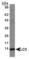
 | Biological Strategies Validation. Western Blot: LC3A Antibody [NBP1-19167] - Total protein from HeLa and Neuro2A cells treated with or without 50 uM chloroquine for 24 hours was separated on a 4-15% gel by SDS-PAGE, transferred to 0.2 um PVDF membrane and blocked in 5% non-fat milk in TBST. The membrane was probed with 2.0 ug/ml anti-LC3A in 1% non-fat milk in TBST and detected with an anti-rabbit HRP secondary antibody using chemiluminescence. Note the detection LC3 II upon chloroquine treatment. |
 | Immunocytochemistry/Immunofluorescence: LC3A Antibody [NBP1-19167] - LC3/MAP1 [NBP1-19167] - LC3 antibody was tested in HeLa cells with Dylight 488 (green). Cells were treated overnight with 50uM chloroquine to induce autophagosome formation. Nuclei and alpha-tubulin were counterstained with DAPI (blue) and Dylight 550 (red). |
 | Immunohistochemistry-Paraffin: LC3A Antibody [NBP1-19167] - LC3/MAP1 [NBP1-19167] - IHC analysis of a formalin fixed and paraffin embedded tissue section of mouse brain using LC3 antibody at 1:300 dilution. The signal was developed using HRP-labelled secondary antibody and DAB reagent, and the sections/nuclei were further counterstained with hematoxylin. Note the diffused cytoplasmic staining of LC3 in all of the cells with highest positivity in various neurons. |
 | Western Blot: LC3/MAP1LC3A Antibody [NBP1-19167] - Human brain lysate. |
 | Immunohistochemistry-Paraffin: LC3/MAP1LC3A Antibody [NBP1-19167] - IHC analysis of a formalin fixed and paraffin embedded tissue section of mouse liver using LC3 antibody at 1:300 dilution. The signal was developed using HRP-labelled secondary antibody and DAB reagent, and the sections/nuclei were further counterstained with hematoxylin. Note the diffused cytoplasmic staining of LC3 in all of the hepatocytes and other liver cells. |
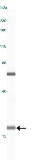 | Simple Western: LC3/MAP1LC3A Antibody [NBP1-19167] - Simple Western lane view shows a specific band for LC3 in 0.5 mg/ml of Neuro2A lysate. This experiment was performed under reducing conditions using the 12-230 kDa separation system. |
カートに商品を
追加しました。
追加しました。
Background
LC3 (microtubule-associated protein light chain 3), the most studied autophagy biomarker, was originally identified as a subunit of microtubule-associated proteins 1A and 1B (MAP1LC3) and was later found to contain similarity to yeast protein Apg8/Aut7/Cvt5. Distributed ubiquitously in eukaryotes, LC3 is expressed as 3 splice variants/isoforms (LC3A, LC3B and LC3C) which undergo post-translational processing, wherein, the unprocessed form of LC3 is proteolytically cleaved by Atg4 protease to form LC3-I with carboxyterminal exposed glycine. During autophagy, this exposed glycine of LC3-I is conjugated by Atg7 (an E1-like activity), Atg3 (an E2-like conjugating activity) and by Atg12-Atg5-Atg16L multimers (E3-like ligase activity) to phosphatidylethanolamine (PE) moiety for generating LC3-II. The lipophilic character of PE group facilitates LC3-II insertion into autophagosomes membranes, and as a result LC3-II is degraded when autophagosomes fuse with lysosomes to form autolysosomes for lysus of intra-autophagosomal components by lysosomal hydrolases. Conversion of LC3I to LC3II when correlated with autophagosome numbers is considered as the best marker of autophagy because LC3-II is the only well-characterized protein which specifically localize to autophagic structures throughout autophagy (from phagophore to lysosomal degradation). LC3 is a great tool in research as autophagy is implicated in numerous physiological/pathological processes including responses to exercise/aging, cancer, metabolic and neurodegenerative disorders, and cardiovascular/pulmonary diseases.カートに商品を
追加しました。
追加しました。
製品情報は掲載時点のものですが、価格表内の価格については随時最新のものに更新されます。お問い合わせいただくタイミングにより製品情報・価格などは変更されている場合があります。
表示価格に、消費税等は含まれていません。一部価格が予告なく変更される場合がありますので、あらかじめご了承下さい。