抗AKT1 [p Ser473]抗体 | Anti-AKT1 [p Ser473] Antibody

掲載日情報:2018/07/09 現在Webページ番号:49379

世界最大級の抗体製品数を取り扱うNovus Biologicals社のAKT1 [p Ser473]に対する抗体(anti-AKT1 [p Ser473] | antibody AKT1 [p Ser473])です。Novus Biologicals社の抗体は数多くの学術論文で使用実績があります。

本製品は研究用です。研究用以外には使用できません。

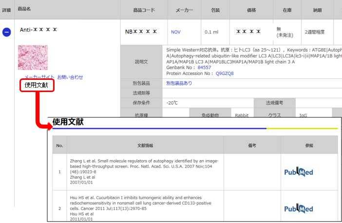
価格表左の「文献」アイコンから、使用文献情報一覧が表示できます。

価格

[在庫・価格 :2025年04月26日 00時00分現在]

※ 表示されている納期は弊社に在庫が無く、取り寄せた場合の納期目安となります。
詳細 商品名
  • 商品コード
  • メーカー
  • 包装
  • 価格
  • 在庫
  • 法規制等
納期 文献数
Anti-Akt1 (pSer473), Phosphorylated, Rabbit-Poly
3~4週間 ※ 表示されている納期は弊社に在庫がなく、取り寄せた場合の目安納期となります。 3
説明文
Keywords:AKT|EC 2.7.11|EC 2.7.11.1|PKBMGC99656|PRKBA|Protein kinase B|Proto-oncogene c-Akt|rac protein kinase alpha|RAC-ALPHA|RAC-alpha serine/threonine-protein kinase|RAC-PK-alpha|RACPKB-ALPHA|v-akt murine thymoma viral oncogene homolog 1
Genbank No: 207
Protein Accession No: P31749
法規制等
保存条件 法規備考
抗原種 Human 免疫動物 Rabbit クラス IgG 標識 Unlabeled
交差性 Human/Mouse/Rat 適用 Dot,ELISA,IC,IF,IHC,Western Blot
クロナリティ Polyclonal フォーマット 性状 Antigen Affinity Purified 吸収処理
掲載カタログ

製品記事
関連記事

[在庫・価格 :2025年04月26日 00時00分現在]

※ 表示されている納期は弊社に在庫が無く、取り寄せた場合の納期目安となります。

Anti-Akt1 (pSer473), Phosphorylated, Rabbit-Poly

文献数: 3

説明文 Keywords:AKT|EC 2.7.11|EC 2.7.11.1|PKBMGC99656|PRKBA|Protein kinase B|Proto-oncogene c-Akt|rac protein kinase alpha|RAC-ALPHA|RAC-alpha serine/threonine-protein kinase|RAC-PK-alpha|RACPKB-ALPHA|v-akt murine thymoma viral oncogene homolog 1
Genbank No: 207
Protein Accession No: P31749
法規制等
保存条件 法規備考
抗原種 Human 免疫動物 Rabbit
交差性 Human/Mouse/Rat 適用 Dot,ELISA,IC,IF,IHC,Western Blot
標識 Unlabeled 性状 Antigen Affinity Purified
吸収処理 クラス IgG
クロナリティ Polyclonal フォーマット
掲載カタログ

製品記事
関連記事


目次に戻る

Image

Western Blot: Akt1 [p Ser473] Antibody [NB600-590] - Lane 1: AKT1 Recombinant Protein. Lane 2: AKT1 Mutant Human Recombinant Protein. Lane 3: AKT1 (phosphatase treated) Human Recombinant Protein. Load: 50 ng per lane. Primary antibody: AKT pS473 antibody at 1:1,000 for overnight at 4C. Secondary antibody: Peroxidase rabbit secondary antibody at 1:40,000 for 30 min at RT. Block: Blocking Buffer for Fluorescent Western Blotting for 30 min at RT. Predicted/Observed size: 56 kDa, 56 kDa for Gli-2. Other band(s): Gli-2 splice variants and isoforms.
Immunocytochemistry/Immunofluorescence: Akt1 [p Ser473] Antibody [NB600-590] - Tissue: cardiomyocytes infected with adenovirus expressing with wild-type AKT. Fixation: 0.5% PFA. Antigen retrieval: not required. Primary antibody: AKT pS473 antibody at 1:40 for 1 h at RT. Secondary antibody: texas-red conjugated rabbit secondary antibody at 1:10,000 for 45 min at RT. Localization: AKT pS473 is nuclear. Staining: AKT pS473 as green fluorescent signal with texas-red conjugated phalloidin (red) to label filamentous actin.
Immunohistochemistry: Akt1 [p Ser473] Antibody [NB600-590] - Tissue: human breast carcinoma. Fixation: formalin fixed paraffin embedded. Antigen retrieval: not required. Primary antibody: Akt pS473 antibody at 100 dilution for 1 h at RT. Secondary antibody: Dako's Techmate streptavidin-biotin reagents at 1:10,000 for 45 min at RT. Localization: Akt pS473 is nuclear and occasionally cytoplasmic.
Western Blot: Akt1 [p Ser473] Antibody [NB600-590] - Lane 1: nuclear extract from cells infected with adenovirus expressing nuclear-targeted AKT kinase. Load: 35 ug per lane. Primary antibody: AKT pS473 antibody at 1:200 dilution for overnight at 4C. Secondary antibody: IRDye800 rabbit secondary antibody at 1:10,000 for 45 min at RT. Block: 5% BLOTTO overnight at 4C. Predicted/Observed size: 56 kDa for AKT pS473. Other band(s): unspecific.
Immunohistochemistry-Paraffin: Akt1 [p Ser473] Antibody [NB600-590] - Rabbit anti-AKT pS473 was used at a 1:100 dilution to detect AKT in human breast tumor tissue. The staining is much stronger than the weak basal level of phosphorylation in normal breast.Signal was developed using Dako's Techmate streptavidinbiotin reagents. Tissue was formalin-fixed and paraffin embedded.
Immunohistochemistry-Paraffin: Akt1 [p Ser473] Antibody [NB600-590] - Rabbit used at a 1:100 dilution to detect AKT in normal human breast tissue. AKT is weakly phosphorylated in normal tissue in the breast. The phosphorylated AKT is clearly localized in the cytoplasm.Signal was developed using Dako's Techmate streptavidin-biotin reagents. Tissue was formalin-fixed and paraffin embedded.


目次に戻る

Background

The serine threonine protein kinase encoded by the AKT1 gene is catalytically inactive in serum starved primary and immortalized fibroblasts. AKT1 and the related AKT2 are activated by platelet derived growth factor. The activation is rapid and specific. In the developing nervous system AKT is a critical mediator of growth factor induced neuronal survival. Survival factors can suppress apoptosis in a transcription independent manner by activating the serine/threonine kinase AKT1, which then phosphorylates and inactivates components of the apoptotic machinery. Multiple alternatively spliced transcript variants have been found for this gene (referenced from entrez gene).

目次に戻る

お問い合わせ先

(テクニカルサポート 試薬担当)

reagent@funakoshi.co.jp

製品情報は掲載時点のものですが、価格表内の価格については随時最新のものに更新されます。お問い合わせいただくタイミングにより製品情報・価格などは変更されている場合があります。
表示価格に、消費税等は含まれていません。一部価格が予告なく変更される場合がありますので、あらかじめご了承下さい。