抗GFP抗体 | Anti-GFP Antibody
掲載日情報:2018/07/09 現在Webページ番号:49265
世界最大級の抗体製品数を取り扱うNovus Biologicals社のGFPに対する抗体(anti-GFP | antibody GFP)です。Novus Biologicals社の抗体は数多くの学術論文で使用実績があります。
※本製品は研究用です。研究用以外には使用できません。
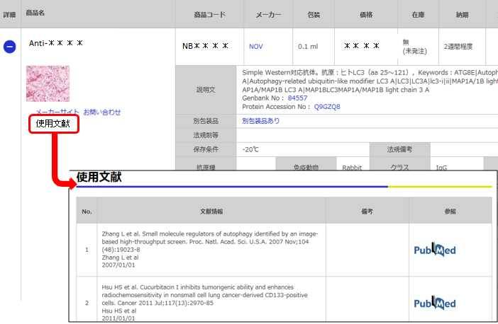
価格表左の「文献」アイコンから、使用文献情報一覧が表示できます。


カートに商品を
追加しました。
追加しました。
価格
[在庫・価格 :2026年02月09日 00時00分現在]
※ 表示されている納期は弊社に在庫が無く、取り寄せた場合の納期目安となります。
| 詳細 | 商品名 |
|
文献数 | ||||||||||||||||||||||||||||||||||||||||||||||||||||||||||||||||||||||||||
|---|---|---|---|---|---|---|---|---|---|---|---|---|---|---|---|---|---|---|---|---|---|---|---|---|---|---|---|---|---|---|---|---|---|---|---|---|---|---|---|---|---|---|---|---|---|---|---|---|---|---|---|---|---|---|---|---|---|---|---|---|---|---|---|---|---|---|---|---|---|---|---|---|---|---|---|---|---|
|
Anti-GFP, Rabbit-Poly |
|
49 | |||||||||||||||||||||||||||||||||||||||||||||||||||||||||||||||||||||||||||
|
|||||||||||||||||||||||||||||||||||||||||||||||||||||||||||||||||||||||||||||
[在庫・価格 :2026年02月09日 00時00分現在]
※ 表示されている納期は弊社に在庫が無く、取り寄せた場合の納期目安となります。
Anti-GFP, Rabbit-Poly
文献数: 49
- 商品コード:NB600-303
- メーカー:NOV
- 包装:0.05ml
- 価格:¥96,000
- 在庫:無(未発注)
- 納期:3~4週間 ※※ 表示されている納期は弊社に在庫がなく、取り寄せた場合の目安納期となります。
- 法規制等:
| 説明文 | レビューあり。Keywords:Enhanced Green Fluorescent Protein|Green Fluorescent Protein|green fluorescent protein (gfp) Protein Accession No: P42212 |
||
|---|---|---|---|
| 法規制等 | |||
| 保存条件 | 法規備考 | ||
| 抗原種 | 免疫動物 | Rabbit | |
| 交差性 | Jellyfish | 適用 | ChIP,ELISA,Electron Microscopy,FCM,IC,IF,IHC,IP,Simple Western,Western Blot |
| 標識 | Unlabeled | 性状 | Antigen Affinity Purified |
| 吸収処理 | クラス | IgG | |
| クロナリティ | Polyclonal | フォーマット | |
| 掲載カタログ |
|
||
| 製品記事 | 抗GFP抗体 | Anti-GFP Antibody |
||
| 関連記事 | |||
カートに商品を
追加しました。
追加しました。
Image
カートに商品を
追加しました。
追加しました。
Background
A highly versatile antibody that gives a stronger signal than other anti-GFP antibodies available. On Western blot the antibody detects the GFP fraction from cell extracts expressing recombinant GFP fusion proteins and has also been shown to be useful on mouse sections fixed with formalin. In immunocytochemistry, the antibody gives a very good signal on recombinant YES-GFP chimeras expressed in COS cells. It is routinely used in immunoprecipitation (IP) and IP-Western protocols and has been used successfully in HRP-immunohistochemistry (1:200) on whole-mount mouse embryos.カートに商品を
追加しました。
追加しました。
製品情報は掲載時点のものですが、価格表内の価格については随時最新のものに更新されます。お問い合わせいただくタイミングにより製品情報・価格などは変更されている場合があります。
表示価格に、消費税等は含まれていません。一部価格が予告なく変更される場合がありますので、あらかじめご了承下さい。


