
抗LC3B抗体 | Anti-LC3B Antibody
掲載日情報:2018/07/09 現在Webページ番号:49178

世界最大級の抗体製品数を取り扱うNovus Biologicals社のLC3Bに対する抗体(anti-LC3B | antibody LC3B)です。Novus Biologicals社の抗体は数多くの学術論文で使用実績があります。
※本製品は研究用です。研究用以外には使用できません。
価格表左の「文献」アイコンから、使用文献情報一覧が表示できます。

カートに商品を
追加しました。
追加しました。
価格
[在庫・価格 :2026年07月15日 20時55分現在]
※ 表示されている納期は弊社に在庫が無く、取り寄せた場合の納期目安となります。
| 詳細 | 商品名 |
|
文献数 | ||||||||||||||||||||||||||||||||||||||||||||||||||||||||||||||||||||||||||||||||||
|---|---|---|---|---|---|---|---|---|---|---|---|---|---|---|---|---|---|---|---|---|---|---|---|---|---|---|---|---|---|---|---|---|---|---|---|---|---|---|---|---|---|---|---|---|---|---|---|---|---|---|---|---|---|---|---|---|---|---|---|---|---|---|---|---|---|---|---|---|---|---|---|---|---|---|---|---|---|---|---|---|---|---|---|---|---|
|
Anti-LC3B, Rabbit-Poly |
|
319 | |||||||||||||||||||||||||||||||||||||||||||||||||||||||||||||||||||||||||||||||||||
|
|||||||||||||||||||||||||||||||||||||||||||||||||||||||||||||||||||||||||||||||||||||


[在庫・価格 :2026年07月15日 20時55分現在]
※ 表示されている納期は弊社に在庫が無く、取り寄せた場合の納期目安となります。
Anti-LC3B, Rabbit-Poly
文献数: 319
- 商品コード:NB600-1384
- メーカー:NOV
- 包装:0.1ml
- 価格:¥115,000
- 在庫:1個
- 納期:3~4週間 ※※ 表示されている納期は弊社に在庫がなく、取り寄せた場合の目安納期となります。
- 法規制等:
| 説明文 | レビューあり。Simple Western対応抗体。抗原:ヒトLC3B(N末端),Keywords:ATG8F|Autophagy-related protein LC3 B|Autophagy-related ubiquitin-like modifier LC3 B|LC3B|lc3b autophagy marker|lc3-i|ii|MAP1 light chain 3-like protein 2|MAP1A/1BLC3|MAP1A/MAP1B LC3 B|map1lc3b|microtubule associated protein 3 b Genbank No: 81631 Protein Accession No: Q9GZQ8 |
||||||
|---|---|---|---|---|---|---|---|
| 別包装品 | 別包装品あり | ||||||
| 法規制等 | |||||||
| 保存条件 | -20℃ | 法規備考 | |||||
| 抗原種 | 免疫動物 | Rabbit | |||||
| 交差性 | Bacteria/Bovine/Canine/Human/Mouse/Porcine/Primate/Rat/Yeast/Zebrafish | 適用 | Electron Microscopy,FCM,IC,IF,IHC,IP,Immunoblotting,Simple Western,Western Blot | ||||
| 標識 | Unlabeled | 性状 | Antigen Affinity Purified | ||||
| 吸収処理 | クラス | IgG | |||||
| クロナリティ | Polyclonal | フォーマット | |||||
| 掲載カタログ |
ニュース2024年8月合併号 p.27
|
||||||
| 製品記事 | 抗LC3B抗体 | Anti- LC3B antibody 抗LC3抗体/抗LC3関連転写因子抗体 抗LC3抗体 オートファジーとLC3研究用試薬 オルガネラ(細胞小器官)マーカー抗体 |
||||||
| 関連記事 | |||||||
カートに商品を
追加しました。
追加しました。
Image
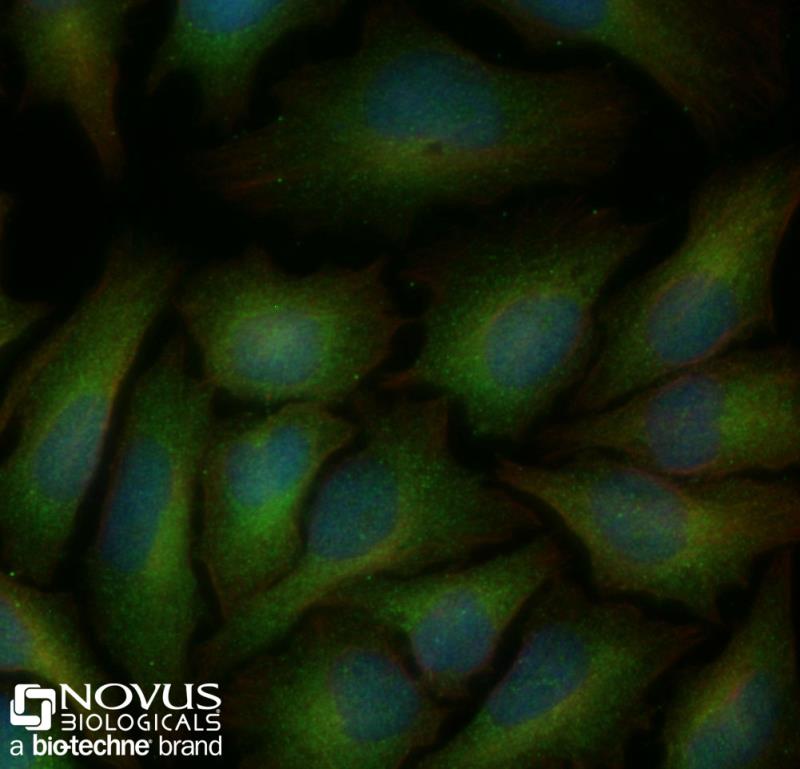
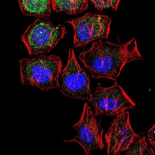 | Immunocytochemistry/Immunofluorescence: LC3B Antibody [NB600-1384] - LC3B/MAP1 [NB600-1384] - IF Confocal analysis of HeLa cells using LC3B antibody (NB600-1384, 1:5). An Alexa Fluor 488-conjugated Goat to rabbit IgG was used as secondary antibody (green). Actin filaments were labeled with Alexa Fluor 568 phalloidin (red). DAPI was used to stain the cell nuclei (blue). |
 | Immunocytochemistry/Immunofluorescence: LC3B Antibody [NB600-1384] - LC3B/MAP1 [NB600-1384] - LC3 antibody was tested in 50uM Chloroquine treated HeLa cells with Dylight 488 (green). Nuclei and alpha-tubulin were counterstained with DAPI (blue) and Dylight 550 (red). An antibody concentration of 0.1 ug/ml was used. Image objective 40x. |
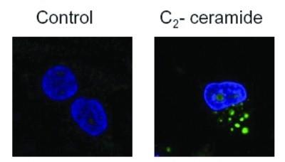 | Biological Strategies Validation. Immunocytochemistry/Immunofluorescence: LC3B Antibody [NB600-1384] - LC3B/MAP1 [NB600-1384] - Analysis using the HRP conjugate of NB600-1384. Staining of treated U373-MG cells using NB600-1384. The nuclei were stained with DAPI. |
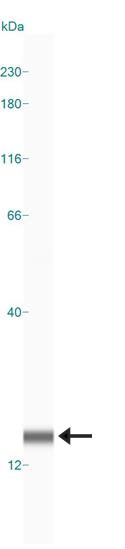
 | Western Blot: LC3B Antibody [NB600-1384] - LC3B/MAP1 [NB600-1384] - analysis of LC3B in IB3-1 whole cell lysate using anti-LC3B antibody. Image from verified customer review. |
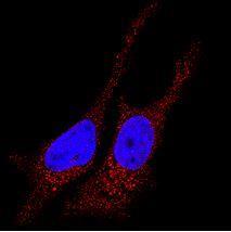 | Immunocytochemistry/Immunofluorescence: LC3B Antibody [NB600-1384] - LC3B/MAP1 [NB600-1384] - analysis of LC3B in HeLa cells using anti-LC3B antibody (red). Nuclei were counterstained with DAPI (blue). |
 | Biological Strategies Validation. Western Blot: LC3B Antibody [NB600-1384] - LC3B/MAP1 [NB600-1384] - Detection of LC3B in treated U87-MG (human glioblastoma astrocytoma) lysates. |
 | Biological Strategies Validation. Western Blot: LC3B Antibody [NB600-1384] - Aldosterone (ALD) treated SV-K1 cell. Western blot shows lysates of SV-K1 cell line untreated (-) or treated with aldosterone. This image was submitted via customer review. |
 | Immunocytochemistry/Immunofluorescence: LC3B Antibody [NB600-1384] - Untreated HeLa cells were fixed for 10 minutes using 10% formalin and then permeabilized for 5 minutes using 1X PBS + 0.05% Triton-X100. The cells were incubated with anti-LC3B at 2 ug/ml overnight at 4C and detected with an anti-rabbit Dylight 488 (Green) at a 1:500 dilution. Alpha tubulin (DM1A) NB100-690 was used as a co-stain at a 1:1000 dilution and detected with an anti-mouse Dylight 550 (Red) at a 1:500 dilution. Nuclei were counterstained with DAPI (Blue). Cells were imaged using a 40X objective. |
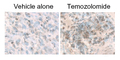
 | Biological Strategies Validation. Immunohistochemistry: LC3B Antibody [NB600-1384] - LC3B/MAP1 [NB600-1384] - Staining of treated U373-MG (human glioblastoma. |
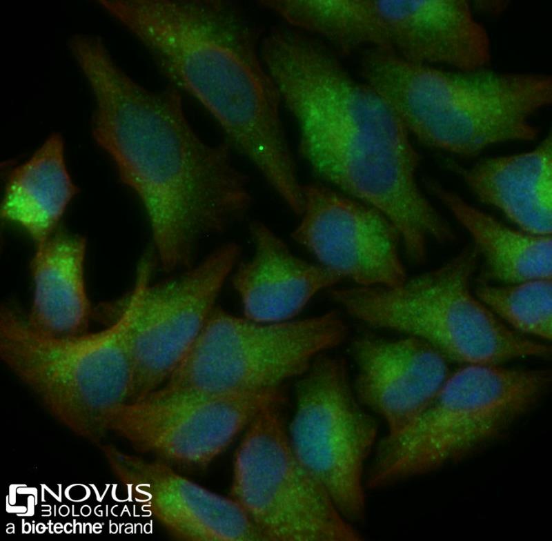 | Immunocytochemistry/Immunofluorescence: LC3B Antibody [NB600-1384] - HeLa cells were treated with 50uM CQ overnight, fixed for 10 minutes using 10% formalin and then permeabilized for 5 minutes using 1X PBS + 0.05% Triton-X100. The cells were incubated with anti-LC3B at 2 ug/ml overnight at 4C and detected with an anti-rabbit Dylight 488 (Green) at a 1:500 dilution. Alpha tubulin (DM1A) NB100-690 was used as a co-stain at a 1:1000 dilution and detected with an anti-mouse Dylight 550 (Red) at a 1:500 dilution. Nuclei were counterstained with DAPI (Blue). Cells were imaged using a 40X objective. |
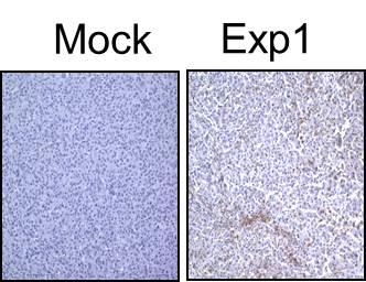 | Immunohistochemistry-Paraffin: LC3B/MAP1LC3B Antibody [NB600-1384] - analysis of U87MG glioma xenografts using anti-LC3B antibdoy. Image from verified customer review. |
 | Immunohistochemistry: LC3B/MAP1LC3B Antibody [NB600-1384] - LC3B staining in treated U87-MG cultured & subcutaneous tumors. |
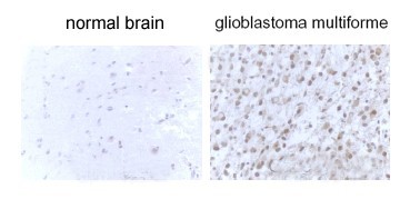 | Immunohistochemistry: LC3B/MAP1LC3B Antibody [NB600-1384] - LC3B staining in gliobastoma multiform tissue. |
 | Simple Western: LC3B/MAP1LC3B Antibody [NB600-1384] - Simple Western lane view shows a specific band for LC3B in 0.5 mg/ml of Neuro2A lysate. This experiment was performed under reducing conditions using the 12-230 kDa separation system. |
カートに商品を
追加しました。
追加しました。
Background
Autophagy is a process of intracellular bulk degradation in which cytoplasmic components, including organelles, are sequestered within double-membrane vesicles that deliver the contents to the lysosome/vacuole for degradation. During macroautophagy, the sequestering vesicles, termed autophagosomes, fuse with the lysosome or vacuole resulting in the delivery of an inner vesicle (autophagic body) into the lumen of the degradative compartment. There are 16 proteins participating in the autophagy pathway in humans. The autophagy protein LC3, a mammalian homologue of Atg8, was originally identified as microtubule-associated protein 1 light chain 3. It is a component of both the MAP1A and MAP1B microtubule-binding domains and the heavy-chain independent regulation of LC3 expression may modify MAP1 microtubule-binding activity during development. LC3 is the only known mammalian protein identified that stably associates with the autophagosome membranes. LC3-I is cytosolic and LC3-II is membrane bound and enriched in the autophagic vacuole fraction. The detection of LC3-I to LC3-II conversion is a useful and sensitive marker for distinguishing autophagy in mammalian cells.カートに商品を
追加しました。
追加しました。
製品情報は掲載時点のものですが、価格表内の価格については随時最新のものに更新されます。お問い合わせいただくタイミングにより製品情報・価格などは変更されている場合があります。
表示価格に、消費税等は含まれていません。一部価格が予告なく変更される場合がありますので、あらかじめご了承下さい。