
抗CD34 (MEC 14.7)抗体 | Anti-CD34 (MEC 14.7) Antibody
掲載日情報:2018/07/09 現在Webページ番号:49130

世界最大級の抗体製品数を取り扱うNovus Biologicals社のCD34 (MEC 14.7)に対する抗体(anti-CD34 (MEC 14.7) | antibody CD34 (MEC 14.7))です。Novus Biologicals社の抗体は数多くの学術論文で使用実績があります。
※本製品は研究用です。研究用以外には使用できません。

追加しました。
価格
[在庫・価格 :2026年04月18日 11時55分現在]
| 詳細 | 商品名 |
|
文献数 | ||||||||||||||||||||||||||||||||||||||||||||||||||||||||||||||||||||||||||||||||||
|---|---|---|---|---|---|---|---|---|---|---|---|---|---|---|---|---|---|---|---|---|---|---|---|---|---|---|---|---|---|---|---|---|---|---|---|---|---|---|---|---|---|---|---|---|---|---|---|---|---|---|---|---|---|---|---|---|---|---|---|---|---|---|---|---|---|---|---|---|---|---|---|---|---|---|---|---|---|---|---|---|---|---|---|---|---|
|
Anti-CD34, Rat-Mono(MEC 14.7) |
|
22 | |||||||||||||||||||||||||||||||||||||||||||||||||||||||||||||||||||||||||||||||||||
|
|||||||||||||||||||||||||||||||||||||||||||||||||||||||||||||||||||||||||||||||||||||


[在庫・価格 :2026年04月18日 11時55分現在]
Anti-CD34, Rat-Mono(MEC 14.7)
文献数: 22
- 商品コード:NB600-1071
- メーカー:NOV
- 包装:0.1ml
- 価格:¥110,000
- 在庫:無(未発注)
- 納期:3~4週間 ※※ 表示されている納期は弊社に在庫がなく、取り寄せた場合の目安納期となります。
- 法規制等:
| 説明文 | Keywords:CD34 antigenhematopoietic progenitor cell antigen CD34|CD34 molecule クローン:MEC 14.7 Genbank No: 947 |
||||||
|---|---|---|---|---|---|---|---|
| 別包装品 | 別包装品あり | ||||||
| 法規制等 | |||||||
| 保存条件 | -20℃ | 法規備考 | |||||
| 抗原種 | 免疫動物 | Rat | |||||
| 交差性 | Mouse | 適用 | ELISA,FCM,IC,IF,IHC,IP,Western Blot | ||||
| 標識 | Unlabeled | 性状 | Protein A/G Affinity Purified | ||||
| 吸収処理 | クラス | IgG | |||||
| クロナリティ | Monoclonal | フォーマット | |||||
| 掲載カタログ |
|
||||||
| 製品記事 | 抗CD34 (MEC 14.7)抗体 | Anti- CD34 (MEC 14.7) antibody |
||||||
| 関連記事 | |||||||
追加しました。
Image
-Immunocytochemistry-Immunofluorescence-NB600-1071-img0003.jpg) | Immunocytochemistry/Immunofluorescence: CD34 Antibody (MEC 14.7) [NB600-1071] - CD34 antibody was tested in WEHI-3 cells with Dylight 488 (green). Nuclei and alpha-tubulin were counterstained with DAPI (blue) and Dylight 550 (red). |
-Immunohistochemistry-Paraffin-NB600-1071-img0014.jpg) | Immunohistochemistry-Paraffin: CD34 Antibody (MEC 14.7) [NB600-1071] - IHC analysis of a formalin fixed and paraffin embedded tissue section of mouse small intestine using rat anti-mouse CD34 (clone MEC 14.7) at 1:100 dilution. The signal was developed using HRP-conjugated anti-rat secondary with DAB reagent which followed counterstaining of nuclei using hematoxylin. This antibody specifically labelled the endothelial cells in blood vessels located primarily in the sub-mucosa, and of that of the mucosa muscularis and the mucosal lacteal. |
-Flow Cytometry-NB600-1071-img0002.jpg) | Flow Cytometry: CD34 Antibody (MEC 14.7) [NB600-1071] - CD34 (MEC 14.7) antibody was tested at 1:250 in WEHI-3 cells with DyLight 488 (green) alongside a matched isotype control (black). |
-Immunohistochemistry-Paraffin-NB600-1071-img0005.jpg) | Immunohistochemistry-Paraffin: CD34 Antibody (MEC 14.7) [NB600-1071] - IHC-P analysis of CD34 on human renal cancer xenograft using CD34 antibody (clone MEC 14.7). The antibody detected the endothelial cells in the tumor vasculature which are originating from the host (mouse). |
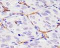
-Immunohistochemistry-Paraffin-NB600-1071-img0006.jpg) | Immunohistochemistry-Paraffin: CD34 Antibody (MEC 14.7) [NB600-1071] - IHC analysis of a human renal cancer tissue section using CD34 antibody (clone MEC 14.7) at 1:100 dilution. The antibody did not detect human CD34 which is an expected outcome for this clone. This section was included in NB600-1071's IHC validation testing as a negative control. |
-Immunohistochemistry-Paraffin-NB600-1071-img0009.jpg) | Immunohistochemistry-Paraffin: CD34 Antibody (MEC 14.7) [NB600-1071] - IHC analysis of a formalin fixed and paraffin embedded tissue section of human renal cancer xenograft using CD34 antibody clone MEC 14.7 at 1:100 dilution. The signal was developed using HRP-conjugated secondary antibody and DAB reagent followed by counterstaining of nuclei with hematoxylin. Notably, the antibody specifically stained the endothelial cells of the tumor vasculature which is originating from the host animal cells (mouse). This observation was verified by including a negative control in this validation testing (see the IHC image of human renal cancer tissue section with negative signal for CD34). |
-Immunohistochemistry-Paraffin-NB600-1071-img0010.jpg) | Immunohistochemistry-Paraffin: CD34 Antibody (MEC 14.7) [NB600-1071] - IHC analysis of a formalin fixed and paraffin embedded tissue section of human renal cancer xenograft using CD34 antibody (clone MEC 14.7) at 1:100 dilution. The staining was detected using HRP-conjugated secondary antibody and DAB reagent followed by counterstaining of nuclei with hematoxylin. Note that this murine CD34 specific antibody stained only the endothelial cells of the tumor vasculature which is originating from the host animal cells (mouse). This observation was verified by including a negative control in this validation testing (see the IHC image of human renal cancer tissue section with negative signal for CD34). |
-Immunohistochemistry-Paraffin-NB600-1071-img0012.jpg) | Immunohistochemistry-Paraffin: CD34 Antibody (MEC 14.7) [NB600-1071] - IHC analysis of a formalin fixed and paraffin embedded tissue section of mouse colon using rat anti-mouse CD34 (clone MEC 14.7) at 1:100 dilution. The signal was developed using HRP-conjugated anti-rat secondary with DAB reagent which followed counterstaining of nuclei using hematoxylin. The antibody specifically generated a staining of the endothelial cells in blood vessels of the colon. |
-Immunohistochemistry-Paraffin-NB600-1071-img0013.jpg) | Immunohistochemistry-Paraffin: CD34 Antibody (MEC 14.7) [NB600-1071] - IHC analysis of a formalin fixed and paraffin embedded tissue section of mouse heart using rat anti-mouse CD34 (clone MEC 14.7) at 1:100 dilution. The signal was developed using HRP-conjugated anti-rat secondary with DAB reagent which followed counterstaining of nuclei using hematoxylin. The antibody specifically generated a staining of the endothelial cells in blood vessels of the heart tissue. |
追加しました。
Background
CD34 Antibody MEC 14.7 (NB600-1071) is a useful reagent for identification and characterization of capillary endothelial cells. Furthermore, it can be used for isolation and characterization of hematopoietic progenitor cells. In Western blot, the reported molecular mass of CD34 is ranging from 100 to 120 kDa, apart from the HEV form (Sgp90) which is about 90 kDa. MEC14.7 can recognize a band of about 100 kDa from the H5V line but a band of 75 to 85 kDa from normal tissue. Since different patterns of glycosylation of the molecule are seen with different cell types, this might explain the wide range of CD34 molecular mass (from 75-85 kDa to 120 kD).追加しました。
製品情報は掲載時点のものですが、価格表内の価格については随時最新のものに更新されます。お問い合わせいただくタイミングにより製品情報・価格などは変更されている場合があります。
表示価格に、消費税等は含まれていません。一部価格が予告なく変更される場合がありますので、あらかじめご了承下さい。